Histopathological Comparison among Biolimus, Zotarolimus and Everolimus-Eluting Stents in Porcine Coronary Restenosis Model
8
0
0
전체 글
(2) Kyung Seob Lim, et al. 745. stents in the porcine coronary restenosis model.. Subjects and Methods Animal preparation and stent implantation The animal study was approved by the Ethics Committee of Chonnam National University Medical School and Chonnam National University Hospital (CNU IACUC-H-2010-18), and conformed to the Guidelines for the Care and Use of Laboratory Animals published by the US National Institutes of Health (NIH Publication No. 85-23, revised 1996). Study animals were castrated male pigs weighing 2025 kg. To prevent acute thrombosis after stenting, premedication with aspirin 100 mg and clopidogrel 75 mg per day was applied for 5 days before the procedure. On the day of the procedure, pigs were anesthetized with zolazepam and tiletamine (2.5 mg/kg, Zoletil50®, Virbac, Caros, France), xylazine (3 mg/kg, Rompun®, Bayer AG, Leverkusen, Germany) and azaperone (6 mg/kg, Stresnil®, Janssen-Cilag, Neuss, Germany). They received supplemental oxygen continuously through oxygen masks. Subcutaneous 2% lidocaine at the cutdown site was administered, the left carotid artery was surgically exposed, and a 7 Fr sheath was inserted. Continuous hemodynamic and surface electrocardiographic monitoring was maintained throughout the procedure. Then, 5000 units of heparin was administered intravenously as a bolus prior to the procedure, the target coronary artery was engaged using standard 7 Fr guide catheters, and control angiograms of both coronary arteries were performed using a nonionic contrast agent in two orthogonal views. The stent was deployed by inflating the balloon and the resulting stent-to-artery ratio was 1.3 : 1. Coronary angiograms were obtained immediately after stent implantation. Thereafter, all equipment was removed and the carotid artery was ligated. Four weeks after stenting, the animals underwent follow-up angiography in the same orthogonal views as before death with 20 mL of a potassium chloride intracoronary injection. The hearts were removed, and the coronary arteries were pres-. sure-perfusion fixed at 110 mm Hg in 10% neutral buffered formalin overnight. Arteries were step-sectioned, processed routinely for light microscopy, and stained for histological analysis. Study groups The pigs were randomly divided into 3 groups (Table 1): group 1 {biolimus A9-eluting stents (BES), BioMatrix®, Biosensors Interventional Technologies Pte Ltd., Singapore, 3.0×18 mm, n=10}, group 2 {zotarolimus-eluting stents (ZES), Endeavor Resolute®, Medtronic CardioVascular, Minneapolis, MN, USA, 3.0×18 mm, n=10}, and group 3 {everolimus-eluting stents (EES), Promus®, Boston Scientific, Natick, MA, USA, 3.0×18 mm, n=10}. A total of 15 pigs were used in this study (15 pigs, 30 coronary arteries, 10 coronary arteries in each group). A BES, a ZES, and an EES were implanted in the left anterior descending artery and left circumflex artery in a randomized manner in each pig. Histopathology and immunohistochemistry analysis Histopathologic evaluations of each artery were performed by an experienced cardiovascular pathologist. The specimens were embedded, and sections of 50 to 100 μm in thickness were obtained at about 1 mm distances apart, and stained with Hematoxylin-Eosin (Fig. 1) and Carstairs’ (Fig. 2) for histological analysis. Measurements of the histopathologic sections were performed using a calibrated microscope, digital video imaging system, and microcomputer program (Visus 2000 Visual Image Analysis System, IMT Tech, CA, USA). Borders were manually traced for the lumen area, the area circumscribed by the internal elastic lamina (IEL), and the innermost border of the external elastic lamina (external elastic lamina area). Morphometric analysis of the neointimal area for a given vessel was calculated as the measured IEL area minus the lumen area. The measurements were made on five cross-sections from the proximal and distal ends, and the three midpoints of each stented segment. Histopathologic stenosis was calculated as 100×{1-(lesion lumen area/lesion IEL area)}.10) Immunohistochemistry (IHC) was conducted through standard procedures, as previously de-. Table 1. Coronary artery morphometric measurements in stented porcine coronary arteries Variables Injury score. BES (n=10). ZES (n=10). EES (n=10). 1.4±0.37. 1.4±0.35. 1.5±0.36. p p=NS. IEL (mm2). 4.3±0.53. 5.1±0.55. 4.4±0.53. p<0.001. Lumen area (mm2). 2.5±0.93. 2.3±1.14. 1.7±1.22. p<0.001. Neointima area (mm2). 1.8±1.03. 2.8±1.00. 2.8±1.23. p<0.001. 40.7±20.80. 55.4±21.23. 64.0±26.00. p<0.001. Fibrin score. 1.7±0.41. 2.0±0.39. 1.8±0.76. p<0.001. Inflammation score. 1.4±0.72. 1.6±0.76. 2.1±0.90. p<0.001. % area stenosis (%). BES: biolimus A9-eluting stents, ZES: zotarolimus-eluting stents, EES: everolimus-eluting stent, IEL: internal elastic lamina, NS: not significant. www.e-kcj.org. http://dx.doi.org/10.4070/kcj.2013.43.11.744.
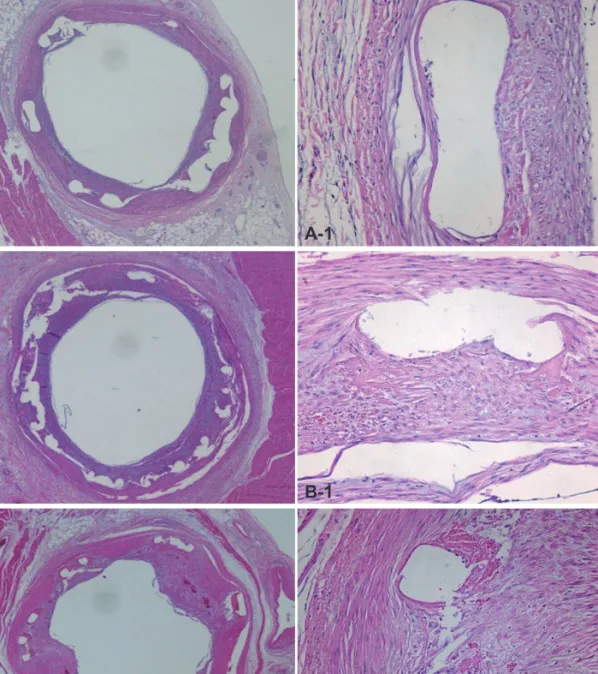
(3) 746 Limus Derivates Eluting Stents. scribed.11) Anti-smooth muscle actin monoclonal antibody (Sigma Aldrich, St. Louis, MO, USA) was used. IHC specimens were analyzed by fluorescence microscopy and digital photography. All histology and IHC results were interpreted by two independent pathologists in a blind manner. Evaluation of the arterial injury The arterial injury at each strut site was determined by the anatomic structures penetrated by each strut. A numeric value was assigned, as previously described by Schwartz et al.:10) 0=no injury; 1= break in the internal elastic membrane; 2=perforation of the media; 3=perforation of the external elastic membrane to the adven-. titia. The average injury score for each segment was calculated by dividing the sum of the injury scores by the total number of struts at the examined section. Evaluation of inflammation scores, neointimal reaction, and fibrin score With regard to the inflammation score for each individual strut, the grading was as follows: 0=no inflammatory cells surrounding the strut; 1=light, noncircumferential lymphohistiocytic infiltrate surrounding the strut; 2=localized, moderate to dense cellular aggregate surrounding the strut noncircumferentially; 3=circumferential dense lymphohistiocytic cell infiltration of the strut. The in-. A . A-1 . B . B-1 . C . C-1 . Fig. 1. Representative images of H&E staining after 4 weeks of stenting. Specimen BES implanted (A: ×20, A-1: ×200), ZES implanted (B: ×20, B-1: ×200), and EES implanted (C: ×20, C-1: ×200). Inflammatory reaction was more severe in the EES stented artery compared to BES and ZES. BES: biolimus A9eluting stents, ZES: zotarolimus-eluting stents, EES: everolimus-eluting stents. http://dx.doi.org/10.4070/kcj.2013.43.11.744. www.e-kcj.org.
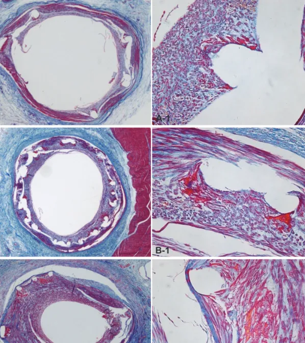
(4) Kyung Seob Lim, et al. 747. flammation score for each cross section was calculated by dividing the sum of the individual inflammation scores by the total number of struts at the examined section.12) Ordinal data for fibrin were collected on each stent section using a scale of 0 to 3, as previously reported.13) Statistical analysis Statistical analysis was performed with the aid of commercially available software {Statistical Package for the Social Sciences (SPSS) Version 15, SPSS Inc., Chicago, IL, USA}. The data were presented as mean value±SD. An unpaired Student’s t-test was used for the comparison of each of the stent groups. Analysis of variance was. used to make comparisons of the three stents groups. A value of p<0.05 was considered statistically significant.. Results Analysis after stenting Two stents were placed for two coronary arteries per swine. A total of thirty stents, including ten BES, ten ZES, and ten EES, were placed in the proximal left anterior descending and proximal circumflex artery for fifteen swine. The mortality rate in this study was zero. There was no significant difference in the stent-to-artery ratio among the three stent groups.. A . A-1 . B . B-1 . C . C-1 . Fig. 2. The Carstair fibrin stain of the low-power fields (magnitude, ×20, ×200) of fibrin infiltration in BES implanted (A and A-1), ZES implanted (B and B-1), and EES implanted (C and C-1). Fibrin deposition surrounding the stent struts was higher in ZES than in BES and EES cases. BES: biolimus A9-eluting stents, ZES: zotarolimus-eluting stents, EES: everolimus-eluting stents. www.e-kcj.org. http://dx.doi.org/10.4070/kcj.2013.43.11.744.
(5) 748 Limus Derivates Eluting Stents. A . B . C . Fig. 3. Representative images of immunohistochemistry using anti-smooth muscle actin monoclonal antibody in the neointima tissue. Immunofluorescence staining showing an expression of α-smooth muscle actin (bright red positive cells, ×200, A: BES, B: ZES, C: EES). BES: biolimus A9-eluting stents, ZES: zotarolimus-eluting stents, EES: everolimus-eluting stents.. Histopathological and immunohistochemistry analysis To exam the characteristics of smooth muscle cells (SMC) in neointima tissue, the stented coronary artery was stained by anti-SMC antibody. Vascular SMC were major components of neointima formation after stenting in all groups (Fig. 3). There was no statistically significant difference in the injury score among the three groups (1.4±0.37 in the BES group vs. 1.4±0.35 in the ZES group vs. 1.5±0.36 in the EES group, p=not significant). There was a statistically significant difference in the IEL among the three groups (4.3±0.53 mm2 in the BES group vs. 5.1±0.55 mm2 in the ZES group vs. 4.4±0.53 mm2 in the EES group, p<0.0001). There was a statistically significant difference in the lumen area among the three groups (2.5±0.93 mm2 in the BES group vs. 2.3±1.14 mm2 in the ZES group vs. 1.7±1.22 mm2 in the EES group, p<0.001). There was a statistically significant difference in the neointima area among the three groups (1.8±1.03 mm2 in the BES group vs. 2.8±1.00 mm2 in the ZES group vs. 2.8±1.23 mm2 in the EES group, p<0.0001). There was a statistically significant difference in percentage area stenosis among the three groups (40.7±20.80% in the BES group vs. 55.4±21.23% in the ZES group vs. 64.0±26.00% in the EES group, p<0.0001). There was a statistically significant difference in the fibrin score among the three groups (1.7±0.41 in the BES group vs. 2.0±0.39 in the ZES group vs. 1.8±0.76 in the EES, p<0.001). There was a statistically significant difference in the inflammation score among the three groups (1.4±0.72 in the BES group vs. 1.6±0.76 in the ZES group vs. 2.1±0.90 in the EES group, p<0.001) (Table 1, Fig. 4).. Discussion This study was conducted in order to compare the histopathological differentiation of the BES, ZES, and EES in the porcine coronary restenosis model. Our study demonstrated that BES was more effective in reducing neointima proliferation compared to ZES and. http://dx.doi.org/10.4070/kcj.2013.43.11.744. EES. In the fibrin score, which indicates delayed arterial healing, ZES was inferior to BES and EES. BES and ZES were more effective in inhibiting the inflammatory reaction compared to EES, according to the inflammation score. The results demonstrated that BES displayed improved histopathological characteristics in the three limus families. Biolimus A9-eluting stents, EES, and ZES were compared to BMS and/or other generation DES in several clinical trials. Two types of second generation stents, ZES and EES, have shown promising results in clinical trials and registries compared with BMS, SES, and paclitaxel eluting stent (PES).8)14)15) Major adverse cardiac events, death, and myocardial infarction were lower for ZES vs. SES and PES in patients with diabetes mellitus.16) In a comparison study between ZES and EES using optical coherence tomography, neointima proliferation was greater in the ZES group than in the EES group at both 3 and 12 months.17) Other clinical trials comparing ZES and EES, however, have found that both stents demonstrated comparable levels of safety and efficacy.18)19) One year clinical results after 3rd generation BES implantation were as safe and efficacious as those after 2nd generation EES implantation.20) Both stents displayed an excellent low rate of target lesion revascularization and an extremely low rate of stent thrombosis.21) In a 4-year long-term follow-up, BES has shown improved safety and efficacy compared with SES.22) In our previous study using BES, BES appeared to be reliable in terms of inflammation at overlapping segments, as well as at nonoverlapping segments.23) In clinical research, BES displayed a lower rate of the composite of major adverse cardiac events in patients with ST-elevation myocardial infarction undergoing primary PCI compared with BMS.24) The major difference between 2nd and 3rd generation stents is the biodegradable polymer used. The polymers are potentially linked with neointima hyperplasia, inflammation, and late stent thrombosis.25)26) The polymer-free BES demonstrated the equivalent early www.e-kcj.org.
(6) Kyung Seob Lim, et al. 749. p=NS. p=NS p=NS. p=NS. 1.5. 1.0. 4. BES. ZES. EES. 5.1 4.4. 4.3. 2. 0.5. B . 0. BES. ZES. p=NS. p<0.0001. 0. 2.8. 2.8 1.8. BES. 2.5. 2.3 1.7. 1 0. BES. ZES. ZES. EES. E . EES. p=NS p<0.05. p<0.0001 3. 80 60 40. 55.4. 64.0. 40.7. Fibrin score. 3 2. 2. p<0.05. p<0.0001. % Area stenosis (%). Neointima area (mm2). D . C . 100. 4. 1. EES. 3. p<0.0001. p<0.0001 5. p<0.01. p=NS 4. Lumen area (mm2). 1.4. 1.4. IEL (mm2). Injury score. 1.5. 0.0. p<0.0001. p<0.0001 6. 2.0. A . p<0.0001. 2. 2.0 1.7. 1.8. 1. 20 0. BES. ZES. EES. F . 0. BES. ZES. EES. p<0.0001 p<0.05. p=NS. Inflammation score. 4. G . 3 2 1 0. 2.1 1.4. BES. 1.6. ZES. EES. Fig. 4. Injury score (A), internal elastic lamina (B), lumen area (C), neointima area (D), % area stenosis (E) fibrin score (F) and inflammation score (G), in the BES, ZES, and EES groups. BES: biolimus A9-eluting stents, ZES: zotarolimus-eluting stents, EES: everolimus-eluting stents.. and superior late inhibition of neointima hyperplasia compared with SES in a porcine model.27) The biolimus-eluting stent with biodegradable polymer (BES) was developed as a third generation DES elutes biolimus A9 from a bioabsorbable polylactic acid (PLA) polymer.28)29) BES releases biolimus A9 into the artery wall while the PLA polymer is absorbed by the contacted coronary vessel tissues. Therefore, this study suggests that the biodegradable polymer of BES achieved superior histopathologic results compared to the permanent polymer of ZES and EES in the porcine coronary restenosis model. Study limitations Our study had some limitations. First, we used normal porcine coronary arteries without atherosclerotic lesions, unlike in human clinical situations with pre-existing atherosclerosis. Second, we exam-. www.e-kcj.org. ined the inflammatory reaction based on H&E stain. IHC techniques were the standard for such studies.30) Third, we did not perform longterm follow-up experiments, such as over 6 months using minipigs. In conclusion, this study shows that BES is more effective in inhibiting neointima hyperplasia compared to ZES and EES. According to fibrin and inflammation score, BES and EES are more effective in decreasing fibrin deposition compared to ZES. Moreover, BES and ZES are more effective in reducing the inflammatory reaction compared to EES. The result demonstrates that BES shows superior histopathological characteristics in BES, ZES, and EES at one month after stenting in the porcine coronary restenosis model.. Acknowledgments This study was supported by a grant of the Korea Healthcare Technology R&D Project (A084869), Ministry of Health and Welfare, http://dx.doi.org/10.4070/kcj.2013.43.11.744.
(7) 750 Limus Derivates Eluting Stents. Republic of Korea, Cardiovascular Research Center, Chonnam National University Hospital and Regeneromics Research Center, Chonnam National University.. References 1. Kushner FG, Hand M, Smith SC Jr, et al. 2009 focused updates: ACC/ AHA guidelines for the management of patients with ST-elevation myocardial infarction (updating the 2004 guideline and 2007 focused update) and ACC/AHA/SCAI guidelines on percutaneous coronary intervention (updating the 2005 guideline and 2007 focused update) a report of the American College of Cardiology Foundation/American Heart Association Task Force on Practice Guidelines. J Am Coll Cardiol 2009;54:2205-41. 2. Palmerini T, Biondi-Zoccai G, Della Riva D, et al. Clinical outcomes with drug-eluting and bare metal stents in patients with ST-segment elevation myocardial infarction: evidence from a comprehensive network meta-analysis. J Am Coll Cardiol 2013;62:496-504. 3. Min GS, Lee JH, Park JH, et al. Long-term safety and efficacy of sirolimus- and Paclitaxel-eluting stents in patients with acute myocardial infarction: four-year observational study. Korean Circ J 2012;42:266-73. 4. Bangalore S, Kumar S, Fusaro M, et al. Short- and long-term outcomes with drug-eluting and bare-metal coronary stents: a mixed-treatment comparison analysis of 117 762 patient-years of follow-up from randomized trials. Circulation 2012;125:2873-91. 5. Jiménez-Quevedo P, Hernando L, Gómez-Hospital JA, et al. Sirolimuseluting stent versus bare metal stent in diabetic patients: the final fiveyear follow-up of the DIABETES trial. EuroIntervention 2013;9:328-35. 6. De Luca G, Dirksen MT, Spaulding C, et al. Meta-analysis comparing efficacy and safety of first generation drug-eluting stents to bare-metal stents in patients with diabetes mellitus undergoing primary percutaneous coronary intervention. Am J Cardiol 2013;111:1295-304. 7. Souza CF, El Mouallem AM, Brito Junior FS, et al. Safety and efficacy of biolimus-eluting stent with biodegradable polymer: insights from EINSTEIN (Evaluation of Next-generation drug-eluting STEnt IN patients with coronary artery disease) Registry. Einstein (Sao Paulo) 2013;11:350-6. 8. Kandzari DE, Leon MB, Meredith I, Fajadet J, Wijns W, Mauri L. Final 5-year outcomes from the endeavor zotarolimus-eluting stent clinical trial program: comparison of safety and efficacy with first-generation drug-eluting and bare-metal stents. JACC Cardiovasc Interv 2013;6: 504-12. 9. Sabate M, Cequier A, Iñiguez A, et al. Everolimus-eluting stent versus bare-metal stent in ST-segment elevation myocardial infarction (EXAMINATION): 1 year results of a randomised controlled trial. Lancet 2012;380:1482-90. 10. Schwartz RS, Huber KC, Murphy JG, et al. Restenosis and the proportional neointimal response to coronary artery injury: results in a porcine model. J Am Coll Cardiol 1992;19:267-74. 11. Jonas M, Edelman ER, Groothuis A, Baker AB, Seifert P, Rogers C. Vascular neointimal formation and signaling pathway activation in response to stent injury in insulin-resistant and diabetic animals. Circ Res 2005;97:725-33.. http://dx.doi.org/10.4070/kcj.2013.43.11.744. 12. Schwartz RS, Edelman E, Virmani R, et al. Drug-eluting stents in preclinical studies: updated consensus recommendations for preclinical evaluation. Circ Cardiovasc Interv 2008;1:143-53. 13. Kolodgie FD, John M, Khurana C, et al. Sustained reduction of in-stent neointimal growth with the use of a novel systemic nanoparticle paclitaxel. Circulation 2002;106:1195-8. 14. Onuma Y, Miquel-Hebert K, Serruys PW; SPIRIT II Investigators. Fiveyear long-term clinical follow-up of the XIENCE V everolimus-eluting coronary stent system in the treatment of patients with de novo coronary artery disease: the SPIRIT II trial. EuroIntervention 2013;8:1047-51. 15. Yamasaki M, Tsujino I, Lima-Filho MO, et al. Comparison of vascular response to the everolimus-eluting stent versus the paclitaxel-eluting stent: intravascular ultrasound results from the SPIRIT III trial. EuroIntervention 2012;8:724-31. 16. Vardi M, Burke DA, Bangalore S, et al. Long term efficacy and safety of zotarolimus-eluting stent in patients with diabetes mellitus: Pooled 5-year results from the ENDEAVOR III and IV trials. Catheter Cardiovasc Interv 2013. [Epub ahead of print] 17. Kim SJ, Lee H, Cho JM, et al. Comparison of zotarolimus-eluting stent and everolimus-eluting stent for vascular healing response: serial 3-month and 12-month optical coherence tomography study. Coron Artery Dis 2013;24:431-9. 18. Park KW, Lee JM, Kang SH, et al. Safety and efficacy of second-generation everolimus-eluting Xience V stents versus zotarolimus-eluting resolute stents in real-world practice: patient-related and stent-related outcomes from the multicenter prospective EXCELLENT and RESOLUTE-Korea registries. J Am Coll Cardiol 2013;61:536-44. 19. Hannan EL, Zhong Y, Wu C, et al. Everolimus-eluting stents and zotarolimus-eluting stents for percutaneous coronary interventions: twoyear outcomes in New York State. Catheter Cardiovasc Interv 2013;81: 1097-105. 20. Smits PC, Hofma S, Togni M, et al. Abluminal biodegradable polymer biolimus-eluting stent versus durable polymer everolimus-eluting stent (COMPARE II): a randomised, controlled, non-inferiority trial. Lancet 2013;381:651-60. 21. Natsuaki M, Kozuma K, Morimoto T, et al. Biodegradable polymer biolimus-eluting stent versus durable polymer everolimus-eluting stent: a randomized, controlled, noninferiority trial. J Am Coll Cardiol 2013;62: 181-90. 22. Stefanini GG, Byrne RA, Serruys PW, et al. Biodegradable polymer drug-eluting stents reduce the risk of stent thrombosis at 4 years in patients undergoing percutaneous coronary intervention: a pooled analysis of individual patient data from the ISAR-TEST 3, ISAR-TEST 4, and LEADERS randomized trials. Eur Heart J 2012;33:1214-22. 23. Park KH, Jeong MH, Kim JM, et al. The impact of triple anti-platelet therapy for endothelialization and inflammatory response at overlapping bioabsorbable polymer coated drug-eluting stents in a porcine coronary model. Int J Cardiol 2013;168:1853-8. 24. Räber L, Kelbæk H, Ostoijc M, et al. Effect of biolimus-eluting stents with biodegradable polymer vs bare-metal stents on cardiovascular events among patients with acute myocardial infarction: the COMFORTABLE AMI randomized trial. JAMA 2012;308:777-87. 25. Vorpahl M, Yazdani SK, Nakano M, et al. Pathobiology of stent throm-. www.e-kcj.org.
(8) Kyung Seob Lim, et al.. bosis after drug-eluting stent implantation. Curr Pharm Des 2010;16: 4064-71. 26. Byrne RA, Joner M, Kastrati A. Polymer coatings and delayed arterial healing following drug-eluting stent implantation. Minerva Cardioangiol 2009;57:567-84. 27. Tada N, Virmani R, Grant G, et al. Polymer-free biolimus a9-coated stent demonstrates more sustained intimal inhibition, improved healing, and reduced inflammation compared with a polymer-coated sirolimus-eluting cypher stent in a porcine model. Circ Cardiovasc Interv 2010;3:174-83.. www.e-kcj.org. 751. 28. Grube E, Buellesfeld L. BioMatrix Biolimus A9-eluting coronary stent: a next-generation drug-eluting stent for coronary artery disease. Expert Rev Med Devices 2006;3:731-41. 29. Ostojic M, Sagic D, Jung R, et al. The pharmacokinetics of Biolimus A9 after elution from the Nobori stent in patients with coronary artery disease: the NOBORI PK study. Catheter Cardiovasc Interv 2008;72: 901-8. 30. Malik N, Gunn J, Holt CM, et al. Intravascular stents: a new technique for tissue processing for histology, immunohistochemistry, and transmission electron microscopy. Heart 1998;80:509-16.. http://dx.doi.org/10.4070/kcj.2013.43.11.744.
(9)
수치

관련 문서
The use of biocompatible DP-everolimus-eluting stents (EES) or BP-biolimus-eluting stents (BES) was associated with superior safety and efficacy outcomes compared with
"Biodegradable polymer drug-eluting stents reduce the risk of stent thrombosis at 4 years in patients undergoing percutaneous coronary intervention: a pooled analysis
Background/Aims: Limited data are available regarding the long-term clinical outcomes of percutaneous coronary intervention (PCI) using second-generation drug-eluting stent
Biodegradable polymer drug-eluting stents reduce the risk of stent thrombosis at 4 years in patients undergoing per- cutaneous coronary intervention: a pooled analysis of
37.7%) during 24-month follow-up. 20 Paclitaxel-eluting balloon was compared with PESs and balloon angioplasty in patients with DES restenosis in ISAR-DESIRE III
EES ⫽ everolimus-eluting stent(s); CABG ⫽ coronary artery bypass graft; MI ⫽ myocardial infarction; PRECOMBAT ⫽ Premier of Randomized Comparison of Bypass Surgery
In the SPIRIT II (Clinical Evaluation of the Xience V Everolimus Eluting Coronary Stent System in the Treatment of Patients With de novo Native Coronary Artery Lesions) and
The aim of this study was to compare the stent designed by Chonnam National University Hospital (designated as CNUH) with commercial cobalt-chromium coro- nary stent in a
