CLINICAL RESEARCH
Everolimus-Eluting Stent Implantation for
Unprotected Left Main Coronary Artery Stenosis
The PRECOMBAT-2 (Premier of Randomized Comparison of Bypass Surgery versus Angioplasty Using Sirolimus-Eluting Stent in Patients with Left Main Coronary Artery Disease) Study
Young-Hak Kim, MD, PHD,* Duk-Woo Park, MD, PHD,* Jung-Min Ahn, MD,*
Sung-Cheol Yun, PHD,* Hae Geun Song, MD,* Jong-Young Lee, MD,*
Won-Jang Kim, MD, PHD,* Soo-Jin Kang, MD, PHD,* Seung-Whan Lee, MD, PHD,*
Cheol Whan Lee, MD, PHD,* Seong-Wook Park, MD, PHD,* Yangsoo Jang, MD, PHD,†
Myung-Ho Jeong, MD, PHD,‡ Hyo-Soo Kim, MD, PHD,§ Seung-Ho Hur, MD, PHD,储 Seung-Woon Rha, MD, PHD,¶ Do-Sun Lim, MD, PHD,# Sung-Ho Her, MD, PHD,**
Ki Bae Seung, MD, PHD,†† In-Whan Seong, MD, PHD,‡‡ Seung-Jung Park, MD, PHD,*
for the PRECOMBAT-2 Investigators Seoul, Gwangju, Daegu, and Daejeon, Korea
JACC: CARDIOVASCULAR INTERVENTIONS CME
This article has been selected as this issue’s CME activity, available online at http://interventions.
onlinejacc.org/by selecting the CME tab on the top navigation bar.
Accreditation and Designation Statement The American College of Cardiology Foundation (ACCF) is accredited by the Accreditation Council for Continuing Medical Education (ACCME) to provide continuing medical education for physicians.
The ACCF designates this Journal-based CME ac- tivity for a maximum of 1 AMA PRA Category 1 Cred- it(s)™. Physicians should only claim credit commensurate with the extent of their participation in the activity.
Method of Participation and Receipt of CME Certificate
To obtain credit for this CME activity, you must:
1. Be an ACC member or JACC: Cardiovascular Interventions subscriber.
2. Carefully read the CME-designated article avail- able online and in this issue of the journal.
3. Answer the post-test questions. At least 2 out of the 3 questions provided must be answered correctly to obtain CME credit.
4. Complete a brief evaluation.
5. Claim your CME credit and receive your certificate electronically by following the instructions given at the conclusion of the activity.
CME Objective for This Article:
1. Describe the MACCE outcomes of patients undergoing unprotected left main revasculariza- tion using EES to SES and to CABG surgery.
2. Compare the composite incidence of death, MI, and stroke in patients undergoing unprotected left main revascularization using EES to SES and to CABG surgery.
3. Compare the rates of ischemia-driven TVR in patients undergoing unprotected left main revas- cularization using EES to SES and to CABG surgery.
CME Editor Disclosure: JACC: Cardiovascular Interventions CME Editor Habib Samady, MB, ChB, FACC, has research grants from the Wallace H.
Coulter Foundation, Volcano Corp., St. Jude Medical, Forrest Pharmaceuticals Inc., and Pfizer Inc.
Author Disclosure: Dr. S. J. Park has received research grants from Cordis and Abbott. Dr. Y. H.
Kim has received honoraria from Boston Scientific. All other authors have reported that they have no relation- ships relevant to the contents of this paper to disclose.
Medium of Participation: Print (article only);
online (article and quiz).
CME Term of Approval:
Issue Date: July 2012
Expiration Date: June 30, 2013
Everolimus-Eluting Stent Implantation for Unprotected Left Main Coronary Artery Stenosis
The PRECOMBAT-2 (Premier of Randomized Comparison of Bypass Surgery versus Angioplasty Using Sirolimus-Eluting Stent in Patients with Left Main Coronary Artery Disease) Study
Objectives This study sought to evaluate the safety and efficacy of second-generation drug-eluting stents (DES) for patients with unprotected left main coronary artery (ULMCA) stenosis.
Background The clinical benefit of second-generation DES for ULMCA stenosis has not been determined.
Methods The authors assessed 334 consecutive patients who received everolimus-eluting stents (EES) for ULMCA stenosis between 2009 and 2010. The 18-month incidence rates of major adverse cardiac or cerebrovascular events (MACCE), including death, myocardial infarction (MI), stroke, or ischemia-driven target vessel revascularization (TVR), were compared with those of a randomized study comparing patients who received sirolimus-eluting stents (SES) (n⫽ 327) or coronary artery bypass grafts (CABG) (n⫽ 272).
Results EES (8.9%) showed a comparable incidence of MACCE as SES (10.8%; adjusted hazard ratio [aHR] of EES: 0.84; 95% confidence interval [CI]: 0.51 to 1.40; p⫽ 0.51) and CABG (6.7%, aHR of EES:
1.40; 95% CI: 0.78 to 2.54; p⫽ 0.26). The composite incidence of death, MI, or stroke also did not differ among patients receiving EES (3.3%), SES (3.7%; aHR of EES: 0.63; 95% CI: 0.27 to 1.47; p⫽ 0.29), and CABG (4.8%; aHR of EES: 0.67; 95% CI: 0.29 to 1.54; p⫽ 0.34). However, the incidence of ischemia-driven TVR in the EES group (6.5%) was higher than in the CABG group (2.6%, aHR of EES:
2.77; 95% CI: 1.17 to 6.58; p⫽ 0.02), but comparable to SES (8.2%, aHR of EES: 1.14; 95% CI: 0.64 to 2.06; p⫽ 0.65). Angiographic restenosis rates were similar in the SES and EES groups (13.8% vs.
9.2%, p⫽ 0.16).
Conclusions Second-generation EES had a similar 18-month risk of MACCE for ULMCA stenosis as first-generation SES or CABG. (Evaluation of Outcomes of EES Implantation for Unprotected Left Main Coronary Artery Stenosis [PRECOMBAT-2];NCT01348022) (J Am Coll Cardiol Intv 2012;5:
708 –17) © 2012 by the American College of Cardiology Foundation
Recent registries and randomized studies have shown that percutaneous coronary intervention (PCI) is safe and effec- tive in patients with unprotected left main coronary artery (ULMCA) stenosis (1–13). These trials have reported that first-generation drug-eluting stents (DES) yielded favorable mid- and long-term outcomes. For example, the left main substudy of the SYNTAX (Synergy Between Percutaneous Coronary Intervention With Taxus and Cardiac Surgery)
randomized study showed that PCI using paclitaxel-eluting stents had similar long-term safety and efficacy as coro- nary artery bypass graft (CABG) surgery in patients with ULMCA stenosis (6). In addition, the recent randomized PRECOMBAT (Premier of Randomized Comparison of Bypass Surgery versus Angioplasty Using Sirolimus-Eluting Stent in Patients with Left Main Coronary Artery Disease) trial showed that PCI with sirolimus-eluting stents (SES)
From the *University of Ulsan College of Medicine, Asan Medical Center, Seoul, Korea; †Yonsei University Severance Hospital, Seoul, Korea; ‡Chonnam National University Hospital, Gwangju, Korea; §Seoul National University Hospital, Seoul, Korea;储Dongsan Medical Center, Daegu, Korea; ¶Korea University Guro Hospital, Seoul, Korea; #Korea University Anam Hospital, Seoul, Korea; **Catholic University of Korea, St. Mary’s Hospital, Daejeon, Korea; ††Catholic University of Korea, St.
Mary’s Hospital, Seoul, Korea; and the ‡‡Chungnam National University Hospital, Daejeon, Korea. This study was supported by funds from the Abbott
Vascular, Santa Clara, California, Healthcare Technology R&D Project, Ministry of Health & Welfare, Republic of Korea (A102065) and Cardiovascular Research Foundation. Dr. S. J. Park has received research grants from Cordis and Abbott.
Dr. Y. H. Kim has received honoraria from Boston Scientific. All other authors have reported that they have no relationships relevant to the contents of this paper to disclose.
Manuscript received November 28, 2011; revised manuscript received May 3, 2012, accepted May 9, 2012.
was noninferior to CABG in terms of 1-year incidence of major adverse cardiac or cerebrovascular events (MACCE) (12). These results have led to recently updated recommen- dations that PCI may be considered an alternative to surgery in patients who are not at high risk (14,15).
Despite these results, certain clinical issues regarding the safety of first-generation DES for treatment of ULMCA stenosis remain unresolved. Although stent thrombosis is rare, its rate continues to increase and has been associated with fatal consequences (16). Moreover, the need for repeat revascularization remains higher after DES than after CABG (6,12,13). Second-generation DES were therefore designed to improve device perfor- mance and clinical safety and effi- cacy compared with first-generation DES. Some studies have reported that second-generation everolimus- eluting stents (EES) are superior to first-generation paclitaxel-eluting stents in unselected patients (17,18).
EES is characterized by thinner stent struts and a lower amount of drug released through a less proin- flammatory durable polymer com- pared with first-generation DES.
However, because no data were available on the use of second- generation DES for patients with ULMCA stenosis, the pro- spective PRECOMBAT-2 (Pre- mier of Randomized Comparison of Bypass Surgery versus Angio- plasty Using Everolimus-Eluting Stent in Patients with Left Main Coronary Artery Disease-2) trial evaluated the safety and efficacy of PCI using everolimus-eluting Xience V stents (Abbott Vascular, Santa Clara, California) for patients with ULMCA stenosis. The results were compared with those of his- torical controls of patients receiving PCI with SES or CABG in the PRECOMBAT randomized study (12).
Methods
Study population. Between May 2009 and September 2010, 392 patients underwent ULMCA stenting with EES in 21 Korean cardiac centers; of these patients, 334 who met the inclusion and exclusion criteria of the PRECOMBAT ran- domized trial (12) were entered into the prospective PRECOMBAT-2 registry study. The inclusion and exclusion criteria of PRECOMBAT have been described (12). In
brief, the trial included patients with angiographic UL- MCA stenosis (⬎50% stenosis), who did not have ST- segment elevation myocardial infarction (MI), cardiogenic shock, another serious comorbidity, or contraindication to DES. The institutional review board of each center ap- proved the use of clinical data for this study, and all patients provided written informed consent.
Procedures. PCI procedures were performed in a standard- ized manner (12). To decrease the influence of selection bias between EES patients and the historical controls of SES and CABG patients, EES were used as the default stent for all patients treated for ULMCA stenosis at the investigating sites during the study period. We attempted to treat all lesions using SES in the PRECOMBAT study and EES in the PRECOMBAT-2 study. The maximum length and diameter of SES were 33 and 3.5 mm, respectively, and the maximum length and diameter of EES were 28 and 4.0 mm, respectively. Use of intravascular ultrasound, adjunc- tive devices, or glycoprotein IIb/IIIa inhibitors was at the operator’s discretion. All patients were prescribed aspirin plus clopidogrel before or during the procedure. After PCI, all patients received 100 mg/day aspirin indefinitely and 75 mg/day clopidogrel for at least 1 year.
Endpoints and definitions. The primary endpoint was MACCE, including death from any cause, MI, stroke, or ischemia-driven target vessel revascularization (TVR) after the procedure. The definitions of endpoints were identical to those in the PRECOMBAT study (12). Deaths were considered cardiac unless an unequivocal, noncardiac cause was established. MI was defined as new Q waves and an increase in creatine kinase-myocardial band concentration to
⬎5 times the upper limit of the normal range, if occurring within 48 h after the procedure, or as new Q waves or an increase in creatine kinase MB concentration to greater than the upper limit of the normal range, plus ischemic symp- toms or signs, if occurring more than 48 h after the procedure. Stroke was defined as a neurological symptom resulting from vascular lesions of the brain, persisting for
⬎24 h. TVR, consisting of repeat revascularization of the treated vessel with either PCI or CABG, was considered ischemia driven if the stenosis of any vessel was at least 50%
of the diameter of the vessel in the presence of ischemic signs or symptoms or if the stenosis was at least 70% of the diameter of the vessel in the absence of ischemic signs or symptoms. Clinically driven TVR was considered when the treated vessels had stenosis of at least 50% in the presence of ischemic signs or symptoms. An independent clinical events committee adjudicated all primary clinical endpoints. Adverse events were assessed in the hospital, at 30 days, and at 6, 9, and 12 months. Patients were asked to return for follow-up angiography 8 to 10 months after the PCI, or earlier if angina symptoms occurred. However, due to the tendency of in-stent restenosis having a benign presentation in patients with ULMCA, angiographic surveillance was less strongly recommended in the
Abbreviations and Acronyms
aHRⴝ adjusted hazard ratio CABGⴝ coronary artery bypass graft
CIⴝ confidence interval DESⴝ drug-eluting stent(s) DSⴝ diameter stenosis EESⴝ everolimus-eluting stent(s)
HRⴝ hazard ratio LADⴝ left anterior descending coronary artery MACCEⴝ major adverse cardiac or cerebrovascular event(s)
MIⴝ myocardial infarction MLDⴝ minimal lumen diameter
PCIⴝ percutaneous coronary intervention SESⴝ sirolimus-eluting stent(s)
TVRⴝ target vessel revascularization ULMCAⴝ unprotected left main coronary artery
PRECOMBAT-2 than in the PRECOMBAT study (19). The updated guideline does not recommend routine angiographic follow-up because of its limited ability to predict stent throm- bosis and good intermediate-term outcomes after ULMCA stenting (15).
Angiographic analysis. Available angiograms were indepen- dently analyzed at the angiographic core laboratory (Car- dioVascular Research Foundation, Seoul, Korea). Quanti- tative angiographic parameters, including minimal lumen diameter (MLD), reference artery diameter, diameter ste- nosis (DS), lesion length, and late luminal loss, were measured at baseline, after the procedure, and during
follow-up in the main branch, including segments of the left main and proximal left anterior descending artery (LAD) and the side branch (usually the left circumflex artery) using dedicated bifurcation angiographic software (CAAS version 5.4, Pie Medical, Maastricht, the Netherlands) (20). When the left main lesion did not involve bifurcation stenosis, analysis included the left main segment. Angiographic restenosis was defined as ⬎50% DS within the stented segment, including the stent and the 5-mm proximal and distal segments, on follow-up angiography. Patterns of angiographic restenosis were quantitatively assessed using the Mehran classification (21).
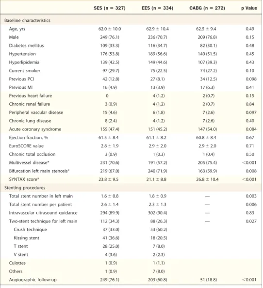
Table 1.Baseline Clinical and Procedural Characteristics of the Patients
SES (nⴝ 327) EES (nⴝ 334) CABG (nⴝ 272) p Value
Baseline characteristics
Age, yrs 62.0⫾ 10.0 62.9⫾ 10.4 62.5⫾ 9.4 0.49
Male 249 (76.1) 236 (70.7) 209 (76.8) 0.15
Diabetes mellitus 109 (33.3) 116 (34.7) 82 (30.1) 0.48
Hypertension 176 (53.8) 189 (56.6) 140 (51.5) 0.45
Hyperlipidemia 139 (42.5) 149 (44.6) 107 (39.3) 0.43
Current smoker 97 (29.7) 75 (22.5) 74 (27.2) 0.10
Previous PCI 42 (12.8) 27 (8.1) 34 (12.5) 0.098
Previous MI 16 (4.9) 13 (3.9) 17 (6.3) 0.41
Previous heart failure 0 4 (1.2) 2 (0.7) 0.15
Chronic renal failure 3 (0.9) 4 (1.2) 2 (0.7) 0.84
Peripheral vascular disease 15 (4.6) 6 (1.8) 7 (2.6) 0.097
Chronic lung disease 8 (2.4) 4 (1.2) 7 (2.6) 0.40
Acute coronary syndrome 155 (47.4) 151 (45.2) 147 (54.0) 0.084
Ejection fraction, % 61.5⫾ 8.4 61.1⫾ 8.2 60.8⫾ 8.4 0.67
EuroSCORE value 2.8⫾ 1.9 2.9⫾ 2.0 2.9⫾ 2.0 0.71
Chronic total occlusion 3 (0.9) 1 (0.3) 1 (0.4) 0.50
Multivessel disease* 231 (70.6) 191 (57.2) 205 (75.4) ⬍0.001
Bifurcation left main stenosis* 219 (67.0) 240 (71.9) 163 (59.9) 0.008
SYNTAX score* 23.8⫾ 9.5 21.1⫾ 8.8 26.8⫾ 10.4 ⬍0.001
Stenting procedures
Total stent number in left main 1.6⫾ 0.8 1.8⫾ 0.9 — 0.003
Total stent number per patient 2.6⫾ 1.4 2.3⫾ 1.3 — 0.006
Intravascular ultrasound guidance 294 (89.9) 302 (90.4) — 0.83
Two-stent technique for left main 112 (34.3) 88 (26.3) — 0.027
Crush technique 37 (33.0) 53 (60.2)
Kissing stent 41 (36.6) 18 (20.5)
T stent 28 (25.0) 7 (8.0)
V stent 4 (3.6) 2 (2.3)
Culottes 1 (0.9) 1 (1.1)
Others 1 (0.9) 7 (8.0)
Angiographic follow-up 249 (76.1) 203 (60.8) 51 (18.8) ⬍0.001
Values are mean⫾ SD or n (%). *p values for post-hoc comparisons: for multivessel disease, ⬍0.001 between SES versus EES, 0.20 between SES versus CABG, and⬍0.001 between EES versus CABG; for bifurcation left main stenosis, 0.17 between SES versus EES, 0.074 between SES versus CABG, and 0.008 between EES versus CABG; and for SYNTAX score,⬍0.001 between SES versus EES, ⬍0.001 between SES versus CABG, and ⬍0.001 between EES versus CABG. EES patients were enrolled in the PRECOMBAT-2 study and SES and CABG patients in the historical controls of PRECOMBAT study.
EES⫽ everolimus-eluting stent(s); CABG ⫽ coronary artery bypass graft; MI ⫽ myocardial infarction; PCI ⫽ percutaneous coronary intervention;
PRECOMBAT⫽ Premier of Randomized Comparison of Bypass Surgery versus Angioplasty Using Sirolimus-Eluting Stent in Patients with Left Main Coronary Artery Disease; PRECOMBAT-2⫽ Premier of Randomized Comparison of Bypass Surgery versus Angioplasty Using Everolimus-Eluting Stent in Patients with Left Main Coronary Artery Disease-2; SES⫽ sirolimus-eluting stent(s).
Statistical analysis. As-treated groups were compared, re- gardless of assigned randomization in the PRECOMBAT study. We included patients comparable to the number of patients in the PRECOMBAT study without sample size justification. Clinical and angiographic characteristics are presented as number and percentage or as mean⫾ SD. For continuous variables, differences among the 3 treatment groups (EES, SES, and CABG) were evaluated by analysis of variance, whereas differences between 2 groups (EES and SES) were analyzed by Student t test. Differences in categorical variables were analyzed using the chi-square test or Fisher exact test, as appropriate. The clinical endpoints were compared using a log-rank test of time to first event
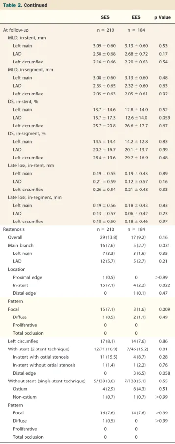
Table 2.Quantitative Angiographic Analysis of the SES and EES Groups
SES EES p Value
Baseline n⫽ 286 n⫽ 311
Lesion length, mm
Main branch 28.5⫾ 15.7 23.5⫾ 15.9 ⬍0.001
Left circumflex 9.8⫾ 12.1 8.3⫾ 10.6 0.26
Reference, mm
Left main 3.64⫾ 0.49 3.71⫾ 0.46 0.12
LAD 3.16⫾ 0.49 3.18⫾ 0.51 0.55
Left circumflex 2.96⫾ 0.50 3.00⫾ 0.55 0.45
MLD, mm
Left main 1.68⫾ 0.63 1.72⫾ 0.55 0.46
LAD 1.66⫾ 0.78 1.77⫾ 0.77 0.087
Left circumflex 1.91⫾ 0.66 2.10⫾ 0.72 0.001
DS, %
Left main 53.8⫾ 16.3 53.5⫾ 14.0 0.83
LAD 47.2⫾ 23.4 44.6⫾ 22.0 0.17
Left circumflex 34.5⫾ 20.78 29.7⫾ 20.4 0.006
After procedure n⫽ 286 n⫽ 311
Stent length, mm
Left main 13.7⫾ 4.4 13.1⫾ 4.5 0.15
LAD 20.3⫾ 14.7 17.8⫾ 14.8 0.045
Left circumflex 10.5⫾ 11.6 6.8⫾ 8.4 ⬍0.001
MLD, in-stent, mm
Left main 3.24⫾ 0.45 3.32⫾ 0.48 0.036
LAD 2.76⫾ 0.49 2.81⫾ 0.56 0.25
Left circumflex 2.44⫾ 0.54 2.41⫾ 0.59 0.64
MLD, in-segment, mm
Left main 3.23⫾ 0.45 3.31⫾ 0.48 0.046
LAD 2.47⫾ 0.55 2.42⫾ 0.60 0.29
Left circumflex 2.24⫾ 0.55 2.23⫾ 0.57 0.86
DS, in-stent, %
Left main 9.4⫾ 9.4 6.1⫾ 9.8 ⬍0.001
LAD 10.1⫾ 9.4 5.9⫾ 9.8 ⬍0.001
Left circumflex 16.6⫾ 13.5 19.0⫾ 14.0 0.040
DS, in-segment, %
Left main 10.1⫾ 9.1 8.4⫾ 8.2 0.017
LAD 16.2⫾ 10.5 15.6⫾ 10.8 0.49
Left circumflex 21.0⫾ 12.9 23.4⫾ 13.0 0.028
Continued in the next column
Table 2.Continued
SES EES p Value
At follow-up n⫽ 210 n⫽ 184
MLD, in-stent, mm
Left main 3.09⫾ 0.60 3.13⫾ 0.60 0.53
LAD 2.58⫾ 0.68 2.68⫾ 0.72 0.17
Left circumflex 2.16⫾ 0.66 2.20⫾ 0.63 0.54
MLD, in-segment, mm
Left main 3.08⫾ 0.60 3.13⫾ 0.60 0.48
LAD 2.35⫾ 0.65 2.32⫾ 0.60 0.63
Left circumflex 2.05⫾ 0.63 2.05⫾ 0.61 0.92
DS, in-stent, %
Left main 13.7⫾ 14.6 12.8⫾ 14.0 0.52
LAD 15.7⫾ 17.3 12.6⫾14.0 0.059
Left circumflex 25.7⫾ 20.8 26.6⫾ 17.7 0.67
DS, in-segment, %
Left main 14.5⫾ 14.4 14.2⫾ 12.8 0.83
LAD 20.2⫾ 16.7 20.1⫾ 13.7 0.99
Left circumflex 28.4⫾ 19.6 29.7⫾ 16.9 0.48
Late loss, in-stent, mm
Left main 0.19⫾ 0.55 0.19⫾ 0.43 0.89
LAD 0.21⫾ 0.59 0.12⫾ 0.57 0.16
Left circumflex 0.26⫾ 0.54 0.21⫾ 0.48 0.33
Late loss, in-segment, mm
Left main 0.19⫾ 0.56 0.18⫾ 0.43 0.83
LAD 0.13⫾ 0.57 0.06⫾ 0.42 0.23
Left circumflex 0.18⫾ 0.50 0.18⫾ 0.46 0.97
Restenosis n⫽ 210 n⫽ 184
Overall 29 (13.8) 17 (9.2) 0.16
Main branch 16 (7.6) 5 (2.7) 0.031
Left main 7 (3.3) 3 (1.6) 0.35
LAD 12 (5.7) 5 (2.7) 0.21
Location
Proximal edge 1 (0.5) 0 ⬎0.99
In-stent 15 (7.1) 4 (2.2) 0.022
Distal edge 0 1 (0.1) 0.47
Pattern
Focal 15 (7.1) 3 (1.6) 0.009
Diffuse 1 (0.5) 2 (1.1) 0.49
Proliferative 0 0
Total occlusion 0 0
Left circumflex 17 (8.1) 14 (7.6) 0.86
With stent (2-stent technique) 12/71 (16.9) 7/46 (15.2) 0.81 In-stent with ostial stenosis 11 (15.5) 4 (8.7) 0.28 In-stent without ostial stenosis 1 (1.4) 1 (2.2) 0.76
Distal edge 0 3 (6.5) 0.058
Without stent (single-stent technique) 5/139 (3.6) 7/138 (5.1) 0.55
Ostium 4 (2.9) 6 (4.3) 0.51
Non-ostium 1 (0.7) 1 (0.7) ⬎0.99
Pattern
Focal 16 (7.6) 14 (7.6) ⬎0.99
Diffuse 1 (0.5) 0 ⬎0.99
Proliferative 0 0
Total occlusion 0 0
Values are mean⫾ SD, n (%), or n/n (%). EES patients were enrolled in the PRECOMBAT-2 study and SES patients in the historical control of PRECOMBAT study.
DS⫽ diameter stenosis; LAD ⫽ left anterior descending coronary artery; MLD ⫽ minimal lumen diameter; other abbreviations as inTable 1.
from the time of the procedure. The Kaplan-Meier method was used to estimate survival. Patients were censored at 18 months or at occurrence of an endpoint. Differences among the groups in risk-adjusted, long-term rates of study out- comes were assessed using multivariable Cox proportional hazards regression with covariates that had a significant effect (p⬍ 0.1) on clinical outcomes. Covariates considered in the Cox models included patient age and sex; presence of hypertension, smoking, hypercholesterolemia, and diabetes mellitus; prior history of MI, stroke, peripheral vascular disease, congestive heart failure, chronic renal failure, chronic lung disease, bifurcation left main stenosis, multi- vessel disease, and PCI; performance of angiographic sur- veillance; and left ventricular ejection fraction. The multi- variate models were confirmed by backward elimination.
The proportional hazards assumption was confirmed by testing of partial (Schoenfeld) residuals, and no relevant violations were found (22). No adjustments were made for multiple comparisons in post hoc secondary analyses. SAS software, version 9.1, was used for all statistical analyses (SAS Institute Inc., Cary, North Carolina).
Results
Study patients. Baseline characteristics and procedural find- ings are shown inTable 1. The 3 groups were generally well matched, except that EES patients were more likely to have bifurcation left main stenosis, single-vessel involvement, and low SYNTAX scores than did SES or CABG patients.
EES patients received more stents per left main lesion, but fewer stents per patient. Angiographic complete revascular- ization was performed in 230 (70.3%) in SES, 240 (71.9%)
in EES, and 185 (68.0%) in CABG patients (p ⫽ 0.59).
Angiographic follow-up was less frequently performed in EES patients than in SES patients at 9.4⫾ 2.1 months and 8.8 ⫾ 2.8 months, respectively (p⫽ 0.014). Baseline angiographic characteristics of the main branch, including the left main and LAD, were well matched in SES and EES patients (Table 2).
However, DS in the side branch and lesion length in the main branch were less severe in EES than SES patients.
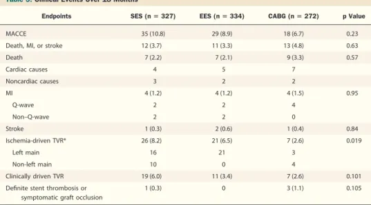
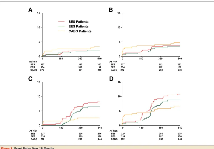
Clinical outcomes. Table 3 and Figure 1 show clinical outcomes over 18 months. Before adjustment, the 3 groups had comparable incidence rates of individual endpoints and the composite of death, MI, or stroke. However, the incidence of ischemia-driven TVR was lower in the CABG than in the SES or EES group, but the clinically driven TVR rate did not differ significantly. The overall incidence of MACCE, the primary endpoint, did not differ among the 3 groups. There were no differences in clinical outcomes between the SES and EES groups. When the risks of events were adjusted using the Cox proportional hazard model, the pattern of differences in clinical outcomes did not change (Fig. 2). In addition, com- pared with CABG, PCI with EES was not associated with the risk of death (hazard ratio [HR]: 0.59; 95% confidence interval [CI]: 0.21 to 1.63; p ⫽ 0.31), composite of death, MI, or stroke (HR: 0.67, 95% CI: 0.29 to 1.54; p ⫽ 0.34), and MACCE (HR: 1.40; 95% CI: 0.78 to 2.54; p⫽ 0.27). But the risk of ischemia-driven TVR was higher in the EES group (HR: 2.77; 95% CI: 1.17 to 6.58; p⫽ 0.021).
Angiographic outcomes after stenting. Table 2 shows the quantitative angiographic outcomes in the SES and EES groups. After the procedure, EES patients achieved lower DS in the main branch, but higher DS in the side branch,
Table 3.Clinical Events Over 18 Months
Endpoints SES (nⴝ 327) EES (nⴝ 334) CABG (nⴝ 272) p Value
MACCE 35 (10.8) 29 (8.9) 18 (6.7) 0.23
Death, MI, or stroke 12 (3.7) 11 (3.3) 13 (4.8) 0.63
Death 7 (2.2) 7 (2.1) 9 (3.3) 0.57
Cardiac causes 4 5 7
Noncardiac causes 3 2 2
MI 4 (1.2) 4 (1.2) 4 (1.5) 0.95
Q-wave 2 2 4
Non–Q-wave 2 2 0
Stroke 1 (0.3) 2 (0.6) 1 (0.4) 0.84
Ischemia-driven TVR* 26 (8.2) 21 (6.5) 7 (2.6) 0.019
Left main 16 21 3
Non-left main 10 0 4
Clinically driven TVR 19 (6.0) 11 (3.4) 7 (2.6) 0.101
Definite stent thrombosis or symptomatic graft occlusion
1 (0.3) 0 3 (1.1) 0.105
Values are n (%), as determined using the Kaplan-Meier method. p values were calculated using the log-rank test. *The p values for post hoc comparisons were 0.41 between SES versus EES, 0.005 between SES versus CABG, and 0.019 for EES versus CABG. EES patients were enrolled in the PRECOMBAT-2 study, and SES and CABG patients in the historical controls of PRECOMBAT study.
MACCE⫽ major adverse cardiac or cerebrovascular event(s); TVR ⫽ target vessel revascularization; other abbreviations as inTable 1.
than SES patients. Quantitative angiographic analyses at follow-up were performed in 73% of SES and 59% of EES patients, but there were no between-group differences in angiographic parameters, including DS, MLD, and late loss in any segment. Subsequently, the rate of overall restenosis did not differ between the 2 groups. However, late loss and restenosis rates in the main branch were slightly lower in EES than in SES patients. In both groups, a focal pattern was predominant in restenotic lesions.
Angiographic and clinical outcomes according to the stent technique. When the stenting technique was separated into the single- and 2-stent techniques, kissing stenting tech- nique and crush technique were most popularly used in SES and EES patients, respectively (Table 1). The restenosis rates in the subgroups stratified by the stent technique are illustrated inFigure 3. Ischemia-driven TVR occurred in 11 (5.3%) SES and 12 (5.1%) EES patients in the single-stent group (log-rank p ⫽ 0.94) and 15 (13.6%) SES and 9
(10.4%) EES patients in the 2-stent group (log-rank p⫽ 0.51). Among them, TVR at left main was included in 5 SES and 12 EES patients in the single-stent group and in 11 SES and 9 EES patients in the 2-stent group.
Discussion
The major finding of this study is that using EES for ULMCA stenosis is as safe and effective as employing SES or CABG, as shown by the 18-month incidence of MACCE. Although the need for repeat revascularization was higher after EES than after CABG, second-generation EES had risks of angiographic and clinical restenosis comparable to those of first-generation SES.
Most studies evaluating the long-term outcomes of PCI using DES for ULMCA stenosis have used first-generation SES or paclitaxel-eluting stents (1–12). Previous random- ized and registry studies found that these DES had similar
15
10
5
0
15
10
5
0
15
10
5
0
15
10
5
0
0 180 360 540
At risk
SES 327 317 300
EES 334 316 191
CABG 272 261 249
0 180 360 540
At risk
SES 327 312 293
EES 334 312 188
CABG 272 258 246
0 180 360 540
At risk
SES 327 296 276
EES 334 299 178
CABG 272 256 244
0 180 360 540
At risk
SES 327 294 275
EES 334 297 176
CABG 272 253 241
A B
C D
SES Patients EES Patients CABG Patients
Figure 1.Event Rates Over 18 Months
(A) Death from any cause; (B) death, MI, or stroke; (C) ischemia-driven TVR; (D) composite primary endpoint of major adverse cardiac or cerebrovascular events.
p Values were calculated using the log-rank test for EES patients in the PRECOMBAT-2, and SES and CABG patients in the historical controls of PRECOMBAT stud- ies. EES⫽ everolimus-eluting stent(s); CABG ⫽ coronary artery bypass graft; MI ⫽ myocardial infarction; PRECOMBAT ⫽ Premier of Randomized Comparison of Bypass Surgery versus Angioplasty Using Sirolimus-Eluting Stent in Patients with Left Main Coronary Artery Disease; PRECOMBAT-2⫽ Premier of Randomized Comparison of Bypass Surgery versus Angioplasty Using Everolimus-Eluting Stent in Patients with Left Main Coronary Artery Disease-2; SES⫽ sirolimus-eluting stent(s); TVR⫽ target vessel revascularization.
long-term safety and efficacy results (7,23). Despite the trend toward higher late loss after paclitaxel-eluting stent implantation, their clinical outcomes, including rates of repeat revascularization, were similar. Nonetheless, PCI for ULMCA stenosis, particularly with bifurcation involve- ment, remains technically challenging and is related to higher rates of stent thrombosis and repeat revasculariza-
tion. Therefore, studies assessing the safety and efficacy of second-generation DES relative to first-generation DES for ULMCA stenosis appear worthwhile. Compared with SES, the reduced strut thickness (81 vs. 140 m) and thinner polymer coating (7.6 vs. 12.6 m), in conjunction with improved biocompatibility of the EES polymer, may favor- ably affect neointimal hyperplasia. In addition, the availabil- ity of a 4.0-mm EES stent might have achieved better stent expansion and drug diffusion. In the PRECOMBAT study, the maximal size of the SES used was 3.5 mm even in the big left main lesions. Our study has the advantage of comparing the outcomes of second-generation EES with those of first-generation SES and CABG. Despite our nonrandomized study design, the 3 patient groups—those treated with EES in the PRECOMBAT-2 trial and those treated with SES and CABG in the PRECOMBAT study—were comparable, without a serious selection bias, since both trials used the same inclusion and exclusion criteria.
We found that the clinical and angiographic outcomes of patients with ULMCA stenosis treated with second- generation EES and first-generation SES were comparable over 18 months. These 2 types of stents were recently compared in patients with unselected lesions. For example, the randomized ESSENCE-DIABETES (Everolimus- Eluting Stent Versus Sirolimus-Eluting Stent Implantation for De Novo Coronary Artery Disease in Patients With Diabetes Mellitus) trial, involving 300 diabetic patients,
Figure 2.Adjusted Hazard Ratios for Outcomes
Adjusted hazard ratios of EES in the PRECOMBAT-2 study compared with SES in the historical control of PRECOMBAT study for the risk of events.
CI⫽ confidence interval; MACCE ⫽ major adverse cardiac or cerebrovascu- lar event(s); other abbreviations as inFigure 1.
Left main
(2.9% SES vs. 0.7% EES, p=0.18)
Left circumflex
(3.6% SES vs. 5.1% EES, p=0.55)
LAD(5.0% SES vs.
2.2% EES, p=0.34)
Left main
(4.2% SES vs. 4.3% EES, p=1.0)
Left circumflex
(16.9% SES vs. 15.2% EES, p=0.81)
LAD(7.0% SES vs.
4.3% EES, p=0.55)
Single-Stent Technique
(N=139 SES, 138 EES)
Two-Stent Technique
(N=71 SES, 46EES)
SES EES
Figure 3.Illustration of Angiographic Restenosis in the Subgroups Stratified By Stenting Technique
Red and blue circles indicate the location of angiographic restenosis in patients treated with SES and EES, respectively. LAD⫽ left anterior descending coronary artery; other abbreviations as inFigure 1.
found a tendency toward lower restenosis rate, but similar clinical outcomes, in the EES compared with the SES group (24). Another large, as yet unpublished trial, SORT OUT IV (the Scandinavian Organisation for Randomised Trials with Clinical Outcome IV) study, involving 2,774 unselected patients, found that EES and SES yielded similar 9-month incidence rates of death, MI, stent throm- bosis, and TVR (25). The similar clinical outcomes of SES and EES in these studies, as well as in ours, may be due primarily to the comparable biological potency of everoli- mus and sirolimus. Alternatively, as suggested in studies comparing SES and paclitaxel-eluting stents for ULMCA stenosis, large caliber in the left main coronary artery may accommodate the impact of intimal tissue growth on net clinical outcomes if similar stenting techniques are used (7,19). Nonetheless, our finding of a slightly lower late loss and restenosis rate in the main branch after EES implan- tation suggests that different DES have different efficacies for ULMCA stenosis. Further studies evaluating the angio- graphic and clinical outcomes achieved using various DES should clarify their differential efficacies.
PCI using EES had a similar incidence of death, MI, or stroke, but a higher incidence of TVR, compared with CABG. This pattern was in agreement with findings comparing PCI using first-generation DES and CABG in the PRECOMBAT and other trials (12,13). However, the rate of clinically driven TVR, which excluded revasculariza- tions for patients without objective recurrence of ischemic symptom or sign, did not differ significantly between EES and CABG patients. This indicates that the “oculostenotic reflex” due to routine angiographic follow-up after stent implantation might have exaggerated the difference of TVR rates between CABG and EES patients. In fact, angio- graphic follow-up was performed in 61% after EES and 19% after CABG procedures. The new practice guidelines do not recommend routine angiographic surveillance even after left main coronary stenting (14,26). Therefore, our findings support the hypothesis of the ongoing randomized Evaluation of XIENCE PRIME versus the EXCEL (Cor- onary Artery Bypass Surgery for Effectiveness of Left Main Revascularization) trial, that PCI using EES is clinically noninferior to CABG.
Study limitations. First, the nonrandomized study design may have introduced biases into the results. Because we used a historical control group of SES patients, the stent tech- niques or medications in EES patients may have been improved or modified. For example, due to the less rigorous recommendation for routine angiographic follow-up, the rate of TVR for restenosis in patients without inducible ischemia may have been lower in EES than in SES patients.
In addition, due to a lack of power calculation, this study may be underpowered in the comparison of clinical end- points across the groups. Second, since we used the same inclusion criteria as the previous randomized study, our
results may have limited generalizability in real-world prac- tice. Third, the shorter follow-up duration in EES patients may have resulted in a lower incidence of adverse events than observed in SES patients. Finally, the 18-month follow-up period was not sufficient to assess the durable safety and efficacy of DES. To compensate for these limitations, a further randomized study with sufficient sample size and follow-up period is warranted.
Conclusions
We found that second-generation EES for ULMCA ste- nosis resulted in a similar risk of death, MI, stroke, or repeat revascularization as first-generation SES or CABG. Further randomized trials are needed to compare PCI using newer- generation DES and CABG in patients with these complex coronary lesions.
Acknowledgments
The authors thank Jun-Hong Kim, MD (Pusan National University Yangsan Hospital, Yangsan), Sang-Gon Lee, MD (Ulsan University Hospital, Ulsan), Hun-Sik Park, MD (Kyungpook National University Hospital, Daegu), Hyeon-Cheol Gwon, MD (Samsung Medical Center, Seoul), Seung-Uk Lee, MD (Kwangju Christian Hospital, Kwangju), Nae-Hee Lee, MD (Soonchunhyang Univer- sity Hospital, Bucheon), Seung-Jea Tahk, MD (Ajou University Hospital, Suwon), Bong-Ki Lee, MD (Kang- won National University Hospital, Chuncheon), Keun Lee, MD (Veterans Hospital, Seoul), Sang-Sig Cheong, MD (Gangneung Asan Hospital, Gangneung), and Kee- Sik Kim, MD (Daegu Catholic University Medical Center, Daegu) for their outstanding contribution to the patients’
enrollment.
Reprint requests and correspondence: Dr. Seung-Jung Park, Heart Institute, Asan Medical Center, University of Ulsan, Songpa-gu, Seoul 138-736, South Korea. E-mail: sjpark@amc.
seoul.kr.
REFERENCES
1. Seung KB, Park DW, Kim YH, et al. Stents versus coronary-artery bypass grafting for left main coronary artery disease. N Engl J Med 2008;358:1781–92.
2. Onuma Y, Girasis C, Piazza N, et al. Long-term clinical results following stenting of the left main stem: insights from RESEARCH (Rapamycin-Eluting Stent Evaluated at Rotterdam Cardiology Hos- pital) and T-SEARCH (Taxus-Stent Evaluated at Rotterdam Cardi- ology Hospital) registries. J Am Coll Cardiol Intv 2010;3:584 –94.
3. Pandya SB, Kim YH, Meyers SN, et al. Drug-eluting versus bare- metal stents in unprotected left main coronary artery stenosis. A meta-analysis. J Am Coll Cardiol Intv 2010;3:602–11.
4. Chieffo A, Magni V, Latib A, et al. 5-year outcomes following percutaneous coronary intervention with drug-eluting stent implanta- tion versus coronary artery bypass graft for unprotected left main coronary artery lesions: the Milan experience. J Am Coll Cardiol Intv 2010;3:595– 601.
5. Naik H, White AJ, Chakravarty T, et al. A meta-analysis of 3,773 patients treated with percutaneous coronary intervention or surgery for unprotected left main coronary artery stenosis. J Am Coll Cardiol Intv 2009;2:739 – 47.
6. Morice MC, Serruys PW, Kappetein AP, et al. Outcomes in patients with de novo left main disease treated with either percutaneous coronary intervention using paclitaxel-eluting stents or coronary artery bypass graft treatment in the Synergy between Percutaneous Coronary Intervention with TAXUS and Cardiac Surgery (SYNTAX) trial.
Circulation 2010;121:2645–53.
7. Mehilli J, Kastrati A, Byrne RA, et al. Paclitaxel- versus sirolimus- eluting stents for unprotected left main coronary artery disease. J Am Coll Cardiol 2009;53:1760 – 8.
8. Buszman PE, Kiesz SR, Bochenek A, et al. Acute and late outcomes of unprotected left main stenting in comparison with surgical revascular- ization. J Am Coll Cardiol 2008;51:538 – 45.
9. Valgimigli M, van Mieghem CA, Ong AT, et al. Short- and long-term clinical outcome after drug-eluting stent implantation for the percuta- neous treatment of left main coronary artery disease: insights from the Rapamycin-Eluting and Taxus Stent Evaluated at Rotterdam Cardi- ology Hospital registries (RESEARCH and T-SEARCH). Circula- tion 2005;111:1383–9.
10. Biondi-Zoccai GGL, Lotrionte M, Moretti C, et al. A collaborative systematic review and meta-analysis on 1278 patients undergoing percutaneous drug-eluting stenting for unprotected left main coronary artery disease. Am Heart J 2008;155:274 – 83.
11. Boudriot E, Thiele H, Walther T, et al. Randomized comparison of percutaneous coronary intervention with sirolimus-eluting stents versus coronary artery bypass grafting in unprotected left main stem stenosis.
J Am Coll Cardiol 2011;57:538 – 45.
12. Park SJ, Kim YH, Park DW, et al. Randomized trial of stents versus bypass surgery for left main coronary artery disease. N Engl J Med 2011;364:1718 –27.
13. Capodanno D, Stone GW, Morice MC, Bass TA, Tamburino C.
Percutaneous coronary intervention versus coronary artery bypass graft surgery in left main coronary artery disease: a meta-analysis of random- ized clinical data. J Am Coll Cardiol 2011;58:1426 –32.
14. Levine GN, Bates ER, Blankenship JC, et al. ACCF/AHA/SCAI guideline for percutaneous coronary intervention: a report of the American College of Cardiology Foundation/American Heart Associ- ation Task Force on Practice Guidelines and the Society for Cardio- vascular Angiography and Interventions. J Am Coll Cardiol 2011;58:
e44 –122.
15. Wijns W, Kolh P, Danchin N, et al., the Task Force on Myocardial Revascularization of the European Society of Cardiology (ESC) and the
European Association for Cardio-Thoracic Surgery (EACTS). Guidelines on myocardial revascularization. Eur Heart J 2010;31:2501–55.
16. Holmes DR Jr., Kereiakes DJ, Garg S, et al. Stent thrombosis. J Am Coll Cardiol 2010;56:1357– 65.
17. Kedhi E, Joesoef KS, McFadden E, et al. Second-generation everolimus-eluting and paclitaxel-eluting stents in real-life practice (COMPARE): a randomised trial. Lancet 2010;375:201–9.
18. Stone GW, Rizvi A, Newman W, et al. Everolimus-eluting versus paclitaxel-eluting stents in coronary artery disease. N Engl J Med 2010;362:1663–74.
19. Lee JY, Park DW, Kim YH, et al. Incidence, predictors, treatment, and long-term prognosis of patients with restenosis after drug-eluting stent implantation for unprotected left main coronary artery disease. J Am Coll Cardiol 2011;57:1349 –58.
20. Ramcharitar S, Onuma Y, Aben JP, et al. A novel dedicated quanti- tative coronary analysis methodology for bifurcation lesions. EuroInt- ervention 2008;3:553–7.
21. Mehran R, Dangas G, Abizaid AS, et al. Angiographic patterns of in-stent restenosis: classification and implications for long-term out- come. Circulation 1999;100:1872– 8.
22. Cain KC, Lange NT. Approximate case influence for the proportional hazards regression model with censored data. Biometrics 1984;40:
493–9.
23. Lee JY, Park DW, Yun SC, et al. Long-term clinical outcomes of sirolimus- versus paclitaxel-eluting stents for patients with unprotected left main coronary artery disease: analysis of the MAIN-COMPARE (Revascularization for Unprotected Left Main Coronary Artery Steno- sis: Comparison of Percutaneous Coronary Angioplasty versus Surgical Revascularization) registry. J Am Coll Cardiol 2009;54:853–9.
24. Kim WJ, Lee SW, Park SW, et al. Randomized comparison of everolimus-eluting stent versus sirolimus-eluting stent implantation for de novo coronary artery disease in patients with diabetes mellitus (ESSENCE-DIABETES): results from the ESSENCE-DIABETES trial. Circulation 2011;124:886 –92.
25. Jensen LO, Thayssen P, Hansen HS, et al. Randomized comparison of everolimus-eluting and sirolimus-eluting stents in patients treated with percutaneous coronary intervention: the Scandinavian Organization for Randomized Trials With Clinical Outcome IV (SORT OUT IV).
Circulation 2012;125:1246 –55.
26. Wijns W, Kolh P, Danchin N, et al. Guidelines on myocardial revascularization. Eur Heart J 2011;31:2501–55.
Key Words: bypass surgery䡲 coronary disease 䡲 left main coronary disease䡲 stents.
To participate in this CME activity by taking the quiz and claiming your CME credit certificate, please go to
http://interventions.onlinejacc.org/
and select the CME tab on the top navigation bar.