Original Article
원고 접수일 2010년 11월 23일, 게재 확정일 2011년 2월 24일 책임저자 김영균
(463-707) 경기도 성남시 분당구 구미동 300, 분당서울대학교병원 치과 구강악안면 외과
Tel: 031-787-7541, Fax: 031-787-4068, E-mail: [email protected]
RECEIVED November 23, 2010, ACCEPTED February 24, 2011 Correspondence to Young-Kyun Kim
Department of Oral and Maxillofacial Surgery, Section of dentistry, Seoul National University Bundang Hospital
300, Gumi-dong, Bundang-gu, Seongnam 463-707, Korea
Tel: 82-31-787-7541, Fax: 82-31-787-4068, E-mail: [email protected]
CC This is an open access article distributed under the terms of the Creative Commons Attribution Non-Commercial License (http://creativecommons.org/licenses/
by-nc/3.0) which permits unrestricted non-commercial use, distribution, and reproduction in any medium, provided the original work is properly cited.
RBM 표면 테이퍼형태 임프란트의 단기간 후향적 임상 평가
김수연ㆍ김영균
분당서울대학교병원 치과, 구강악안면외과
Abstract
Short-Term Retrospective Clinical Study of Resorbable Blasting Media Surface Tapered Implants
Soo-Yeon Kim, Young-Kyun Kim
Department of Oral and Maxillofacial Surgery, Section of Dentistry, Seoul National University Bundang Hospital
Purpose: The aim of the present study was to evaluate the clinical outcome of resorbable blasting media surface tapered implant.
Methods: 169 OsstemⓇ GS III dental implants in 73 patients who received implant treatments at Seoul National University Bundang Hospital, were included in this study. The incidence of biological and prosthetical complications has been carefully analysed for each implant.
Results: The short-term implant survival rate was 97.63%, success rate 94.7%. The prevalence of biological complications was 15.38% and the prevalence of prosthetic complications was 13.04%. The mean value of crestal bone loss was 0.28±0.57 mm. The relationship between loading periods and marginal bone loss was small and not statistically significant. In mandible, marginal bone loss was larger than in maxilla, no statistically significant. Also, length and diameter of implant had no relationship with marginal bone loss.
Conclusion: We suggest that this implant system could achieve successful and stable results.
Key words: Complications, RBM, Success, Survival, Tapered
서 론
Resorbable blasting media (RBM) 표면의 테이퍼형태 임프란트 (Osstem GSIII
Ⓡ)는 internal hex 연결 방식의 submerged type 이며, tapered body형태로 우수한 초기 고정력 및 장기 안정성을 구현하고자 하였다. Tapered body 임프란트는 초기 삽입이 용이
하고, 술식이 간단하며 골 삭제량이 많지 않은 장점을 가진다.
또한, RBM 표면의 테이퍼형태 임프란트(Osstem GSIII
Ⓡ)는 corkscrew thread와 micro thread로 강력한 self threading효 과와 변연골에서의 응력집중 감소를 유도한다.
본 연구에서는 RBM 표면의 테이퍼형태 임프란트(Osstem
GSIII
Ⓡ)의 임상검사와 방사선학적 평가를 포함한 연구를 통하여
Table 2. Length and diameter of implant
Length (mm) N Diameter (mm) N
7 15 <4.0 14
8.5 22 4.0 51
10 40 4.5 23
11.5 49 5.0 77
13 39
15 4
Total 169 Total 169
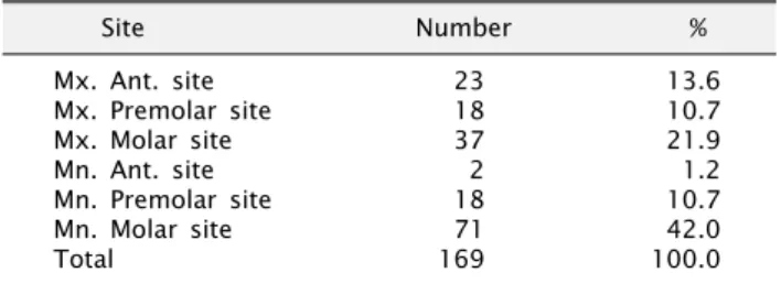
Table 1. Fixture location
Site Number %
Mx. Ant. site 23 13.6
Mx. Premolar site 18 10.7
Mx. Molar site 37 21.9
Mn. Ant. site 2 1.2
Mn. Premolar site 18 10.7
Mn. Molar site 71 42.0
Total 169 100.0
임프란트의 생존율과 합병증의 발생률을 조사하고, 임프란트 실 패와 합병증 발생에 미치는 요인을 평가해 보고자 하였다.
연구방법
본 연구는 분당서울대학교병원에서 2008년 8월부터 2010년 7월까지 73명의 환자에서 169개의 GSIII
Ⓡ(Osstem, Seoul, Korea) 임프란트 시술을 받은 환자를 대상으로 하였다. 남자가 37명, 여자가 36명이었다. 전신질환을 보유한 환자들은 고혈압 11명, 당뇨 2명, 당뇨 및 고혈압 2명이었으나 모두 내과적으로 잘 조절되었다. 의무기록지 및 방사선 사진을 후향적으로 분석하 여 임프란트의 초기안정성과 이차안정성, 골유착실패, 술 후 및 보철물 장착 후 합병증, 변연골 흡수량과 임프란트 성공률 및 생존율을 조사하였다.
1. 임프란트 시술 정보
의무기록지를 검토하여 연령, 성별, 식립 부위, 임프란트 직경 및 폭경 분포, 동반된 외과적 술식, 임프란트 초기 및 이차고정 등을 조사하였다.
2. 합병증
술 후 치유과정에서 발생된 생물학적 및 보철적 합병증을 조사 하였다.
3. 성공률 및 생존율
최종 경과관찰 시점에 기능하고 있는 경우를 생존율에 포함시 켰고 임프란트 성공율은 통증, 유동성, 주위연조직염증, 임프란트 주변 방사선투과상이 없으면서 최종 경과관찰 시점에서 변연골 흡수량이 1.0 mm 이하인 경우를 성공 범주에 포함시켰다[1].
4. 변연골 흡수량
최종경과관찰 시 변연골 흡수량을 치근단 방사선 사진에서 측 정한 후, 식립 부위와 임프란트의 직경 및 폭경에 따른 변연골 흡수량을 비교하였다. 변연골 흡수량의 측정은 술 후 치근단 방사 선사진과 최종경과 관찰 시 촬영한 사진을 비교하여, 식립된 임프 란트의 길이를 기준으로 확대율을 산출한 후 임프란트 근심측과 원심측에서의 골흡수량의 평균값을 구하였다. 식립 부위와 임프 란트의 직경 및 폭경에 따른 변연골 흡수량을 비교하였다.
보철물 장착 후 하중이 가해진 시기에 따라 3군으로 분류하여 변연골 흡수량을 측정하였다. 1군은 2주 이내 하중, 2군은 2∼6 주, 3군은 6주 후 하중이 가해진 증례들이었다.
5. 통계
임프란트의 직경 및 길이에 따른 변연골 소실량과 3가지 하중
군에 따른 변연골 소실량, 부위에 따른 변연골 소실량을 비교하기 위해 Kruskall-Wallis test를 사용했다. 통계학적 분석은 SPSS (SPSS Inc., Chicago, IL, USA)를 통해 이루어졌다.
결 과
환자들의 나이는 18세부터 77세까지로 평균 52.5세였으며, 임프란트 식립 후부터 최종보철물 장착 시까지의 기간은 평균 4.3개월이었으며, 치유기간(임프란트 식립 후부터 이차수술 또는 첫 인상채득일)은 상악에서 평균 3.4개월이었고, 하악에서는 평 균 1.6개월이었다.
1. 부위별 분포(Table 1)
하악 대구치부에서 71개의 가장 많은 임프란트를 식립하였고, 상악 대구치부에서는 37개, 상악 전치부에 23개, 상악 소구치부와 하악 소구치부에 각각 18개 그리고 하악 전치부에서 2개의 순서였 다.
2. 임프란트의 길이 및 폭경(Table 2)
길이 11.5 mm의 임프란트가 47개로 가장 많이 식립되었으며, 폭경은 5.0 mm의 임프란트가 77개로 가장 많았다.
3. 일차 및 이차 안정성
Osstell mentor
Ⓡ(Osstell Mentors
Ⓡ, Integration diag-
nostics AB, Savedalen, Sweden)를 이용하여 일차 안정성이
측정된 임프란트는 162개였으며, 평균값은 68.5±14.55였다. 이
차 안정성이 측정된 임프란트는 146개였으며, 평균값은 74.6±
Table 3. Biological complication distribution
Complications N
Wound dehiscence 19
Infection 2
Osseointegration failure 4
Pain 2
Total 27
Table 4. Prosthetic complications distribution
Prosthetic complications N %
Galvanic 1 4.8%
Loose contact 6 28.6%
Food impaction 7 33.3%
Fractured porcelain 5 23.8%
Cheek biting 2 9.5%
Total 21 100%
Table 5. Crestal bone loss (mm) according to loading time (P>
0.05)
Group Crestal bone loss (mean)
1 0.27±0.57
2 0.33±0.42
3 0.27±0.55
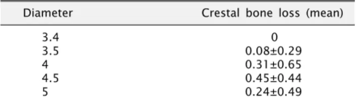
Table 6. Crestal bone loss (mm) according to diameter (P>0.05)
Diameter Crestal bone loss (mean)
3.4 0
3.5 0.08±0.29
4 0.31±0.65
4.5 0.45±0.44
5 0.24±0.49
Table 7. Crestal bone loss (mm) according to length (P>0.05)
Length Crestal bone loss (mean)
7 0.27±0.58
8.5 0.57±0.90
10 0.20±0.46
11.5 0.25±0.37
12 0
13 0.13±0.46
Table 8. Crestal bone loss (mm) according to site (P>0.05)
Site Crestal bone loss (mean)
Mx. Ant. site 0.16±0.37
Mx. Premolar site 0.22±0.43
Mx. Molar site 0.15±0.39
Mn. Ant. Site 0
Mn. Premolar site 0.55±0.87
Mn. Molar site 0.32±0.53
12.01이었다.
4. 동반된 외과적 술식
외과적 술식이 동반된 경우는 총 138개의 임프란트였으며, 그 중 Guided Bone Regeneration술식이 124건이었으며, Sinus Lifting술식이 14건이었다.
5. 합병증(Table 3, 4)
식립된 169개의 임프란트 중 창상열개로 인한 덮개나사 노출 19개, 동통 4개, 감염 2개였으며 4개의 임프란트가 제거되었다.
상부 보철물 장착 후 합병증은 음식물 침착이 7개, 접촉점 느슨해 짐(contact loss)가 6개, 도재파절(porcelain fracture)이 5개였 고, 총 21개의 임프란트에서 보철적 합병증이 발생하였다.
6. 변연골 소실(Table 5, 6, 7, 8)
하중기간에 따른 평균 변연골 소실량의 차이는 매우 작았으며, 이 차이는 통계학적으로 유의하지 않게 나타났다. 1군에서 0.27±0.57 mm, 2군에서 0.33±0.42 mm, 3군에서 0.27±
0.55 mm로 나타났다. 부위에 따른 평균 변연골 소실량은 상악 전치부에서는 0.16±0.37 mm, 상악 소구치부에서는 0.22±
0.43 mm, 상악 대구치부에서는 0.15±0.39 mm, 하악 소구치부 에서는 0.55±0.87 mm, 하악 대구치부에서는 0.32±0.53 mm 로 나타났다. 식립된 모든 임프란트의 변연골 소실량은 0.28±
0.57 mm였다. 상악 대구치부에서 가장 작은 값이었고, 상악 전치부, 상악 소구치부, 하악 대구치부, 하악 소구치부의 순서였으 며, 상악보다 하악에서 더 큰 값을 나타냈다. 하지만, 통계학적으 로 유의성 있는 차이는 없었다.
직경 4.5 mm의 임프란트에서 0.45±0.44 mm로 가장 큰 변연골 소실량을 나타냈고, 직경 3.4 mm의 임프란트에서는 0±0 mm로 가장 적은 변연골 소실량을 나타냈지만, 직경과 변연골 소실량 사이에 통계학적인 유의성은 없었다.
길이 8.5 mm의 임프란트에서 0.57±0.90 mm로 가장 큰 변연골 소실량을 나타냈으며, 길이 13 mm의 임프란트에서 0.13±0.46 mm의 변연골 소실량을 보이며, 길이가 긴 임프란트 에서 더 적은 변연골 소실량을 보이는 경향이 있었다. 하지만 길이에 따른 평균 변연골 소실량 사이에 통계학적인 유의성은 없었다.
7. 생존율 및 성공율
임프란트를 제거한 경우는 3명의 환자에서 4개의 고정체였다.
최종경과관찰 시의 생존율은 97.63%였다. 최종경과 관찰 시점에 기준치 이상의 변연골 소실이 일어난 경우가 5개로 임프란트 성공 률은 94.7%였다.
총 4개의 임프란트에서 골유착이 실패하였다. 39세 여자환자에
서 #13과 #11부위에 식립된 임프란트의 초기고정이 불량했으며,
골유착 실패로 식립 5개월만에 제거하였다. 60세 남자환자에서
#21부위에 식립된 임프란트는 초기고정값 44였으며, 식립 3개월 만에 제거되었다. 34세 여자환자에서 #46부위에 식립된 임프란트 는 즉시하중이 가해졌으며, 초기고정값은 75로 비교적 우수하였 으나 골유착 실패로 제거하였다.
고 찰
Resonance frequency analysis (RFA)는 임프란트의 안정성 및 골융합 정도를 평가하는 수단이다[2]. RFA는 주변골이 아닌 주로 임프란트의 길이 및 임프란트와 골 계면의안정성과 관련이 있다[3]. RFA값의 측정에 관한 연구는 아직까지 불충분하지만 파괴적이지 않은 기계적 진동에 의해서 임프란트의 안정성을 측정 하는 중요한 방법으로 여겨지고 있다[4,5]. 본 연구에서는 Osstell mentor
Ⓡ(Osstell Mentors
Ⓡ, Integration diagnostics AB, Savedalen, Sweden)장치를 이용하여 임프란트의 안정성을 평 가하였다. 일차 안정성이 측정된 임프란트는 162개였으며, 평균 값은 68.5±14.55였으며, 이차 안정성이 측정된 임프란트는 146 개로 평균값은 74.6±12.01로 양호하게 나타났다.
Wennerberg 등[6]은 RBM 표면 임프란트가 removal torque value, 골 접촉률 등에서 기계연마표면(machined surface) 임프 란트보다 우수한 결과를 보인다고 보고하였다. Blasting 방법은 표면에 분사되는 입자의 영향으로 표면적이 증가되어 요철 효과로 골파괴의 결합력이 증가되며 거친 표면에 세포의 반응이 활성화되 는 장점을 지닌다고 알려져 있다.
Muftu와 Chapman[7]은 tapered implant 식립 시 지대주 나사풀림(abutment loosening)이 발생할 위험이 줄어드는 것으 로 보인다고 발표한 바 있다. Makkonen 등[8]은 경부에 micro- thread를 갖는 Astra
Ⓡtech implant (Astra Tech AB, Molndal, Sweden)로 5년 간의 후향적 연구에 대한 결과를 보고하였다.
총 33명의 환자에서 하악에 155개의 임프란트를 식립하였으며, 평균 변연골 흡수량은 0.48 mm, 생존율은 98.7%를 보이는 성공 적인 결과를 발표하였다. Diss 등[9]도 상악동 골이식과 동반된 Astra
Ⓡimplant식립에 대한 결과를 보고한 바 있다. 총 20명의 환자에서 35개의 임프란트를 식립한 후 1년 동안 관찰한 결과, 방사선학적으로 신생골이 평균 3.2±1.5 mm (근심부에서 3.5±1.4 mm, 원심부에서 2.9±1.6 mm)가 생성되었으며, 임프 란트 기능에서도 우수한 결과를 보였다고 발표하였다. Wilke 등[10]은 임프란트 표면이 어떤 형태로든 거칠어지면, 임프란트와 골 사이의 계면간 전단강도에 대한 저항이 증가한다고 하였다.
그리고 Hansson과 Norton[11]은 임프란트 경부에 있는 micro- thread가 피질골에서의 정점 계면간 전단 강도를 감소시켰다고 하였다. 또한 그는 표면 거칠기 혹은 microthread 같은 저항 형태가 임프란트 표면과 변연골 사이의 연동력에 대항하는 저항성
을 증가시킨다고 가정하였다.
Ormianer과 Palti[12]는 tapered multi-threaded implants의 장기간 임상 평가를 시행하였다. 60명의 환자들에서 총 218개의 임프란트가 식립되었으며 술 후 평균 관찰 기간은 67.5개월, 보철 물 장착 후 평균 관찰기간은 60개월이었으며 4개의 임프란트가 골유착에 실패하였으나 즉시 굵은 임프란트로 대체한 후 보철치료 를 완료하였다. 98%의 임프란트는 1 mm 미만의 변연골 흡수를 보였고 2%의 임프란트 주변에서 1 mm의 골흡수가 관찰되었다.
하중 5년 후 임프란트의 누적 생존율은 98.2%, 보철물의 누적 생존율은 96.3%로 매우 우수한 성적을 보였다고 발표하였다.
본 연구에서 평균 변연골 소실량은 1군에서 0.27±0.57 mm, 2군에서 0.33±0.42 mm, 3군에서 0.27±0.55 mm로 나타났으 며, 상악 전치부에서는 0.16±0.37 mm, 상악 소구치부에서는 0.22±0.43 mm, 상악 대구치부에서는 0.15±0.39 mm, 하악 소구치부에서는 0.55±0.87 mm, 하악 대구치부에서는 0.32±
0.53 mm로 나타났다. 식립된 모든 임프란트의 변연골 소실량은 0.28±0.57 mm이었다. 또한, 대체적으로 직경이 큰 임프란트에 서 변연골 소실이 더 크게 나타나는 경향이 있었으나, 통계학적 유의성은 없었으며, 길이가 짧은 임프란트에서 변연골 소실이 더 크게 나타났으나, 이 또한 통계학적인 유의성은 보이지 않았다.
Jung 등[13]은 하중기간에 따른 변연골 흡수 양상에 대해 보고 한 바 있다. 이 연구에서 즉시하중군과 지연하중군 간 시간에 따른 변연골 수준 변화 양상 비교시 유의성 있는 차이를 나타내었 다( P <0.001). 이는 보철물 장착 시점이 다르기 때문이며 6개월 평가 시기 이후로는 두 군 간의 차이를 보이지 않았으며 안정된 변연골 수준을 보여주었다고 발표하였다.
구치부의 임프란트 식립은 전치부위에 비해 더 주의가 요한다 고 보고되었는데 이는 구치부위에 대개 골질이 떨어지고 골양 또한 부족한 경우가 대부분이기 때문이다[14]. Naert 등[15]에 의하면 임프란트에 지대주 장착 후 첫 6개월 동안 하악보다 상악 에서 평균 0.31 mm 많이 변연골의 흡수가 일어났다고 보고하고 있다. 그러나 Wyatt과 Zarb[16]은 전치부와 구치부에서 악궁 간 혹은 악궁 내에서 모두 변연골 흡수량에 유의차가 없음을 보고하였으며, 본 연구에서도 식립 부위에 따른 변연골 흡수량에 서 통계학적으로 유의한 차이를 보이지 않았다.
본 연구에서는 GSIII
Ⓡsystem이 사용되었으며 표면처리는 흡 수성분사입자(RBM) 방식이었으며, tapered body로 경부에 mi- cro thread를 갖는 형태이다. 이 중 실패한 임프란트는 모두 조기실패(early failure)였으며, 원인으로는 초기고정불량, 골질 불량, 치유기간부족, 과부하, 감염, 식립오류 등의 소인들이 관여 한 것으로 추정되었고, 모두 임프란트를 재식립함으로써 치료를 종료할 수 있었다.
단기간의 경과관찰이면서 후향적 단일기관 연구인 것이 본 연
구의 문제점이긴 하지만 비교적 양호한 결과를 보였다. 단일기관
연구의 단점을 극복하기 위해서는 여러 기관이 참여하는 전향적 임상연구가 필요하다고 생각된다.
결 론
RBM 표면의 테이퍼형태 임프란트(Osstem GSIII
Ⓡ) 식립 결과 성공율은 94.7%, 생존율은 97.63%, 평균 변연골 흡수량은 평균 0.28±0.57 mm로 안정적인 결과를 보였다. 따라서 GSIII
Ⓡ임프 란트의 식립은 충분히 안정적이며 예지성있는 결과를 나타낼 수 있을 것이라고 사료된다.
References