R E S E A R C H Open Access
Retrospective clinical study of ultrawide implants more than 6 mm in diameter
Jeong-Kui Ku
1, Yang-Jin Yi
2,3, Pil-Young Yun
1and Young-Kyun Kim
1,3*Abstract
Background: The prognosis of wide implants tends to be controversial. While wider implants were initially expected to result in a larger osseointegration area and have higher levels of primary stability, they were reported to have a relatively high rate of failure. The clinical outcome of ultrawide implants of more than 6 mm in diameter was evaluated through a retrospective study.
Methods: The investigation was conducted on patients who had received ultrawide implant ( ≥6 mm diameter) placements in Seoul National University Bundang Hospital from January 2008 to December 2013. Complications were investigated during the maintenance period, and marginal bone loss was measured using periapical
radiography. Primary stability immediately after the implant placement and second stability after second surgery or during impression were measured using Osstell
®Mentor (Osstell, Sweden) as an implant stability quotient (ISQ).
Results: Fifty-eight implants were placed in 53 patients (30 male, 23 female), and they were observed for an average of 50.06 ± 23.49 months. The average ISQ value increased from 71.22 ± 10.26 to 77.48 ± 8.98 ( P < 0.005). The primary and secondary stability shows significantly higher at the mandible than at the maxilla ( P < 0.001). However, mean survival rate shows 98.28 %. Average marginal bone loss of 0.018 and 0.045 mm were measured at 12 and 24 months after the loading and 0.14 mm at final follow-up date (mean 46.25 months), respectively. Also in this study, the bone loss amount was noticeably small compared to regular implants reported in previous studies.
Conclusions: The excellent clinical outcome of ultrawide implants was confirmed. It was determined that an ultrawide implant can be used as an alternative when the bone quality in the posterior teeth is relatively low or when a previous implant has failed.
Keywords: Ultrawide implant, Diameter, Outcome
Background
Implant placement has become a universal type of dental treatment, and diverse studies have been conducted on implants. However, the prognosis of wide implants tends to be controversial. Haas et al. [1] contended that neither diameter nor length of implants affected their survival rate, and Bischof et al. noted that neither diam- eter nor length of implants remarkably affected the implant stability quotient (ISQ) [2]. According to the 2015 systematic review of implant diameter, the diameter
of implants in the posterior maxilla makes a secondary contribution to their long-term survival, and the factors making the most important contribution include rough- ness of the implant surface, torque in implantation, initial stability, surgical types, and preoperative and postopera- tive oral hygiene management and maintenance [3].
While wider implants were initially expected to result in a larger osseointegration area and have higher levels of primary stability, they were reported to have a rela- tively high rate of 5-year failure (9 –24 %) [4–6]. Small and Tarnow reported that implants of ≥5 mm in diam- eter caused buccal alveolar bone resorption and gingival recession due to excessive pressure on buccal bone while they were placed [7]. In contrast, He et al. [8] reported clinically successful outcome of immediate re-insertion of a wider implant when the previous implantation was
* Correspondence: [email protected]
1
Department of Oral and Maxillofacial Surgery, Section of Dentistry, Seoul National University Bundang Hospital, Gumi-ro 173-82, Bundang-gu, Seongnam City, Gyeonggi-do 13620, South Korea
3
Department of Dentistry and Dental Research Institute, School of Dentistry, Seoul National University, Daehak-ro 101, Jongno-gu, Seoul 03080, South Korea
Full list of author information is available at the end of the article
© 2016 The Author(s). Open Access This article is distributed under the terms of the Creative Commons Attribution 4.0 International License (http://creativecommons.org/licenses/by/4.0/), which permits unrestricted use, distribution, and reproduction in any medium, provided you give appropriate credit to the original author(s) and the source, provide a link to the Creative Commons license, and indicate if changes were made.
a failure, and Nelissen et al. [9] reported that implants of 4.5 mm in diameter were more stable than those of 3.75 mm in diameter and brought about clinically favor- able effects in a short period (6 months) of follow-up.
It is known that implant surgery is risky in the maxil- lary and mandibular posterior regions, which are charac- terized by strong occlusion, poor bone quality, and lack of remaining bone quantity in many cases [10, 11]. In particular, mechanical load may act very unfavorably on the posterior maxilla, which has a thinner cortical bone layer and lower bone density than the posterior man- dible. It is therefore necessary to give priority to large- diameter implants [12, 13]. Bihan et al. [14] observed lit- tle difference in ISQ between 3.8- and 4.6-mm diameter implants, both of which were inserted into cancellous bone, and some researchers reported that while implant length affected initial stability at a region with poor bone quality, wide implants were less likely to have specific effects on initial stability than general ones [15].
Vandeweghe et al. reported that the rate of 1-year survival for wide and short implants of 8–9 mm in diameter and 7–9 mm in length was higher in the maxilla (97.8 %) than that in the mandible (90.9 %) [16].
The purpose of this study was to evaluate the clinical prognosis of ultrawide implants of ≥6 mm in diameter, which were inserted into the maxillary and mandibular posterior regions, in terms of location, implant diameter, implant placement types, and prosthetics types, through a retrospective study.
Methods
This study was conducted with the approval of the Institutional Review Board of Seoul National University Bundang Hospital (IRB No. B-1308-216-105).
The research was conducted in the patients who had visited the department of oral and maxillofacial surgery in Seoul National University Bundang Hospital mainly for tooth loss from January 2008 to December 2013 and received treatment with an ultrawide implant of ≥6 mm in diameter (Superline, Dentium Co., Seoul, Korea). We examined complications after implant placement and prosthetic restoration and used radiographs to estimate the ratio of the length of an actual implant fixture to that of the fixture in a periapical view, taking the magni- fication rate into account, and determine the resorption on the mesial and distal alveolar bones of the implant.
Osstell Mentor (Osstell®, Gothenburg, Sweden) was used to measure both primary stability at implant place- ment and secondary stability after the secondary operation or at the time of the first impression. The Kaplan-Meier method was used to determine the survival rate, and the difference between primary and secondary stability in ISQ was analyzed using paired t test. The differences by surgical procedure, the variation in ISQ by the maxilla/
mandible, prosthetic types (single or splinted crown), implant diameter, the survival rate, and peri-implant marginal bone loss were analyzed using independent- sample t test. The Kruskal-Wallis test was used to assess the differences in primary and secondary stability, mar- ginal bone loss, prosthesis type, survival rate, and success rate by implant length, and Spearman’s rank correlation coefficient was used to determine correlation between implant length and primary and secondary stability.
The success rate was estimated among those implants with ≤0.2-mm vertical bone resorption on an annual basis and without mobility, pain, discomfort, or infection [17].
Results
Fifty-eight implants were placed in 53 patients (30 males and 23 females) and were followed up for an average of 46.25 months after prosthesis loading. The complica- tions included peri-implant gingivitis (one case; 1.7 %) and ≥0.2-mm marginal bone resorption on an annual basis (3; 5.2 %); of these, one case (1.7 %) was accompanied by temporomandibular disorder (TMD) and another one involved implant removal due to failed osseointegration. In the follow-up, the mean survival rate for implants was esti- mated at 98.28 ± 0.13 % and the success rate at 94.83 ± 0.22 % on the basis of Albrektsson’s success criteria.
The variation in peri-implant marginal bone loss was determined using periapical radiography 12 and 24 months after implant loading following prosthesis installation as well as during the follow-up. Bone resorp- tion was estimated at an average of 0.018 and 0.045 mm for 12 and 24 months after prosthesis installation, respect- ively, and at an average of 0.14 ± 0.47 mm for an average of 46.25 months after functional loading.
Of the three implants with ≥0.2-mm marginal bone loss on an annual basis during the follow-up, one was removed due to peri-implantitis and, consequently, an average of 2.0-mm vertical bone resorption for 32 months of func- tional loading. For another implant, abutment screw frac- ture and alveolar bone loss occurred in 23.5 months of functional loading. Follow-up has been made after pros- thesis removal and bone graft, and it has been functional for 42.75 months after functional loading. For the remaining one, no marginal bone loss had been observed up to 24 months of functional loading, but an average level of marginal bone loss (2.74 mm) was observed in 40 months; therefore, it is under maintenance treatment.
Osstell Mentor was used to measure both primary and
secondary stability in 47 of 47 patients. The mean ISQ
increased statistically, significantly from 71.22 ± 10.26 for
primary stability to 77.48 ± 8.98 for secondary stability
(P < 0.005). Mandibular implants had statistically, signifi-
cantly higher levels of both primary and secondary sta-
bility than the maxillary ones (P < 0.005). An average of
0.01-mm peri-implant marginal bone loss was observed
in 24 months in both the maxilla and the mandible, and one failure was found in the maxilla; therefore, neither the implant survival rate—100 % for the mandible and 95.83 % for the maxilla—nor the success rate—91.67 and 97.06 %, respectively—was statistically significant. In the maxilla, peri-implant marginal bone loss was estimated at an average of 0.04 ± 0.142 mm for 23 implants in 12 months and at an average of 0.11 ± 0.363 mm for 17 implants in 24 months. In the mandible, no bone resorp- tion was found for 31 implants in 12 months and for 25 implants in 24 months. During the follow-up, the rate of
≥0.2-mm marginal bone loss on an annual basis was sta- tistically, insignificantly higher in the mandible (8.33 %) than in the maxilla (2.94 %) (Table 1).
Forty-eight implants were restored using a single crown and the remaining 10 using a splinted crown.
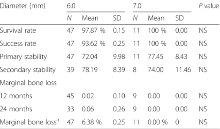
Forty-seven implants of 6 mm in diameter and 11 of 7 mm in diameter were placed, with the length of the implants ranging from 7 to 12 mm (Table 2). Neither prosthesis type nor implant diameter made statistically significant difference in the implant survival or success rate, primary or secondary stability, or marginal bone loss in 12 and 24 months (Tables 3 and 4).
A longer implant tended to show a lower level of pri- mary and secondary stability. Kruskal-Wallis test revealed that implant length was statistically, significantly corre- lated with primary and secondary stability (P < 0.05).
Mann-Whitney U test showed that implants of 7.0 mm in length had statistically, significantly higher levels of pri- mary (P < 0.05) and secondary (P = 0.005) stability than those of 12.0 mm and that there was no statistically sig- nificant difference in primary and secondary stability among the other cases of length. However, there were a relatively small number of 7- and 12-mm implants with
statistical significance: 6 implants of 7 mm, 21 of 8 mm, 23 of 10 mm, and 8 of 12 mm (Table 5).
Twenty-six implants were placed using one-stage sur- gical protocol and 32 using two-stage protocol. For ini- tial stability, one-stage protocol resulted in significantly higher ISQ than the two-stage protocol (P < 0.01). There was no statistically significant difference between the one- and two-stage protocol in secondary stability after the mean healing period of 19.13 weeks for one-stage protocol and 17.16 weeks for two-stage protocol, in the survival rate, or in marginal bone loss after functional loading (Table 6).
Discussion
The correlation between implant length/diameter and the bone quality and implant stability remains contro- versial. Many researchers reported that larger-diameter implants, which had a larger area of contact with the supporting bone and put less stress distribution on peri- implant bone, could more favorably secure high levels of initial stability [18]. The biomechanical analysis also showed that the larger the implant diameter, the greater the removal torque, demonstrating that wide implants are more stable than narrow ones in general [19]. Degidi et al. [20] contended that the bone quality was weakly Table 1 For detect differences of survival rate, success rate,
marginal bone loss, and stability between the maxilla and the mandible
Maxilla Mandible P value
N Mean SD N Mean SD Sig.
(two-tailed) Survival rate 24 95.83 % 0.20 33 100 % 0.00 NS Success rate 24 91.67 % 0.28 34 97.06 % 0.17 NS Primary stability 24 67.42 8.87 34 77.06 8.58 +++
Secondary stability 21 71.52 8.11 26 82.29 6.48 +++
Marginal bone loss
12 months 23 0.04 0.14 31 0.00 0.00 NS 24 months 17 0.11 0.36 25 0.00 0.01 NS Marginal bone
loss
24 8.33 % 0.28 34 2.94 % 0.17 NS
NS nonsignificant
+++Independent-samples t test is significant at the 0.005 level (two-tailed)
Table 2 Number of implant by length and diameter of implant
Size of implants Diameter (mm) Total
6.00 7.00
Length (mm) 7.00 6 0 6
8.00 19 2 21
10.00 16 7 23
12.00 6 2 8
Total 47 11 58
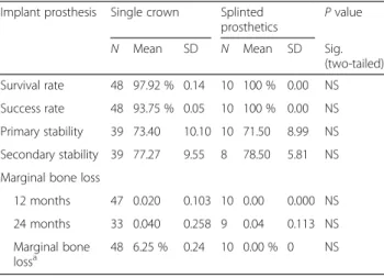
Table 3 For detect differences of survival rate, success rate, marginal bone loss, and stability between types of prosthetics Implant prosthesis Single crown Splinted
prosthetics
P value
N Mean SD N Mean SD Sig.
(two-tailed) Survival rate 48 97.92 % 0.14 10 100 % 0.00 NS Success rate 48 93.75 % 0.05 10 100 % 0.00 NS Primary stability 39 73.40 10.10 10 71.50 8.99 NS Secondary stability 39 77.27 9.55 8 78.50 5.81 NS Marginal bone loss
12 months 47 0.020 0.103 10 0.00 0.000 NS 24 months 33 0.040 0.258 9 0.04 0.113 NS Marginal bone
loss
a48 6.25 % 0.24 10 0.00 % 0 NS
NS nonsignificant
aAbove annually 0.2 mm until final follow-up date
correlated with ISQ, which was significantly affected by implant diameter and length. Wide implants were re- portedly less likely to have their initial stability affected specifically by a low bone quality than general ones [18].
In this study, the maxilla with relatively lower bone density showed a lower level of primary and secondary stability than the mandible. In the maxilla, however, the levels of both primary and secondary stability—67.42 ± 8.87 and 71.52 ± 8.11, respectively—were higher than those of minimal ISQ (65) for immediate or early load- ing [21]. No significant difference was found in the im- plant survival rate or marginal bone loss by arch, and it was believed that the gap in the bone quality between the maxilla and the mandible had no significant effect on the clinical prognosis of ultrawide implants.
The ultrawide Superline (Dentium, Suwon, Korea) in this study, which is characterized by internal connection, an SLA surface, and tapered shape, is up to 7.0 mm in diameter and can be inserted into the sites containing low-quality bone or into those where general-diameter implants failed to be placed. Bihan et al. [14] observed
little difference in ISQ between 3.8- and 4.6-mm-diam- eter implants, both of which were inserted into the can- cellous bone, and no difference was found in primary or secondary stability between the 6- and 7-mm-diameter implants in this study.
Some researchers reported that the implants of
≥5 mm in diameter could cause buccal alveolar bone re- sorption and gingival recession and be less likely to sur- vive due to excessive pressure on buccal bone while they were placed [21]. However, the implants of 6 and 7 mm in diameter in this study were very likely to survive and no more than three (5.17 %) of them were found to cause severe marginal bone loss of ≥0.2 mm on an annual basis during the follow-up. In this study, wide implants were se- lectively applied to the sites containing low-quality bone or to previously failed sites, and care was taken not to put
≥35 Ncm torque in placing implants; therefore, excessive pressure was not put on the buccal bone, causing alveolar bone resorption, which was not excessive, contrary to the research conducted before 2000 [22].
Peri-implant marginal bone resorption is correlated with excessive stress on bone tissues [23]. A 3D geometric analysis showed that offset placement failed to reduce ten- sile force, which could rather be reduced by the decrease in inclination of the cusp as well as by wide implants (>5 mm) [24]. Lateral force, which is put on the placed large-diameter implant, can relieve the load put on the peri-implant marginal bone and put less load on the im- plant than vertical force [25]. A larger-diameter implant lets less stress concentrated on peri-implant cortical bone in the neck due to lateral force in chewing [26]. This is why a small amount of marginal bone loss was observed around ultrawide implants in this study.
The patients with advanced bone loss were suspected of parafunction, such as bruxism, rejected the advice on wearing a night guard during the follow-up, and experi- enced repetitive abutment screw fracture, with one im- plant removed due to failed osseointegration. Patients Table 4 For detect differences of survival rate, success rate,
marginal bone loss, and stability between diameter of implants
Diameter (mm) 6.0 7.0 P value
N Mean SD N Mean SD
Survival rate 47 97.87 % 0.15 11 100 % 0.00 NS Success rate 47 93.62 % 0.25 11 100 % 0.00 NS Primary stability 47 72.04 9.98 11 77.45 8.43 NS Secondary stability 39 78.19 8.39 8 74.00 11.46 NS Marginal bone loss
12 months 45 0.02 0.10 9 0.00 0.00 NS
24 months 33 0.06 0.26 9 0.00 0.00 NS
Marginal bone loss
a47 6.38 % 0.25 11 0.00 % 0 NS
NS nonsignificantaAbove annually 0.2 mm until final follow-up date
Table 5 For detect differences of survival rate, success rate, marginal bone loss, and stability among length of implants
Length (mm) 7.0 8.0 10.0 12.0 P
value
N Mean SD N Mean SD N Mean SD N Mean SD
Survival rate 6 100 % 0.00 21 100 % 0.00 23 100 % 0.00 8 87.50 % 0.35 NS
Success rate 6 100 % 0.00 21 95.24 % 0.22 23 95.65 % 0.21 8 87.50 % 0.35 NS
Primary stability 6 77.33 9.77 21 72.14 10.10 23 75.52 9.27 8 65.25 7.40 +
Secondary stability 6 84.42 3.50 17 76.85 8.59 17 78.97 8.45 7 69.43 9.48 +
Marginal bone loss
12 months 5 0.00 0.00 21 0.02 0.10 21 0.00 0.00 7 0.08 0.20 NS
24 months 4 0.00 0.00 16 0.00 0.00 16 0.03 0.09 6 0.25 0.60 NS
Marginal bone loss
a6 0.00 % 0.00 21 4.76 % 0.22 23 4.35 % 0.22 8 12.50 % 0.35 NS
NS nonsignificant
aAbove annually 0.2 mm until final follow-up date
+Kruskal-Wallis test is significant at the 0.05 level (two-tailed)
with bruxism require a careful follow-up because they can ultimately experience peri-implant bone loss or im- plant failure [27].
Since a high level of initial stability permits implant surgery using one-stage protocol, it is natural that im- plants with one-stage protocol are initially more stable than those with two-stage protocol. No remarkable dif- ference was observed in secondary stability between one- and two-stage protocol after a proper healing period and no statistically significant difference was found in the survival rate or marginal bone loss between them. It is presumed, therefore, that if a surgical situation is taken into account in choosing an implant surgery method and if enough time is given for osseointegration, the implant surgery method will have no special effect on the clinical prognosis.
Many researchers reported that a longer implant was usually more stable and more successful [28–32]. In this study, a longer and wider implant generally tended to have a lower level of primary and secondary stability, with the secondary stability being lower than the pri- mary. In particular, Mann-Whitney U Test showed that implants of 7.0 mm in length had statistically signifi- cantly higher levels of primary and secondary stability than those of 12.0 mm. However, since 7- and 12-mm implants were relatively fewer than the 8- and 10-mm implants, further research should be conducted in a larger sample of implants.
Conclusions
Ultrawide implants of ≥6 mm in diameter showed an ex- cellent survival rate (98.28 %) and a very small amount of marginal bone loss (0.14-mm marginal bone resorp- tion for 46.25 months on average). The mean survival rate was 98.28 ± 0.13 %, with the removal of 1 out of the 58 implants. The mean success rate was 94.83 ± 0.22 % and the factors affecting implant failure included ≥0.2-
mm marginal bone resorption on an annual basis (5.2 %), TMD (1.7 %), and peri-implant gingivitis (1.7 %).
Significant differences were found neither in the volume of marginal bone resorption nor in the implant survival rate by the length of implants, surgical types, location of arch, or prosthetic types. However, a longer implant tended to show a lower level of primary and secondary stability.
It is possible to apply ultrawide implants to the maxillary and mandibular posterior regions with poor bone quality as well as to the previously failed sites, and an implant sur- gery method suitable for an anatomical situation at the time of operation, enough time for osseointegration, and prosthetic maintenance are expected to help bring about clinically favorable effects.
Acknowledgements
This work was supported by the Convergence Research Program from the School of Dentistry and College of Medicine, Seoul National University (grant no. 860 –20140121).
Authors ’ contributions
KJK participated in writing the manuscript and data collection. YYJ performed all the prosthodontic treatment. YPY participated in the study design and performed the statistical analysis. KYK participated in the design and coordination and helped to draft the manuscript. All authors read and approved the final manuscript.
Competing interests
The authors declare that they have no competing interests.
Author details
1
Department of Oral and Maxillofacial Surgery, Section of Dentistry, Seoul National University Bundang Hospital, Gumi-ro 173-82, Bundang-gu, Seongnam City, Gyeonggi-do 13620, South Korea.
2Department of Prosthodontics, Section of Dentistry, Seoul National University Bundang Hospital, Gumi-ro 173-82, Bundang-gu, Seongnam City, Gyeonggi-do 13620, South Korea.
3Department of Dentistry and Dental Research Institute, School of Dentistry, Seoul National University, Daehak-ro 101, Jongno-gu, Seoul 03080, South Korea.
Received: 21 May 2016 Accepted: 27 June 2016
References
1. Haas R, Mensdorff-Pouilly N, Mailath G, Watzek G (1996) Survival of 1,920 IMZ implants followed for up to 100 months. Int J Oral Maxillofac Implants 11:581 –588
2. Bischof M, Nedir R, Szmukler-Moncler S, Bernard J-P, Samson J (2004) Implant stability measurement of delayed and immediately loaded implants during healing. Clin Oral Implants Res 15:529 –539
3. Javed F, Romanos GE (2015) Role of implant diameter on long-term survival of dental implants placed in posterior maxilla : a systemic review. Clin Oral Invest 19:1 –10
4. Ivanoff CJ, Grondahl K, Sennerby L, Bergström C, Lekholm U (1999) Influence of variations in implant diameters: a 3- to 5-year retrospective clinical report. Int J Oral Maxillofac Implants 14:173 –180
5. Attard NJ, Zarb GA (2013) Implant prosthodontic management of partially edentulous patients missing posterior teeth: the Toronto experience.
J Prosthet Dent 89:352 –559
6. Eckert SE, Meraw SJ, Weaver AL, Lohse CM (2001) Early experience with wide-platform Mk II implants. Part I: implant survival. Part II: evaluation of risk factors involving implant survival. Int J Oral Maxillofac Implants 16:208 –216 7. Small PN, Tarnow DP (2000) Gingival recession around implants: a 1-year
longitudinal prospective study. Int J Oral Maxillofac Implants 15:527 –532 Table 6 For detect differences of survival rate, success rate,
marginal bone loss and stability between surgery protocol
Surgery protocol 1-stage 2-stage P value
N Mean SD N Mean SD
Survival rate 26 100 % 0.00 32 96.88 % 0.18 NS Success rate 26 96.15 % 0.20 32 93.75 % 0.25 NS Primary stability 26 76.81 7.80 32 70.03 10.42 * Secondary stability 15 79.97 7.99 32 76.31 9.30 NS Marginal bone loss
12 months 25 0.00 0.00 31 0.01 0.06 NS
24 months 21 0.02 0.07 21 0.07 0.32 NS
Marginal bone loss
a26 3.85 % 32 32 6.25 % 0.42 NS
aNS nonsignificant
aIndependent-samples t test is significant at the 0.05 level (two-tailed)
*Independent-samples t test is significant at the 0.01 level (two-tailed)
8. He J, Shang YW, Deng CF, Shang DH, Zhang C, Wang DN, Zhao BH (2014) A clinical retrospective analysis of dental implants replaced in previously failed site. Shanghai Kou Qiang Yi Xue 23:196 –200
9. Nelissen RC, den Besten CA, Mylanus EA, Hol MK (2016) Stability, survival and tolerability of a 4.5-mm-wide bone-anchored hearing implant: 6-month data from a randomized controlled clinical trial. Eur Arch Otorhinolaryngol 273:105 –111
10. Muftu A, Chapman RJ (1998) Replacing posterior teeth with freestanding implants: four-year prosthodontic results of a prospective study. J Am Dent Assoc 129:1097 –1102
11. Truhlar RS, Orenstein IH, Morris HF, Ochi S (1997) Distribution of bone quality in patients receiving endosseous dental implants. J Oral Maxillofac Surg 55:38 –45
12. Roos J, Sennerby L, Lekholm U, Jemt T, Grondahl K, Albrektsson T (1997) A qualitative and quantitative method for evaluating implant success: a 5-year retrospective analysis of the Branemark implant. Int J Oral Maxillofac Implants 12:504 –514
13. Jaffin RA, Berman CL (1991) The excessive loss of Branemark fixtures in type IV bone: a 5-year analysis. J Periodontol 62:2 –4
14. Bilhan H, Geckili O, Mumcu E, Bozdag E, Sunbuloglu E, Kutay O (2010) Influence of surgical technique, implant shape and diameter on the primary stability in cancellous bone. J Oral Rehabil 37:900 –907
15. Barikani H, Rashtak S, Akbari S, Badri S, Daneshparvar N, Rokn A (2013) The effect of implant length and diameter on the primary stability in different bone types. J Dent (Tehran) 10:449 –455
16. Vandeweghe S, De Ferrerre R, Tschakaloff A, De Bruyn H (2011) A wide- body implant as an alternative for sinus lift or bone grafting. J Oral Maxillofac Surg 69:67 –74
17. Zarb GA, Albrektsson T (1998) Consensus report: towards optimized treatment outcomes for dental implants. J Prosthet Dent 80:641 18. Bardyn T, Gedet P, Hallermann W, Buchler P (2009) Quantifying the
influence of bone density and thickness on resonance frequency analysis:
an in vitro study of biomechanical test materials. Int J Oral Maxillofac Implants 24:1006 –1014
19. Degidi M, Daprile G, Piattelli A (2012) Primary stability determination by means of insertion torque and RFA in a sample of 4,135 implants. Clin Oral Implants Res 14:501 –507
20. Isoda K, Ayukawa Y, Tsukiyama Y, Sogo M, Matsushita Y, Koyano K (2012) Relationship between the bone density estimated by cone-beam computed tomography and the primary stability of dental implants. Clin Oral Implants Res 23:832 –836
21. Glauser R, Sennerby L, Meredith N, Ree A, Lundgren AK, Gottlow J, Hammerle CHF (2004) Resonance frequency analysis of implants subjected to immediate functional occlusal loading. Successful versus failing implants.
Clin Oral Implants Res 15:428 –434
22. Ivanoff CJ, Sennerby L, Johansson C, Rangert B, Lekholm U (1997) Influence of implant diameters on the integration of screw implants. An experimental study in rabbits. Int J Oral Maxillofac Surg 26:141 –148
23. Isidor F. Influence of forces on peri-implant bone. Clin Oral Implants Res 2006;17(Suppl 2):8-18.
24. Hansson S, Werke M (2013) The implant thread as a retention element in cortical bone: the effect of thread size and thread profile: a finite element study. J Biomech 36:1247 –1458
25. Sato Y, Shindoi N, Hosokawa R, Tsuga K, Akagawa Y (2000) A biomechanical effect of wide implant placement and offset placement of three implants in the posterior partially edentulous region. J Oral Rehabil 27:15 –21 26. Santiago JFJ, Pellizzer EP, Verri FR, de Carvalho PS (2013) Stress analysis in
bone tissue around single implants with different diameters and veneering materials: a 3-D finite element study. Mater Sci Eng C Mater Biol Appl 33:4700 –4714
27. Baggi L, Cappelloni I, Di Girolamo M, Maceri F, Vairo G (2008) The influence of implant diameter and length on stress distribution of osseointegrated implants related to crestal bone geometry: a three-dimensional finite element analysis. J Prosthet Dent 100:422 –431
28. Choi YS (2014) Clinical assessment to the dental treatment of bruxism:
literature review. J Dent Rehabil Appl Sci 30:36 –44
29. Lee JY, Lee WC, Kim MS, Kim JE, Shin SW (2012) The influence of implant diameter, length and design changes on implant stability quotient (ISQ) value in artificial bone. J Korean Acad Prosthodont 50:292 –298 30. Sim CP, Lang NP (2000) Factors influencing resonance frequency
analysis assessed by Osstell mentor during implant tissue integration: I.
Instrument positioning, bone structure, implant length. Clin Oral Implants Res 21:598 –604
31. Krennmair G, Waldenberger O (2004) Clinical analysis of wide-diameter frialit-2 implants. Int J Oral Maxillofac Implants 19:710 –715
32. Telleman G, Raghoebar GM, Vissink A, den Hartog L, Huddleston Slater JJ, Meijer HJ (2011) A systematic review of the prognosis of short (<10 mm) dental implants placed in the partially edentulous patient. J Clin Periodontol 38:667 –676
Submit your manuscript to a journal and benefi t from:
7 Convenient online submission 7 Rigorous peer review
7 Immediate publication on acceptance 7 Open access: articles freely available online 7 High visibility within the fi eld
7 Retaining the copyright to your article