대한임상신경생리학회지 11(1):37~39, 2009 ISSN 1229-6414
Copyright 2009 by The Korean Society for Clinical Neurophysiology 37
소세포 폐암 환자에서 발생한 종양신경항체가 음성인 감각신경세포병증 1예
충북대학교 의과대학 신경과학교실
이상수·이형석
A Case of Sensory Neuronopathy without Onconeuronal Antibodies in a Small Cell Lung Carcinoma Patient
Sang-Soo Lee, M.D., Hyung-Suk Lee, M.D.
Department of Neurology, Chungbuk National University College of Medicine, Cheongju, Korea Received 2 October 2008; received in revised form 27 February 2009; accepted 9 March 2009.
Key Words: Paraneoplastic polyneuropathy, Small cell carcinoma
Address for correspondence;
Sang-Soo Lee, M.D.
Department of Neurology,
Chungbuk National University College of Medicine, 410 Sungbong-ro, Heungduk-gu, Cheongju-si, Chungbuk 361-711, Korea
Tel: +82-43-269-6336 Fax: +82-43-275-7591 E-mail: [email protected]
Serum anti-Hu antibodies are regarded as markers of paraneoplastic sensory neuronopathy (PSN) and small cell lung carcinoma (SCLC).1 However, the value of anti-Hu antibodies could have been over- estimated, because PSN patients without anti-Hu antibodies are less likely to be reported unless they present with other anti-neuronal antibodies. There is no significant difference in the clinical features between PSN patients with and without anti-Hu an- tibodies, except for a trend among anti-Hu antibody positive patients to develop evidence of involvement of CNS.2 Anti-amphiphysin antibodies react with a 128-kd protein in the synaptic vesicles. Anti-am-
phiphysin I antibodies are known to be present in patients with SCLC irrespective of the presence of a paraneoplastic neurological disorders.3
CASE REPORT
A 57-year-old man with a 2-month history of a tingling sensation in the hands was admitted. Over the 3 weeks, the tingling sensation extended to the lower extremities. He smoked for 30 years. His past medical history was unremarkable. He had neither dry eyes nor dry mouth. He was alert and cranial nerves were intact. He had minimal weakness of the proximal lower limb muscles. The tendon reflexes were completely absent. Position and vibratory sen- sation in the distal limbs were seriously impaired.
Hypalgesia in the distal legs and allodynia even on the trunk were noted. The distribution of altered sensation area was not symmetrical. Romberg test was positive, but cerebellar function was normal.
The results of the following tests were normal, non-
이상수·이형석
Korean J Clin Neurophysiol / Volume 11 / June, 2009 38
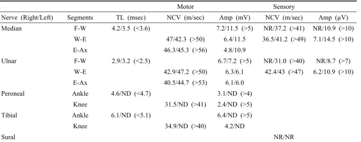
Table 1. Data of nerve conduction studies
Motor Sensory
Nerve (Right/Left) Segments TL (msec) NCV (m/sec) Amp (mV) NCV (m/sec) Amp (µV)
Median F-W 4.2/3.5 (<3.6) 7.2/11.5 (>5) NR/37.2 (>41) NR/10.9 (>10)
W-E 47/42.3 (>50) 6.4/11.5 36.5/41.2 (>49) 7.1/14.5 (>10)
E-Ax 46.3/45.3 (>56) 4.8/10.9
Ulnar F-W 2.9/3.2 (<2.5) 6.7/7.2 (>5) NR/31.0 (>40) NR/8.7 (>7)
W-E 42.9/47.2 (>50) 6.3/6.1 42.4/43 (>47) 6.2/10.9 (>10)
E-Ax 40.5/44.7 (>53) 6.1/6.0
Peroneal Ankle 4.6/ND (<4.7) 3.1/ND (>4)
Knee 31.5/ND (>41) 2.4/ND (>5)
Tibial Ankle 6.1/ND (<5.1) 6.4/ND (>5)
Knee 34.9/ND (>40) 4.2/ND
Sural NR/NR
TL; terminal latency, NCV; nerve conduction velocity, AMP; amplitude, F-W; finger-wrist, NR; no response, W-E; wrist- elbow, E-Ax; elbow-axilla, ND; not done (reference value of normal limit within parentheses).
specific, or negative: routine chemical batteries;
simple chest X-ray; serum vitamin B12; thyroid study; urinalysis; serum protein and immunoelec- trophoresis; C-reactive protein; hepatitis Bs anti- gen; cryoglobulin, rheumatoid arthritis factor; au- toantibodies (anti-double stranded DNA; fluorescent anti-nuclear; anti-Ro; anti-La; anti-neutrophil cytoplasmic; anti-Hu; and anti-amphiphysin). The CSF examination showed elevated protein concen- tration (159 mg%) without cellular reaction. Nerve conduction abnormalities were in keeping with sen- sory neuropathy with some motor involvement (Table 1). The EMG did not reveal any abnormality.
Although he had neither anti-onconeuronal anti- bodies nor abnormality in chest X-ray, a history of smoking made us perform a chest CT. It revealed multiple conglomerated lymph node enlargements in the mediastinum and right hilar area. The broncho- scopic biopsy showed a small cell lung carcinoma.
The sensory deficits progressed proximally further into the trunk and he could not stand without aid. In spite of steroid medication, chemotherapy and radi- ation therapy for SCLC, he died 1 year after the diagnosis.
DISCUSSION
Our patient had all characteristics of definite par- aneoplastic subacute sensory neuronopathy with minimal motor nerve involvement. The criteria of definite PSN include subacute onset with a Rankin score of at least 3 before 12 weeks of evolution, on- set of numbness, and often pain, marked asymmetry of symptoms at onset, involvement of the arms, pro- prioceptive loss in the areas affected, electro- physiological studies that show marked, but not re- stricted, involvement of the sensory fibers with ab- sent sensory nerve action potentials in at least one of nerves studied.4
Approximately 20% of sensory neuronopathy are paraneoplastic; the remainder are associated with systemic immune disorders, or toxin exposure or re- main idiopathic.5 Paraneoplastic sensory neuronop- athy is uncommon, affecting less than 1% of patients who have SCLC.5 The underlying neoplasm of para- neoplastic sensory neuronopathy is SCLC in 80% to 90% of cases, but subacute sensory neuronopathy may also occur with breast cancer, ovarian cancer, sarcoma or Hodgkin’s disease. In nearly all patients, the neurologic syndrome and seropositivity for onco- neuronal antibodies precede diagnosis of the tumor.
소세포 폐암 환자에서 발생한 종양신경항체가 음성인 감각신경세포병증 1예
Korean J Clin Neurophysiol / Volume 11 / June, 2009 39
Neuropathies which occur within a few years of the tumor evolve rapidly and correspond mostly to in- flammatory disorders. The sensory neuropathy- ganglinopathy that is related to Sjögren disease and an idiopathic variety do not have the onconeuronal antibodies, making its presence a reliable marker for lung cancer. As dysimmune neuropathies are prob- ably paraneoplastic in a limited number of cases, patients with these disorders should probably not be investigated systematically for carcinoma in the ab- sence of anti-onconeuronal antibodies, except when the neuropathy is associated with encephalomyelitis and probably with vasculitis.6 Nevertheless, it can- not be stated that seronegativity in a patient with sensory neuronopathy excludes cancer. Well-char- acterized paraneoplastic antibodies in sensory neu- ronopathy are anti-Hu antibody, anti-amphiphysin antibody, and anti-CV2 antibody.7 It was known that the specificity of Anti-Hu antibody was 99%
and the sensitivity was 82%.2 Anti-amphiphysin an- tibodies are associated with various paraneoplastic syndromes including subacute sensory neuropathy, paraneoplastic encephalomyelitis and stiff-man syndrome. The incidence of anti-amphiphysin anti- bodies in patient with cancer, but without paraneo- plastic disorders, is probably low. Therefore, what- ever their pathogenic role, they are useful tools for the diagnosis of tumors in patients with suspected paraneoplastic disorders. Regarding the anti-CV2 antibody, we could not check it because it was not available in Korea.
The paraneoplastic antibodies appear to be a use-
ful tool for diagnosing a neurological disorder as paraneoplastic and indicating the probable type of underlying tumor. However, our case also suggests that the diagnostic approach to detection of under- lying tumor in Anti-Hu antibody negative patients with sensory neuronopathy should be similar to that recommended for those patients with Anti-Hu anti- body or other onconeuronal antibodies and directed to the lung.
REFERENCES
1. Dalmau J, Graus F, Rosenblum MK, Poser JB. Anti-Hu-asso- ciated paraneoplastic encephalomyelitis/sensory neuronopathy.
A clinical study of 71 patients. Medicine 1992;71:59-72.
2. Molinuevo JL, Graus F, Serrano C, Reñe R, Guerrero A, IllaI.
Utility of anti-Hu antibodies in the diagnosis of paraneoplastic sensory neuronopathy. Ann Neurol 1998;44:976-980.
3. Saiz A, Dalmau J, Butler MH, Chen Q, Delattre JY, Camilli PD, et al. Anti-amphiphysin I antibodies in patients with para- neoplastic neurological disorders associated with small cell lung carcinoma. J Neurol Neurosurg Psychiatry 1999;66:214- 217.
4. Graus F, Delattre JY, Antoine JC, Dalmau J, Giometto B, Grisold W, et al. Recommended diagnostic criteria for para- neoplastic neurological syndromes. J Neurol Neurosurg Psy- chiatry 2004;75:1135-1140.
5. Elrington GM, Murray NM, Spiro SG, Newsom-Davis J.
Neurological paraneoplastic syndromes in patients with small cell lung cancer. A prospective survey of 150 patients. J Neurol Neurosurg Psychiatry 1991;54:764-767.
6. Antoine JC, Mosnier JF, Absi L, Convers P, Honnorat J, Michel D. Carcinoma associated paraneoplastic peripheral neu- ropathies in patients with and without anti-onconeural anti- bodies. J Neurol Neurosurg Psychiatry 1999;67:7-14.
7. de Beukelaar JW, Sillevis Smitt PA. Managing paraneoplastic neurological disorders. Oncologist 2006;11:292-305.