ISSN 1225-6552, eISSN 2287-7630 https://doi.org/10.7853/kjvs.2019.42.4.169
< Original Article >
Veterinary Service
Available online at http://kjves.org
*Corresponding author: Jong-Hyun Kim, Tel. +82-55-772-2366, Fax. +82-55-772-2308, E-mail. [email protected]
비접촉 조건에서의 Naegleria fowleri 에 의한 표적세포의 세포독성
강창근
1ㆍ홍일화
1ㆍ김종현
1,2*경상대학교 동물의학연구소1, 경상대학교 수의과대학 기생충학연구실2
Cytotoxicity of target cell against Naegleria fowleri under non-contact condition
Changgeun Kang1, Il-Hwa Hong1, Jong-Hyun Kim1,2*
1Institute of Animal Medicine, Gyeongsang National University, Jinju 52828, Korea
2Laboratory of Parasitology, College of Veterinary Medicine, Gyeongsang National University, Jinju 52828, Korea (Received 7 February 2019; revised 11 September 2019; accepted 30 September 2019)
Abstract
Naegleria fowleri, a pathogenic free-living amoeba, leads to a fatal infection known as primary amebic meningoencephalitis (PAM) in human and animals. PAM is an acute, fulminant, necrotizing, and hemor- rhagic disease that leads to death in approximately seven days. In this study, we investigate the cytotox- icity of target cells and the secreted molecules of N. fowleri under the non-contact condition. The target cell (U87MG cell) treated with N. fowleri lysates showed no morphological changes and no cytotoxicity.
By contrast, the U87MG cells co-cultured with N. fowleri trophozoites under the non-contact condition induced morphological changes and reduction in number. When U87MG cells were co-cultured with N.
fowleri trophozoites under the non-contact condition for 30 min, 2 hr, and 4 hr, the levels of cytotox- icity of target cells were 32.3, 35.5, and 37.8%, respectively. Particularly, when the ratio of amoeba to target cells is 10 to 1, the level of cytotoxicity of target cells was 49.7% at 30 min. To show the proteins secreted from N. fowleri under the non-contact condition, we carried out 2D electrophoresis and observed 6 major proteins. Finally, these results suggest that the molecules released from N. fowleri un- der the non-contact condition induce the cell death and this process is an important step in pathogenesis of N. fowleri.
Key words : Naegleria fowleri, Cytotoxicity, Non-contact condition, U87MG cells
서 론
자유생활아메바(free-living amoeba)는 토양과 담수 에서 자유생활을 하며, 자연 환경에서는 세균들을 포 식하며 살아가는 아메바들로써 현재까지는 자유아메 바(Naegleria spp.), 가시아메바(Acanthamoeba spp.),
Balamuthia 등이 보고되고 있다(Visvesvara 등, 2007).
특히, 병원성이 강한 Naegleria fowleri는 사람과 동물 들에게 원발성 아메바성 수막뇌염(primary amoebic
meningoencephalitis; PAM)을 일으킨다(Carter, 1970;
Schuster와 Visvesvara, 2004). N. fowleri는 비강을 통해 침입하며, 비인후 점막을 뚫고 들어와 혈관-뇌 장벽을 뚫고, 뇌신경으로 빠르게 침입한다(Carter, 1968; An- derson과 Jamieson, 1972). 원발성 아메바성 수막뇌염 은 매우 빠르고, 치명적인 질병으로 보통 감염된 지 일주일에서 이주일 이내 사망하게 된다(Visvesvara 등, 2007). 특히, 원발성 아메바성 수막뇌염은 진단이 매 우 어렵고 아직까지 적절한 치료제가 없기 때문에 치 사율이 98%에 이른다(Apley 등, 1970; Shin과 Im, 2004; Visvesvara 등, 2007).
N. fowleri의 병원성 유발기전은 아직 정확히 밝혀 져 있지는 않았지만, 표적세포에 부착이 일어난 다음 표적세포의 사멸을 유도하는 세포병변 효과가 일반적 이다(Jonckheere, 1980). N. fowleri의 병인기전에서 표 적세포에 부착은 아메바가 비강을 통해 중추신경계로 들어갈 수 있는 가장 중요한 과정이다. N. fowleri 뿐 만 아니라 근연종인 가시아메바(Acanthamoeba spp.)의 경우에도 먼저 표적세포에 부착을 한 뒤, 여러 가지 병인인자들을 분비하여 표적세포가 사멸되도록 유도 한다(Jonckheere, 1980; Ferrante와 Bates, 1988; Khan 등, 2000). Han 등(2004)은 아메바의 부착에 있어서 integrin-like protein과 protein kinase C, 그리고 fi- bronectin의 중요성에 대해 보고한바 있다. 이전까지 보고된 바에 따르면, 접촉 의존성 과정으로는 Nfa1 단 백질이 중요한 역할을 한다고 생각된다. Nfa1 단백질 은 아메바의 위족에 위치하며, 아메바가 식세포 작용 을 하는 과정에서 food-cup 구조를 형성하는데 매우 중요한 역할을 한다고 보고되었다(Shin 등, 2001;
Kang 등, 2005). 특히, N. fowleri에 항-Nfa1 항체를 처 리하였을 때, 아메바가 표적세포를 죽이는 세포독성 효과가 현저하게 감소한다는 연구결과는 표적세포에 부착에 있어서 매우 중요한 기능을 담당하는 Nfa1 단 백질이 N. fowleri의 병원성에 크게 기여한다는 것을 입증한다(Jeong 등, 2004; Oh 등, 2005). 또한 아메바의 운동성에 관여하는 위족의 형성과 고온에 대한 내열 성과 연관이 있는 열충격단백질-70 (HSP-70)도 Nfa1 단백질과 더불어 N. fowleri의 식세포 작용에 중요한 역할을 한다고 보고되었다(Song 등, 2008).
이처럼 N. fowleri가 표적세포를 공격하는데 있어서 부착과정이 가장 중요하고, 병인과정의 시작점이 된 다 하더라도, 아메바에 의해 분비되는 phospholipases, neuraminidases, pore forming enzymes 같은 병인인자도 표적세포를 사멸시키는데 매우 중요한 요소이다(Cursons 등, 1978; Jonckheere, 1980; Marciano-Cabral과 John, 1983; Eisen과 Franson, 1987; Barbour와 Marciano- Cabral, 2001; Herbst 등, 2004). 이러한 비접촉 과정은
N. fowleri에서 분비되는 여러 수용성 용해 물질들을
통한 표적세포의 사멸 과정을 말한다. 본 연구에서는N. fowleri가 표적세포를 사멸시키는 과정에서 접촉 과
정(contact pathways) 뿐만 아니라 비접촉 과정(non-con- tact pathways)의 중요성에 대해 알아보고자 한다. 따 라서 본 연구에서는 비접촉 상태에서의 N. fowleri에 의한 표적세포의 세포독성 효과를 알아보고 이때 분 비되는 물질들에 대해 알아보고자 한다.재료 및 방법
N. fowleri 배양 및 lysate의 준비
병원성이 강한 N. fowleri (ATCC No. 30215)를 75 cm2 culture flask (Nunc. Danmark)에 15 mL의 Nelson 배지를 이용하여 37°C 항온기에서 무균적으로 배양하 였다(Willaert, 1971). 배양된 N. fowleri 영양형(tropho- zoites)을 800×g에서 10분 동안 원심분리하여 침전물 을 얻고, phosphate-bufferedsaline (PBS)으로 두 번 씻 어준다. 모아진 N. fowleri 영양형을 freeze-thawing 방 법을 이용하여 lysate를 만든다(Shin과 Im, 1992).
표적세포 배양
사람교모세포주인 U87MG 세포는 60 mm petri dish (Nunc, Denmark)에 10 mL의 Dulbecco’s modified ea- gle’s medium (DMEM; Sigma, USA)과 10% fetal bo- vine serum (Gibco BRL, USA), 1% penicillin/strep- tomycin (Gibco BRL)을 첨가하여 37°C의 온도로 5%
CO2 항온기에서 무균적으로 배양하였다.
비접촉 배양 시스템
6 well culture plate (Nunc, Danmark)에 3×10⁵ cell/well로 각각의 세포를 배양하고, 0.2 m pore membrane을 갖는 25 mm tissue culture insert (Nunc, Denmark)를 이용하여 비접촉 배양 조건을 만든 후, membrane 윗부분에 N. fowleri를 각각 6×10⁵ troph- ozoites/well, 1.5×10⁶ trophozoites/well 그리고 3×10⁶ trophozoites/well의 농도로 5% CO2 항온기에서 37°C 의 온도로 배양하였다.
형태학적 관찰
37°C 항온기에서 배양한 6 well culture plate를 30 분, 2시간, 4시간째에 꺼내어 광학현미경으로 표적세 포의 형태학적 모양을 관찰하였다.
LDH release assay를 이용한 세포독성 실험
세포독성을 측정하기 위하여 lactate dehydrogenase (LDH) release assay를 시행하였다. 96 well plate에 배 양된 N. fowleri의 형태학적 관찰과 수를 측정한 뒤,
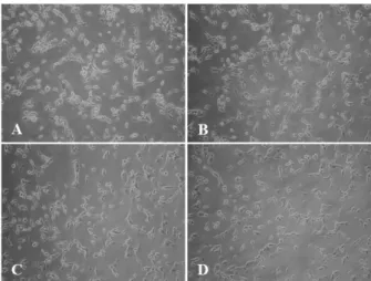
Fig. 1. Light microscopic images. (A) U87MG cells (3×10⁵ cells/well) without N. fowleri lysates were used as negative control.
The U87MG cells were treated with N. fowleri lysates at concen- trations of 1 mg/mL for 30 min (B), 2 hrs (C), and 4 hrs (D), re- spectively (×200).
세포독성 실험을 진행하였다. 실험을 위해 96 well as- say plate (Nunc, Danmark)를 준비하고, 여기에 96 well culture plate에서 배양한 N. fowleri를 6,000 rpm에서 3 분간 원심분리 한 뒤, 상청액 45 L를 96 well assay plate에 넣어주었다. 여기에 CytoTox96 non-radioactive cytotoxicity assay kit (Promega, USA)에 있는 recon- stituted assay buffer를 45 L 첨가한 후, 실온에서 30 분간 보관하였다. 발색정도에 따라 2∼3시간 정도까 지 반응시키고, 발색을 정지하기 위해 stop solution 45
L를 첨가하여 보관하였다. 각 well의 세포독성 결과 는 ELISA reader를 이용하여 490 nm에서 흡광도를 측 정하였다. 이때 세포독성 공식은 다음과 같다.
세포독성(%)={(실험군의 흡광도–대조군의 흡광도) /대조군의 흡광도}×100
2차원 전기영동
2차원 전기영동은 다음과 같이 시행하였다. 1차원 isoelectro focusing (IEF)은 immobilized linear PH 4∼7 gradient (IPG) strips (Bio-Rad, USA)을 사용하였고 protean IEF cell (Bio-Rad)을 이용하여 20°C에서 수행 하였다. 단백질을 농축하여 7 M urea, 2 M thiourea, 4% CHAPS w/v, 20 mM DTT, bromophenol blue와 섞 어 최종적으로 350 L가 되도록 하였다. 16시간 동안 passive rehydration 과정을 거친 후, 300 V에서 1시간, 4,000 V에서 2시간, 4,000 V에서 6시간 30분의 조건으 로 IEF를 수행하였다. IEF가 끝난 후 strips은 6 M urea, pH 8.8, 0.375 M Tris, 2% SDS, 그리고 20%
glycerol이 포함된 equilibration 완충액에서 알칼리화 시킨 후, 10% polyacrylamide gel로 2차원 전기영동을 수행하였다. SDS-PAGE로부터 electro blotting 하기 위 해 PVDF membrane에 transfer시키고, Coomassie blue 로 염색하였다.
통계 처리
대조군과 실험군 사이의 통계학적 유의성 검정은 Student’s t-test를 사용하였으며 P값이 0.05 이하인 것 만 유의한 것으로 하였다.
결 과
N. fowleri lysate에 의한 표적세포의 형태학적 변화 및 세포독성
N. fowleri lysate가 표적세포에 어떠한 영향을 미치 는지 알아보고자 N. fowleri lysate를 추출하여 U87MG 세포에 1 mg/mL의 농도로 처리하였다. N. fowleri lysate 처리 후, 30분, 2시간, 4시간째에 U87MG 세포의 형태 학적 변화를 광학현미경을 이용하여 관찰하였다. 관 찰 결과, N. fowleri lysate에 의한 표적세포의 형태학 적 변화는 크게 관찰되지 않았다(Fig. 1). 또한 N.
fowleri lysate에 의한 표적세포의 세포독성을 확인하
기 위해 LDH release assay를 실시하였다. N. fowleri 및 U87MG 세포를 단독으로 배양한 대조군에서는 30 분, 2시간, 4시간 모두에서 세포독성이 거의 나타나지 않았다. N. fowleri lysate를 1 mg/mL의 농도로 처리한 그룹에서는 30분, 2시간, 4시간에서의 세포독성이 각 각 3.7%, 5.2%, 5.6%로 측정되었으며, 이는 N. fowleri 및 U87MG 세포를 단독으로 배양한 대조군에 비해 세 포독성이 약 2배정도 증가되었다(*P<0.05) (Fig. 2).비접촉 상태에서의 N. fowleri 에 의한 표적세포의 형태학적 변화 및 세포독성
비접촉 병인기전에 의한 N. fowleri의 표적세포의 변화를 알아보고자 tissue culture insert 장치를 이용한
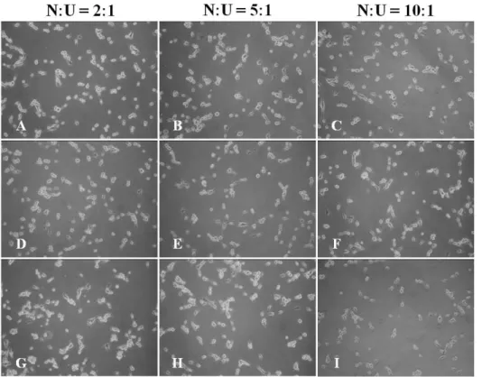
Fig. 3. Light microscopic images.
The U87MG cells were co-cul- tured with N. fowleri trophozoites under the non-contact condition for 30 min (A∼C), 2 hrs (D∼F), and 4 hrs (G∼I), respectively (×200). N, N. fowleri; U, U87MG cells.
Fig. 2. The cytotoxicity of U87MG cells against N. fowleri lysate by LDH release assay. The U87MG cells were treated with N. fowleri ly- sates for 30 min, 2 hrs, and 4 hrs, respectively. The groups of N. fowl- eri alone and U87MG cells alone were used as the controls.
비접촉 배양 상태에서 U87MG 세포와 N. fowleri를 함 께 배양하였다. N. fowleri와 표적세포를 함께 배양 후, 30분, 2시간, 4시간째에 U87MG 세포의 형태학적 변 화를 광학현미경을 이용하여 관찰하였다. N. fowleri 영양형 수에 따라 U87MG 세포의 사멸이 크게 증가하 는 것을 관찰할 수 있었으며 특히, 30분이라는 짧은 배양시간에서도 세포 사멸이 급속도로 빠르게 시작되 는 것을 관찰하였다(Fig. 3). 또한 LDH release assay를 통해 표적세포의 세포독성을 관찰한 결과, N. fowleri
와 표적세포를 2:1의 비율로 30분, 2시간, 4시간 동안 배양하였을 때, 세포독성은 각각 32.3%, 35.5%, 37.8%
로 나타났다(Fig. 4). N. fowleri lysate를 1 mg/mL로 처 리한 그룹과 비교하였을 때 비접촉 상태에서 N. fowl-
eri를 표적세포와 함께 배양한 그룹에서는 세포독성이
매우 크게 증가되었다(*P<0.05). 특히, N. fowleri와 표적세포를 10:1의 비율로 함께 배양하였을 경우에는 30분에서 49.7% 세포독성이 일어났다(Fig. 4).비접촉 상태에서 분비되는 N. fowleri의 단백질 분석
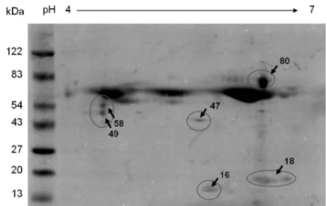
비접촉 상태에서 분비되는 N. fowleri의 단백질을 확인하기 위해 비접촉 배양 상태에서 N. fowleri와 U87MG 세포를 함께 배양한 후, 상청액을 모아 2차원 전기영동을 시행하였다. 2차원 전기영동 결과, 16, 18, 47, 49, 58, 80 kDa의 주요 단백질이 특징적으로 관찰 되었다(Fig. 5).
고 찰
N. fowleri는 사람 및 동물에서 원발성 아메바성 수 막뇌염(PAM)을 일으키는 병원성 원충이다. 비강을 통
Fig. 4. The cytotoxicity of U87MG cells against N. fowleri by LDH release assay. The U87MG cells were co-cultured with N. fowleri trophozoites and were treated with N. fowleri lysates under the non-contact condition for 30 min, 2 hrs, and 4 hrs, respectively. The group of N. fowleri lysate was used as the control group. N, N. fowleri;
U, U87MG cells.
Fig. 5. Protein spots of supernatants collected from U87MG cells co-cultured with N. fowleri for 1 hr under the non-contact condition post 2D electrophoresis. The spots of supernatants were electro- transferred to PVDF membrane and were stained with comassie blue post 2D electrophoresis.
해 침입하여 후신경과 뇌막을 침범하면서 수막뇌염으 로 발전시켜 감염 후 10일 이내에 사망에 이르게 한 다(Carter, 1968; Anderson과 Jamieson, 1972). 일반적으 로 감염 후 증상은 메스꺼움, 구토, 고열, 사지의 기능 장애 및 뇌염을 동반하며 질병의 진행 정도가 매우 빠 르고 감염부위의 특수성 때문에 진단이 어려워 치사 율이 98%에 이르지만, 아직까지도 적절한 치료와 예 방법은 미비한 상태이다(Shin과 Im, 2004; Visvesvara 등, 2007).
N. fowleri의 최초 병원성 유발기전은 표적세포에 대한 아메바의 접촉이며, 이때 N. fowleri는 food-cup structure 또는 amoebastome이라는 구조물 만들어 식세 포작용을 통해 표적세포를 파괴한다. 기존 연구에서 는 표적세포로의 부착과 연관된 N. fowleri의 주요 물 질들로 integrin-like protein 및 protein kinase C가 보고 되었고, 세포융해에 관여하는 병인 요소로 phospholi- pases, neuraminidases, pore forming protein 등이 보고 된바 있다(Cursons 등, 1978; Jonckheere, 1980; Eisen 과 Franson, 1987; Barbour와 Marciano-Cabral, 2001;
Han 등, 2004; Herbst 등, 2004). 이처럼 N. fowleri의 병인기전에서 접촉 의존성 과정은 아메바 세포질의 분출형태인 food-cup 구조나 amoebastome이 주로 관 여하여 세포를 탐식하는 과정이다. 하지만 본 연구에 서는 비접촉 과정에서의 다양한 수용성 용해 물질들 에 의한 세포사멸 과정에 주안점을 두고 비접촉 상태 에서의 N. fowleri에 의한 표적세포의 세포독성 효과를 알아보고자 하였다.
N. fowleri가 표적세포에 접촉하기 이전에 분비되는 물질들에 의해 표적세포들이 어떤 영향을 받는지 알
아보기 위해 N. fowleri와 표적세포를 tissue culture in- sert를 이용하여 비접촉 상태를 만들고 이때 표적세포 의 변화를 관찰하였다. 표적세포는 30분이라는 매우 짧은 시간에서도 세포사멸이 유도되었으며, 특히 아 메바와 표적세포를 10:1의 비율로 배양하였을 경우에 는 30분에서 절반에 가까운 세포가 파괴되는 결과를 보였다. 4시간 동안 고농도의 N. fowleri lysate를 처리 하였을 때에도 대조군에 비해 세포독성의 변화가 없 었던 것과 비교해 봤을 때, 아메바가 표적세포와 함께 있는 상황에서 분비되는 병인 물질에 의해서는 매우 짧은 시간에도 표적세포의 사멸을 유도되었으며, 이 는 매우 놀라운 결과라고 생각된다. 이처럼 비접촉 상 태에서 강한 세포독성을 유발하는 N. fowleri의 단백질 을 확인하기 위해 시행한 2차원 전기영동 결과에서도 6가지의 중요 단백질들을 확인할 수 있었다.
본 연구를 통해, N. fowleri가 표적세포에 접촉하는 과정이 없이도 자체 분비 물질들을 통해 표적세포의 세포사멸을 유도시킬 수 있다는 결과를 얻었다. 특히, 30분이라는 매우 짧은 시간에서도 세포의 사멸이 진 행된다는 사실은 표적세포가 인접한 상황에서 N.
fowleri가 분비하는 물질이 세포사멸에 있어서 매우
강한 독성을 미친다는 것을 잘 보여준다. 또한 N.fowleri lysate를 처리한 세포독성 결과와 tissue culture
insert를 이용한 비접촉 상태에서의 결과가 큰 차이를 보이는 이유는 lysate는 아메바가 휴지기(resting) 상태 에서 분비하는 물질인 반면, 비접촉 상태에서 아메바 가 분비하는 물질은 표적세포가 N. fowleri로 분비하는 다양한 사이토카인(cytokine)이나 케모카인(chemokine) 과 같은 자극 물질에 의해 N. fowleri가 상호 자극을받은 상태에서 분비한 물질이기 때문에 이와 같은 차 이를 보이는 것이라고 생각된다.
N. fowleri는 식세포작용과 같은 접촉에 의해 표적 세포를 탐식할 뿐만 아니라 비접촉 상태에서 분비하 는 물질에 의해서도 강력한 세포사멸을 유도한다는 결과를 얻을 수 있었으며, 향후 본 연구를 통해 얻은
N. fowleri의 주요 단백질들의 추가적인 분석과 기능을
확인하는 연구가 필요하겠다. 이상의 연구결과는 N.fowleri의 병인 과정을 이해하는데 도움을 주며, 원발
성 아메바성 수막뇌염의 효과적인 진단법 및 치료제 개발의 기초자료로 활용될 수 있을 것이다.결 론
병원성 N. fowleri의 비접촉성 병인기전에 의해 일 어나는 표적세포(U87MG 세포)의 세포독성과 이때 분 비되는 N. fowleri의 물질에 대해 알아보고자 하였다.
첫째, N. fowleri lysate에 의한 표적세포의 형태학적 변화 및 세포독성은 관찰되지 않았다. 둘째, 비접촉 상태에서의 표적세포의 변화를 알아보고자 tissue cul- ture insert를 이용하여 U87MG 세포와 N. fowleri를 함 께 배양하였고, 이때 32.3∼49.7%의 세포독성이 관찰 되었다. 셋째, 비접촉 상태에서 분비되는 N. fowleri의 단백질을 확인하기 위한 2차원 전기영동 결과, 6가지 의 주요 단백질이 특징적으로 관찰되었다. 이를 통해, 비접촉 상태에서 N. fowleri가 분비하는 병인 물질에 의해 표적세포의 세포사멸이 유도된다는 것을 확인하 였다.
감사의 글
본 연구는 2015년도 경상대학교 발전기금재단 재원 으로 수행되었습니다.
REFERENCES
Anderson K, Jamieson A. 1972. Primary amoebic meningoen- cephalitis. Lancet 19: 379.
Apley J, Clarke SK, Roome AP, Sandry SA, Saygi G, Silk B, Warhurst DC. 1970. Primary amoebic meningoencephali- tis in Britain. Br Med J 7: 596-599.
Barbour SE, Marciano-Cabral F. 2001. Naegleria fowleri amoe-
bae express a membrane-associated calcium-independent phospholipase A. Biochim Biophys Acta 1530: 123-133.
Carter RF. 1968. Primary amoebic meningoencephalitis: clinical, pathological and epidemiological features of six fatal cases. J Pathol Bacteriol 96: 1-25.
Carter RF. 1970. Description of a Naegleria species isolated from two cases of primary amoebic meningoencephalitis and of the experimental pathological changes induced by it.
J Pathol 100: 217-244.
Cursons RT, Brown TJ, Keys EA. 1978. Virulence of pathogenic free-living amebae. J Parasitol 64: 744-745.
De Jonckheere JF. 1980. Growth characteristics, cytopathic effect in cell culture, and virulence in mice of 36 type strains belonging to 19 different Acanthamoeba spp. Appl Environ Microbiol 39: 681-685.
Eisen D, Franson RC. 1987. Acid-active neuraminidases in the growth media from cultures of pathogenic Naegleria fowleri and in sonicates of rabbit alveolar macrophages.
Biochim Biophys Acta 924: 369-372.
Ferrante A, Bates EJ. 1988. Elastase in the pathogenic free-living amebae Naegleria and Acanthamoeba spp. Infect Immun 56: 3320-3321.
Han KL, Lee HJ, Shin MH, Shin HJ, Im KI, Park SJ. 2004. The involvement of an integrin-like protein and protein kin- ase C in amoebic adhesion to fibronectin and amoebic cytotoxicity. Parasitol Res 94: 53-60.
Herbst R, Marciano-Cabral F, Leippe M. 2004. Antimicrobial and pore-forming peptides of free-living and potentially highly pathogenic Naegleria fowleri are released from the same precursor molecule. J Biol Chem 279: 25955- 25958.
Jeong SR, Kang SY, Lee SC, Song KJ, Im KI, Shin HJ. 2004.
Decreasing effect of an anti-Nfa1 polyclonal antibody on the in vitro cytotoxicity of pathogenic Naegleria fowleri.
Korean J Parasitol 42: 35-40.
Kang SY, Song KJ, Jeong SR, Kim JH, Park S, Kim K, Kwon MS, Shin HJ. 2005. Role of the Nfa1 protein in patho- genic Naegleria fowleri co-cultured with CHO target cells. Clin Diagn Lab Immunol 12: 873-876.
Khan NA, Jarrol EL, Panjwani N, Cao Z, Paget TA. 2000.
Proteases as markers for differentiation of pathogenic and non-pathogenic species of Acanthamoeba. J Clin Microbiol 43: 391-395.
Marciano-Cabral F, John DT. 1983. Cytopathogenicity of Naegleria fowleri for rat neuroblastoma cell cultures:
scanning electron microscopy study. Infect Immun 40:
1214-1217.
Oh YH, Jeong SR, Kim JH, Song KJ, Kim K, Park S, Sohn S, Shin HJ. 2005. Cytopathic changes and pro-in- flammatory cytokines induced by Naegleria fowleri trophozoites in rat microglial cells and protective effect of an anti-Nfa1 antibody. Parasite Immunol 27: 453-459.
Schuster FL, Visvesvara GS. 2004. Free-living amoebae as oppor- tunistic and non-opportunistic pathogens of humans and animals. Int J Parasitol 34: 1001-1027.
Shin HJ, Cho MS, Jung SY, Kim HI, Park S, Kim HJ, Im KI.
2001. Molecular cloning and characterization of a gene encoding a 13.1 kDa antigenic protein of Naegleria fowleri. J Eukaryot Microbiol 48: 713-717.
Shin HJ, Im KI. 1992. Analysis of antigenic specificities of Naegleria fowleri using EITB. Yonsei Rep Trop Med 23: 9-13.
Shin HJ, Im KI. 2004. Pathogenic free-living amoebae in Korea.
Korean J Parasitol 42: 93-119.
Song KJ, Song KH, Kim JH, Sohn HJ, Lee YJ, Park CE, Shin
HJ. 2008. Heat shock protein 70 of Naegleria fowleri is important factor for proliferation and in vitro cytotoxicity. Parasitol Res 103: 313-317.
Visvesvara GS, Moura H, Schuster FL. 2007. Pathogenic and op- portunistic free-living amoebae: Acanthamoeba spp., Balamuthia mandrillaris, Naegleria fowleri, and Sappinia diploidea. FEMS Immunol Med Microbiol 50: 1-26.
Willaert E. 1971. Isolement et culture in vitro des amibes de genre Naegleria. Ann Soc Belg Med Trop 51: 701-708.