• 검색 결과가 없습니다.

Impact of Plaque Composition on Long-Term Clinical Outcomes in Patients with Coronary Artery Occlusive Disease

N/A
N/A
Protected

Academic year: 2022

Share "Impact of Plaque Composition on Long-Term Clinical Outcomes in Patients with Coronary Artery Occlusive Disease"

Copied!
7
0
0

로드 중.... (전체 텍스트 보기)

전체 글

(1)

Korean Circulation Journal

Introduction

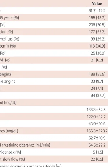
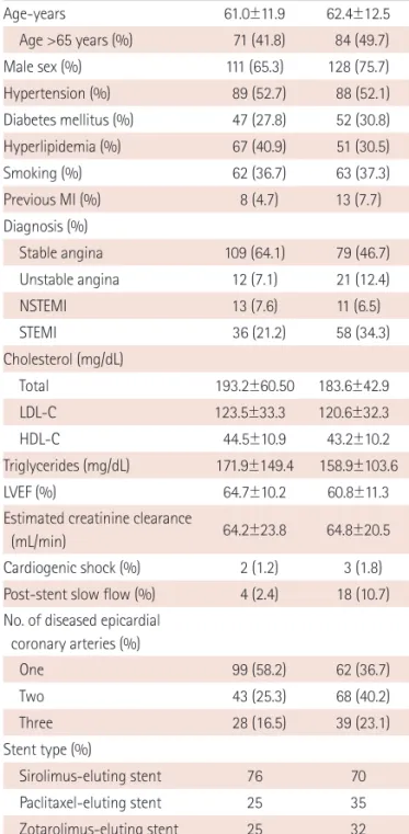
Atherosclerotic plaque responsible for coronary heart disease is

http://dx.doi.org/10.4070/kcj.2013.43.6.377 Print ISSN 1738-5520 • On-line ISSN 1738-5555

Impact of Plaque Composition on Long-Term Clinical Outcomes in Patients with Coronary Artery Occlusive Disease

Ki Hong Kim, MD 1 , Wan Ho Kim, MD 2 , Hyun Woong Park, MD 1 , In Girl Song, MD 1 , Dong Ju Yang, MD 1 , Young Hoon Seo, MD 1 , Hyung Bin Yuk, MD 1 , Yo Han Park, MD 1 , Taek Geun Kwon, MD 1 ,

Charanjit S Rihal, MD 3 , Amir Lerman, MD 3 , Moo-Sik Lee, MD 4 , and Jang-Ho Bae, MD 1

1

Division of Cardiology, Konyang University Hospital, Daejeon,

2

Cardiology, Andong Sungso Hospital, Andong, Korea

3

Cardiology, Mayo Clinic, Rochester, MN, USA

4

Division of Epidemiology, Konyang University, Daejeon, Korea

Background and Objectives: It is unclear which plaque component is related with long-term clinical outcomes in patients with coronary artery occlusive disease (CAOD). We assessed the relationship between plaque compositions and long-term clinical outcomes in those patients.

Subjects and Methods: The study subjects consisted of 339 consecutive patients (mean 61.7±12.2 years old, 239 males) who underwent coronary angiogram and a virtual histology-intravascular ultrasound examination. Major adverse cardiac and cerebrovascular events (MACCE), including all-cause death, non-fatal myocardial infarction, cerebrovascular events, and target vessel revascularization were evaluated during a mean 28-month follow-up period.

Results: Patients with high fibrofatty volume (FFV, >8.90 mm

3

, n=169) had a higher incidence of MACCE (25.4% vs. 14.7%, p=0.015), male sex (75.7% vs. 65.3%, p=0.043), acute coronary syndrome (53.3% vs. 35.9%, p=0.002), multivessel disease (62.7% vs. 41.8%, p<0.001) and post-stent slow flow (10.7% vs. 2.4%, p=0.002) than those with low FFV (FFV≤8.90 mm

3

, n=170). Other plaque composition factors such as fibrous area/volume, dense calcified area/volume, and necrotic core area/volume did not show any impact on MACCE. Cardiogenic shock {hazard ratio (HR)=8.44; 95% confidence interval (CI)=3.00-23.79; p<0.001} and FFV (HR=1.85; 95% CI=1.12-3.07; p=0.016) were the independent predictors of MACCE by Cox regression analysis. Thin-cap fibroatheroma, necrotic core area, and necrotic core volume were not associated with MACCE.

Conclusion: FFV of a culprit lesion was associated with unfavorable long-term clinical outcomes in patients with CAOD. (Korean Circ J 2013;43:377-383)

KEY WORDS: Intravascular ultrasonography; Plaque, atherosclerotic; Coronary artery disease.

Received: April 15, 2013 Revision Received: May 20, 2013 Accepted: May 31, 2013

Correspondence: Jang-Ho Bae, MD, Division of Cardiology, Konyang Uni- versity Hospital, 158 Gwanjeodong-ro, Seo-gu, Daejeon 302-718, Korea Tel: 82-42-600-9400, Fax: 82-42-600-9420

E-mail: [email protected]

• The authors have no financial conflicts of interest.

This is an Open Access article distributed under the terms of the Creative Commons Attribution Non-Commercial License (http://creativecommons.

org/licenses/by-nc/3.0) which permits unrestricted non-commercial use, distribution, and reproduction in any medium, provided the original work is properly cited.

heterogeneous in their composition, containing a variable amount of lipid, scar tissue, calcium, neovessels, inflammatory cells, and th- rombotic material.

1)

Various imaging techniques are currently under investigation for the tissue characterization of coronary plaque. Vir- tual histology-intravascular ultrasound (VH-IVUS) is one of the most promising techniques available. Indeed, VH-IVUS has been shown to have a 93-97% ex vivo and 87-92% in vivo accuracy rate for the characterization of four different types of atherosclerotic plaque (fi- brous, fibrofatty, dense calcium, and necrotic core).

2-8)

Until now, there have been few reports demonstrating an associ- ation between plaque composition and long-term clinical outcomes.

Recently, there was a report that thin-cap fibroatheroma (TCFA), high

plaque burden (PB), and small luminal area were responsible for long-

term major adverse cardiac events (MACE) in non-culprit lesions

(2)

with acute coronary syndrome (ACS).

9)

Other reports have shown only necrotic core and calcium were significantly greater in the non- culprit lesions of patients with a future MACE.

10)

However, these reports evaluated the plaque composition of non- culprit lesions in ACS. Therefore, we sought to evaluate the impact of in vivo plaque composition of culprit lesions using VH-IVUS on long-term clinical outcomes in patients who had coronary artery occlusive disease (CAOD) and had undergone percutaneous coro- nary intervention (PCI).

Subjects and Methods

Study population

All consecutive patients were prospectively enrolled in the Kon- yang University Hospital VH-IVUS registry (n=339). They had under- gone successful PCI and VH-IVUS study between July 2006 and July 2008. They were followed up for a mean 28 months for major ad- verse cardiac and cerebrovascular events (MACCE). Exclusion crite- ria for VH-IVUS were severe vessel tortuousness or severe luminal narrowing with calcification precluding the insertion of an IVUS catheter. Stent selection and the use of a glycoprotein IIb/IIIa inhi- bitor were all up to the operator and physicians’ discretion. Patient demographics and laboratory data were obtained before the PCI &

VH-IVUS study. The study was approved by the hospital ethics com- mittee of the Konyang University Hospital.

Intravascular ultrasound examination and analysis

The VH-IVUS study, using a dedicated VH-IVUS console (Volcano Therapeutics, Rancho Cordova, CA, USA), was performed on native de novo target lesions (significance of the stenosis defined by an angiographic diameter stenosis >70%) in patients undergoing clini- cally indicated PCI after intracoronary administration of 100 to 200 µg nitroglycerin. A 20-MHz, 2.9 F monorail, electronic Eagle Eye Gold IVUS catheter (Volcano Therapeutics, Rancho Cordova, CA, USA) was advanced into the target lesion after wiring (n=242, 71.4%) or small sized (1.5 mm diameter) ballooning (n=97, 28.6%) in order to min- imize the effect of ballooning on plaque morphology. Automatic pullback at 0.5 mm/s was conducted onto an aorto-ostial junction.

The VH-IVUS image was recorded on a DVD-ROM for offline analysis at a later stage. The VH-IVUS uses spectral analysis of IVUS radio- frequency data to construct a tissue map.

Qualitative and quantitative analyses of gray scale IVUS images were performed according to the criteria of the American College of Cardiology’s Clinical Expert Consensus Document on IVUS.

3)

Minimal luminal area (MLA) was identified as if there were several slices with equal lumen size, and that with the largest external elastic mem- brane and plaque cross sectional area was selected. A small MLA

was defined as an area less than 4 mm

2

. Proximal and distal refer- ence was defined by the site with the largest lumen proximal and distal to a stenosis, but usually within 10 mm of the stenosis with no major intervening branches, respectively.

Spectral analysis of intravascular ultrasound radiofrequency data

These analyses were done on the culprit lesion with customized software (IVUS Lab; Volcano Therapeutics, Rancho Cordova, CA, USA) by examiners (Bae JH and Kwon TG) who were unaware of the clinical characteristics of the patients. For both the lumen and the media-adventitia interface, automatic border detection was per- formed at the predefined lesion segment. Then, the border detec- tion was manually corrected again in the lesion after automatic bor- der detection. After confirming the border detection, the software automatically calculates and shows the results. For each frame, vir- tual histological findings were expressed in colors, as previously de- scribed (green for fibrous, green-yellow for fibrofatty, white for dense calcified, and red for necrotic core area). In addition, the area (mm

2

) and percentage of each tissue component of plaque was ex- pressed as well as conducting a volumetric analysis (mm

3

). The pre- dictive accuracy of this method with tissue mapping has been vali- dated.

4)

Outcomes

Clinical follow-up data were obtained from outpatient records or telephone interviews.

A MACCE was defined as the composite of all-cause death, non- fatal myocardial infarction (MI), cerebrovascular events, or target vessel revascularization. Non-fatal MI was defined as the presence of clinical symptoms, electrocardiographic change, or abnormal im- aging findings of MI, combined with an increase in creatine kinase- MB fraction or troponin T/I >99th percentile of the upper normal limit that was not related to an interventional procedure.

11)

Cerebro- vascular events were defined as a stroke, transient ischemic attack, and reversible ischemic neurological deficit, as determined by a neu- rologist and confirmed on imaging. Target vessel revascularization was defined as the PCI of any segment of the epicardial coronary artery, including the target lesion. Low left ventricular ejection frac- tion (LVEF) was defined as LVEF less than 40%.

Statistical analysis

All analyses were performed with Statistical Package for the Social

Sciences (SPSS, version 18.0; SPSS Inc., Chicago, IL, USA). Values are

expressed as mean±SD. We used the Student t-tests, chi-square test,

and Fisher’s exact test to compare the mean or frequency of vari-

ables by the groups. Cumulative event rates were estimated using

(3)

the Kaplan-Meier method. Relevant baseline clinical and VH-IVUS characteristics showing a significant difference (p<0.05) were in- cluded in the multivariate analysis. Multivariate logistic regression analysis was performed to assess the predictors of long-term MAC- CE. Cox regression analysis with a forward stepwise method was performed to evaluate the independent factors of long-term clini- cal outcomes in study patients.

Results

Patient demographics and predictors for major adverse cardiac and cerebrovascular events

Between July 2006 and July 2008, a total of 339 patients with CA- OD were enrolled after they had undergone a successful PCI (Table 1). The median age was 63.0 years, 70.5% were men, 29.2% had dia- betes mellitus, 52.2% had hypertension, 34.8% had dyslipidemia,

and 36.9% were smokers. The mean follow-up was 28 months.

Each of the plaque compositions in MLA {fibrous area (FA), fibro- fatty area (FFA), dense calcium area (DCA) and necrotic core area (NCA)} and the entire culprit lesion {fibrous volume (FV), fibrofatty

Table 1. Demographic, clinical, and procedural characteristics of the patients

Variable Value

Age-years 61.7±12.2

Age >65 years (%) 155 (45.7)

Male sex (%) 239 (70.5)

Hypertension (%) 177 (52.2)

Diabetes mellitus (%) 99 (29.2)

Hyperlipidemia (%) 118 (36.9)

Smoking (%) 125 (36.9)

Previous MI (%) 21 (6.2)

Diagnosis (%)

Stable angina 188 (55.5)

Unstable angina 33 (9.7)

NSTEMI 24 (7.1)

STEMI 94 (27.7)

Cholesterol (mg/dL)

Total 188.3±52.5

LDL-C 122.0±32.7

HDL-C 43.9±10.6

Triglycerides (mg/dL) 165.3±128.2

LVEF 62.7±10.9

Estimated creatinine clearance (mL/min) 64.5±22.2

Cardiogenic shock (%) 5 (1.5)

Post-stent slow flow (%) 22 (6.5)

No. of diseased epicardial coronary arteries (%)

One 161 (47.5)

Two 111 (32.7)

Three 67 (19.8)

MI: myocardial infarction, (N)STEMI: (Non) ST-segment elevation myocardial infarction, LDL-C: low density lipoprotein-cholesterol, HDL-C: high density lipoprotein-cholesterol, LVEF: left ventricle ejection fraction

Table 2. Univariate analysis for MACCE

Variable Odds

ratio

95%

confidence interval p

Age >65 years 1.80 1.05-3.08 0.041

Male sex 0.92 0.52-1.64 0.768

Diabetes mellitus 1.42 0.81-2.49 0.235

Hypertension 0.71 0.42-1.21 0.224

Hyperlipidemia 0.99 0.56-1.74 1.000

Smoking 0.60 0.34-1.08 0.093

Previous MI 1.65 0.61-4.41 0.396

Clinical presentation-ACS 1.14 0.67-1.94 0.683

Multi-vessel disease 1.39 0.81-2.38 0.277

eGFR <30 mL/min 0.94 0.10-8.60 1.000

LVEF <40% 0.78 0.45-1.34 0.407

Post-stent slow flow 2.45 0.98-6.10 0.057

Cardiogenic shock 16.81 1.85-152.98 0.006

TCFA 0.81 0.47-1.42 0.488

MLA <4.0 mm

2

1.35 0.76-2.39 0.326

Plaque rupture 0.95 0.34-2.60 1.000

Plaque burden ≥70% 1.65 0.91-2.98 0.118

Plaque length (mm) 1.00 0.95-1.05 0.945

Total plaque area at MLA (mm

2

) 1.00 0.95-1.06 0.983 Total plaque volume (mm

3

) 1.00 0.99-1.00 0.465

Large FA 1.01 0.59-1.71 1.000

Large FFA 1.72 0.99-2.98 0.058

Large DCA 0.64 0.38-1.10 0.131

Large NCA 0.88 0.52-1.50 0.685

High PV 1.24 0.73-2.11 0.498

High FV 1.15 0.68-1.96 0.684

High FFV 1.98 1.15-3.42 0.015

High DCV 1.02 0.60-1.74 1.000

High NCV 1.46 0.85-2.49 0.177

High corrected PV 1.12 0.68-2.01 0.586

High corrected FV 1.19 0.69-2.04 0.584

High corrected FFV 1.74 1.01-3.02 0.055

High corrected DCV 0.93 0.55-1.60 0.891

High corrected NCV 1.42 0.83-2.45 0.218

MACCE: major adverse cardiac and cerebrovascular events, MI: myocardial infarction, ACS: acute coronary syndrome, eGFR: estimated glomerular fil- tration rate, LVEF: left ventricle ejection fraction, TCFA: thin-cap fibroather- oma, MLA: minimal lumen area, FA: fibrous area, FFA: fibrofatty area, DCA:

dense calcium area, NCA: necrotic core area, PV: plaque volume, FV: fibrous

volume, FFV: fibrofatty volume, DCV: dense calcium volume, NCV: necrotic

core volume

(4)

volume (FFV), dense calcium volume (DCV), and necrotic core volume (NCV)} were divided into two groups according to median value to evaluate the impact of each plaque composition on long-term clini- cal outcomes. We also evaluated MACCE according to the median value of corrected plaque compositions, adjusted with different le- sion length (volume/lesion length). The univariate logistic analysis showed that age (≥65 year old), cardiogenic shock, and FFV were significant predictors for MACCE (Table 2). TCFA, high PB, and small MLA were not significant predictors for MACCE. Another plaque composition, such as FA, FV, FFA, DCA, DCV, NCA, NCV, and each corrected plaque composition did not have any impact on MACCE.

Multivariate logistic analysis was evaluated with reliable variables including patient age, cardiogenic shock, high FFV, ACS, low LVEF, post-stent no reflow, and multivessel disease. The independent pre- dictors of long-term MACCE were cardiogenic shock, FFV, and pa- tient age (Table 3). A Cox regression analysis also showed that car- diogenic shock {hazard ratio (HR)=8.44; 95% confidence interval (CI) I=3.00-23.79; p<0.001) and high FFV (HR=1.85; 95% CI=1.12-3.07;

p=0.016} were the only significant predictors for MACCE (Table 4).

Therefore, the investigators divided the study subjects according to FFV to evaluate the impact of plaque compositions on long- term clinical outcomes.

Subgroup analysis for stable angina and acute coronary syndrome

Subgroup analysis was performed according to clinical diagnosis

because ACS is one of the main risk factors of MACCE. Univariate analysis showed that previous MI, high PB (≥70%), and high FFV were significant predictors of MACCE in patients with stable angina, however, patient age, male sex, diabetes, smoking, small MLA, and larger NCA were significant predictors in patients with ACS. Multi- variate logistic analysis showed that small MLA and larger NCA were

Table 3. Multivariate analysis of predictors of MACCE

Variable Odds

ratio

95%

confidence interval p

Cardiogenic shock 16.36 1.73-154.42 0.015

High FFV 1.91 1.04-3.52 0.038

Age >65 years 1.81 1.01-3.26 0.048

Acute coronary syndrome 0.73 0.39-1.34 0.301

Low LVEF 0.82 0.46-1.48 0.516

Post-stent no reflow 1.44 0.45-4.61 0.540

Multivessel disease 1.15 0.63-2.09 0.660

MACCE: major adverse cardiac and cerebrovascular events, FFV: fibrofatty volume, LVEF: left ventricular ejection fraction

Table 4. Cox proportional hazards regression analysis of predictors of MACCE

Variable Hazard

ratio

95%

confidence interval p

Cardiogenic shock 8.44 3.00-23.79 <0.001

High FFV 1.85 1.12-3.07 0.016

Age >65 years 1.61 0.98-2.65 0.062

MACCE: major adverse cardiac and cerebrovascular events, FFV: fibrofatty volume

Table 5. Characteristics of patients by FFV

Variable Low FFV High FFV p

Age-years 61.0±11.9 62.4±12.5 0.270

Age >65 years (%) 71 (41.8) 84 (49.7) 0.157

Male sex (%) 111 (65.3) 128 (75.7) 0.043

Hypertension (%) 89 (52.7) 88 (52.1) 1.000

Diabetes mellitus (%) 47 (27.8) 52 (30.8) 0.633

Hyperlipidemia (%) 67 (40.9) 51 (30.5) 0.052

Smoking (%) 62 (36.7) 63 (37.3) 1.000

Previous MI (%) 8 (4.7) 13 (7.7) 0.368

Diagnosis (%) 0.006

Stable angina 109 (64.1) 79 (46.7)

Unstable angina 12 (7.1) 21 (12.4)

NSTEMI 13 (7.6) 11 (6.5)

STEMI 36 (21.2) 58 (34.3)

Cholesterol (mg/dL)

Total 193.2±60.50 183.6±42.9 0.097

LDL-C 123.5±33.3 120.6±32.3 0.423

HDL-C 44.5±10.9 43.2±10.2 0.267

Triglycerides (mg/dL) 171.9±149.4 158.9±103.6 0.362

LVEF (%) 64.7±10.2 60.8±11.3 0.001

Estimated creatinine clearance

(mL/min) 64.2±23.8 64.8±20.5 0.810

Cardiogenic shock (%) 2 (1.2) 3 (1.8) 0.249

Post-stent slow flow (%) 4 (2.4) 18 (10.7) 0.002 No. of diseased epicardial

coronary arteries (%) <0.001

One 99 (58.2) 62 (36.7)

Two 43 (25.3) 68 (40.2)

Three 28 (16.5) 39 (23.1)

Stent type (%) 0.479

Sirolimus-eluting stent 76 70

Paclitaxel-eluting stent 25 35

Zotarolimus-eluting stent 25 32

Others 30 29

Stent diameter (mm) 3.4±3.0 3.3±0.4 0.808

Stent length (mm) 21.4±5 24.4±5.5 <0.001

FFV: fibofatty volume, MI: myocardial infarction, (N)STEMI: (Non) ST-segment

elevation myocardial infarction, LDL-C: low density lipoprotein-cholesterol,

HDL-C: high density lipoprotein-cholesterol, LVEF: left ventricle ejection frac-

tion

(5)

significant predictors for MACCE only in patients with ACS.

Patient characteristics by fibrofatty volume

High FFV (FFV>8.90 mm

3

, n=169) and low FFV (FFV≤8.90 mm

3

, n=

170) were classified according to median value (8.90 mm

3

). The pa- tients with high FFV had a higher incidence of male gender (75.7%

vs. 65.3%, p=0.043), ACS (53.3% vs. 35.9%, p=0.002), and multi- vessel disease (62.7% vs. 41.8%, p<0.001) than patients with low

FFV. The high FFV group had a higher rate of post-stent slow flow (10.7% vs. 2.4%, p=0.002), lower LVEF (60.8±11.3% vs. 64.7±10.2%, p=0.001), and longer stent length (24.4±5.5 mm vs. 21.4±5.0 mm, p<0.001) than the low FFV group. There were no significant differ- ences in age, risk factors for coronary artery disease, lipid profile, stent type, and stent diameter (Table 5).

Virtual histology-intravascular ultrasound findings by fibrofatty volume

At the MLA, the external elastic membrane (EEM) area, lumen area, plaque and media (P&M) area, PB, FA and FFA were significantly larger and higher in the high FFV group than in the low FFV group. Re- modeling index and positive remodeling were not significantly dif- ferent between the two groups (Table 6).

For the entire lesion length (21.4±8.3 mm vs. 16.3±6.5 mm, p<

0.001), EEM volume (382.2±174.0 mm

3

vs. 197.2±87.2 mm

3

, p<

0.001), lumen volume (141.7±69.5 mm

3

vs. 87.0±38.1 mm

3

, p<

0.001), P&M volume (240.5±113.6 mm

3

vs. 110.9±54.0 mm

3

, p<

0.001), FV (97.6±52.7 mm

3

vs. 34.4±19.9 mm

3

, p<0.001), FFV (28.2±20.6 mm

3

vs. 4.1±2.4 mm

3

, p<0.001), DCV (11.8±10.9 mm

3

vs. 7.6±8.1 mm

3

, p<0.001), and NCV (26.3±21.3 mm

3

vs. 15.5±13.7 mm

3

, p<0.001) were significantly higher in the high FFV group than the low FFV group. The high FFV group also had significant cor- rected higher total plaque and media volume (114.8±35.7 mm

3

/cm vs. 69.8±24.8 mm

3

/cm, p<0.001), corrected FV (46.9±19.4 mm

3

/cm vs. 22.2±11.9 mm

3

/cm, p<0.001), corrected FFV (14.3±10.2 mm

3

/ cm vs. 2.9±2.3 mm

3

/cm, p<0.001), and corrected NCV (12.2±8.7 mm

3

/cm vs. 9.5±7.5 mm

3

/cm, p=0.002) per fixed segment length, except corrected DCV.

Clinical outcomes by fibrofatty volume

A composite rate of MACCE was 25.4% in the high FFV group and 14.7% in the low FFV group (p=0.015), but all-cause death (11.2%

vs. 5.3%, p=0.051), nonfatal MI (0.6% vs. 0.6%, p=1.000), cerebro- vascular events (1.8% vs. 0.0%, p=0.123) and TVR (11.8% vs. 8.8%, p=0.378) were not significantly different between the two groups Table 6. Gray-scale and virtual histology-intravascular ultrasound findings

by FFV

Variable Low FFV High FFV p

Site of MLA

EEM CSA (mm

2

) 12.2±3.9 18.0±5.4 <0.001

Lumen CSA (mm

2

) 3.8±1.2 4.1±1.2 0.014

Plaque and media CSA (mm

2

) 8.4±3.4 13.9±4.9 <0.001 Plaque burden (%) 67.5±9.7 76.0±6.7 <0.001

Remodeling index 0.97±0.19 0.99±0.18 0.189

Positive remodeling (>1.05),

n (%) 47 (27.6) 57 (33.7) 0.341

MLA <4.0 mm

2

, n (%) 120 (70.6) 96 (56.8) 0.009 Fibrous CSA (mm

2

) 3.14±1.75 6.29±2.95 <0.001 Fibrofatty CSA (mm

2

) 0.43±0.51 2.05±1.71 <0.001 Dense calcium CSA (mm

2

) 0.58±0.58 0.56±0.53 0.700 Necrotic core CSA (mm

2

) 1.41±1.14 1.54±1.19 0.333 Volumetric analysis

EEM volume (mm

3

) 197.2±87.2 382.2±174.0 <0.001 Lumen volume (mm

3

) 87.0±38.1 141.7±69.5 <0.001 Plaque and media volume

(mm

3

) 110.9±54.0 240.5±113.6 <0.001

Lesion length (mm) 16.3±6.5 21.4±8.3 <0.001 Fibrous volume (mm

3

) 34.4±19.9 97.6±52.7 <0.001 Fibrofatty volume (mm

3

) 4.1±2.4 28.2±20.6 <0.001 Dense calcium volume (mm

3

) 7.6±8.1 11.8±10.9 <0.001 Necrotic core volume (mm

3

) 15.5±13.7 26.3±21.3 <0.001 Volumetric analysis adjusted

by lesion length

Corrected plaque and media

volume (mm

3

/cm) 69.8±24.8 114.8±35.7 <0.001 Corrected fibrous volume

(mm

3

/cm) 22.2±11.9 46.9±19.4 <0.001

Corrected fibrofatty volume

(mm

3

/cm) 2.9±2.3 14.3±10.2 <0.001

Corrected dense calcium volume

(mm

3

/cm) 4.5±4.2 5.4±4.4 0.088

Corrected necrotic core volume

(mm

3

/cm) 9.5±7.5 12.2±8.7 0.002

EEM: external elastic membrane, CSA: cross-sectional area, MLA: minimal lumen area

Table 7. Long-term clinical outcomes by FFV

Variable Low FFV High FFV p

MACCE (%) 25 (14.7) 43 (25.4) 0.015

All-cause death (%) 9 (5.3) 19 (11.2) 0.051

Sudden cardiac death 3 (1.8) 7 (4.1) 0.218

Non-cardiac death 6 (3.5) 12 (7.1) 0.154

Non-fatal MI (%) 1 (0.6) 1 (0.6) 1.000

Cerebrovascular events (%) 0 (0.0) 3 (1.8) 0.123

TVR (%) 15 (8.8) 20 (11.8) 0.378

FFV: fibrofatty volume, MACCE: major adverse cardiac and cerebrovascular

events, MI: myocardial infarction, TVR: target vessel revascularization

(6)

(Table 7). The Kaplan-Meier curve showed a significantly higher MACCE rate in the high FFV group (p=0.012) (Fig. 1).

Discussion

To the best of our knowledge, this is the first study showing the impact of VH-IVUS findings at a culprit lesion on long-term clini- cal outcomes in patients who had CAOD and underwent PCI.

The main finding of this study is that high FFV in a culprit lesion was an important predictor of long-term clinical outcomes. Previous studies showed that the FFV of culprit lesions in patients who had undergone primary PCI was an important factor in long-term clini- cal outcomes.

12)

In the present study, subjects with stable angina as well as ACS were included and FFV was also an important predic- tor of long-term clinical outcomes, but not other plaque composi- tions including NCV and DCV. Corrected high FFV adjusted by lesion length also had a similar tendency to increase the incidence of MACCE, although it was not statistically significant (p=0.055).

One study showed that NCA and DCA were greater in the non- culprit lesions of patients who had diabetes and metabolic syndrome with MACE than without MACE.

10)

However, this study showed no significant differences in plaque compositional area, including FA, FFA, DCA, and NCA for long-term MACCE. The reason could possible be because the study included only ACS patients, and the result was from non-culprit lesions.

The PROSPECT study showed TCFA, high PB, and small MLA were related with high MACE in non-culprit lesions in ACS.

9)

Our study showed that TCFA was correlated with ACS, but was not related with MACCE. In addition, high PB and small MLA did not affect MACCE.

The high FFV group had larger MLA and had a worse MACCE rate than the low FFV group. This difference could result from the prob- lems of culprit or non-culprit lesions and whether they included stable angina or not.

Previous studies have demonstrated that plaque composition has several clinical significances, such as positive remodeling, plaque vulnerability, slow flow phenomenon, risk factor for sudden coro- nary death and cardiovascular risk factors.

13-17)

Several studies have revealed that positive remodeling lesions have a large lipid bur- den,

7)18)19)

but also less necrotic core percent area at the MLA site, compared with intermediate/negative remodeling lesions.

15)

It has been shown that a large amount of calcium deposition is related to negative remodeling,

19)20)

whereas other studies identified calcium deposition as an indicator of positive remodeling.

18)21)

The associa- tion between coronary artery remodeling and plaque composition remains controversial. In this study, coronary artery remodeling was not found to be correlated with MACCE.

Plaque composition is associated with plaque disruption and th- rombosis, which leads to acute coronary events.

22-25)

Lesions with a large lipid core have a higher risk for disruption than sclerotic pla- ques.

25-27)

However, there were differences in clinical outcomes ac- cording to the coronary plaque composition. Bae et al.

13)

reported that patients with slow flow were associated with more FV and FFV, but not NCV, at the time of the primary PCI for acute MI. Nakamura et al.

14)

also reported a trend towards a larger percentage of FFV in the no-reflow group than in the normal-reflow group in a VH-IVUS study. Our study also showed that there was a significant correlation between high FFV and post-stent slow flow. To clarify this result, further investigation is required.

Our study has some limitations. We arbitrarily divided the study population into two groups according to plaque composition, be- cause the present study focused only on the clinical outcomes ac- cording to coronary plaque composition. Furthermore, until now, there is no cut-off value or appropriate value for the absolute pla- que composition. There may be the possibility of selection bias.

However, multivariate analysis showed that FFV was a significant in- dependent predictor of MACCE. Subgroup analysis depending on clinical manifestations such as ACS showed that FFV was not a sig- nificant predictor of MACCE. These conflicts should be resolved in future studies. Second, the current VH-IVUS tree is not able to dif- ferentiate intraluminal thrombus from other plaque components.

28)

Thrombus may be misclassified as fibrous plaque (fibrofatty depen- dent on age), proportionally increasing this plaque component at the expense of others.

29)

Therefore, we analyzed the lesions, exclud- ing the plaque cavity, in order to avoid any possible thrombus, po- tentially leading to an underestimation of NCA. Finally, we did not analyze the impact of lesion type of the culprit lesion on MACCE, Fig. 1. Comparison of Kaplan-Meier curves for major adverse cardiac and

cerebrovascular events (MACCE) according to fibrofatty volume (FFV). A log-rank showed a significant difference (p=0.012) between the low FFV (FFV≤8.90 mm

3

) and high FFV groups (FFV>8.90 mm

3

).

1.0

0.8

0.6

0.4

Follow up duration (months)

Cu m ul at iv e M AC CE f re e su rv iv al ra te

0 10 20 30 40 Low FFV (n=170)

High FFV (n=169)

Mean follow up duration

=28.0±11.6 months

Log rank p=0.012

(7)

because there are still some ambiguous issues with the lesion type, such as lumen containing necrotic core angle, which is a main im- portant characteristic of TCFA. Similarly, the amount of calcium can be misinterpreted due to some artifact usually expressed as a quadrangle.

In conclusion, the present study suggests that VH-IVUS analysis for plaque composition in a culprit lesion may be useful to predict the risk of long-term clinical outcomes in patients who have under- gone PCI.

References

1. Shah PK. Mechanisms of plaque vulnerability and rupture. J Am Coll Cardiol 2003;41(4 Suppl S):15S-22S.

2. DeMaria AN, Narula J, Mahmud E, Tsimikas S. Imaging vulnerable plaque by ultrasound. J Am Coll Cardiol 2006;47(8 Suppl):C32-9.

3. Fujii K, Carlier SG, Mintz GS, et al. Association of plaque characteriza- tion by intravascular ultrasound virtual histology and arterial remod- eling. Am J Cardiol 2005;96:1476-83.

4. Nair A, Kuban BD, Tuzcu EM, Schoenhagen P, Nissen SE, Vince DG. Cor- onary plaque classification with intravascular ultrasound radiofre- quency data analysis. Circulation 2002;106:2200-6.

5. Nasu K, Tsuchikane E, Katoh O, et al. Accuracy of in vivo coronary pla- que morphology assessment: a validation study of in vivo virtual his- tology compared with in vitro histopathology. J Am Coll Cardiol 2006;

47:2405-12.

6. Rodriguez-Granillo GA, McFadden EP, Valgimigli M, et al. Coronary plaque composition of nonculprit lesions, assessed by in vivo intracor- onary ultrasound radio frequency data analysis, is related to clinical presentation. Am Heart J 2006;151:1020-4.

7. Rodriguez-Granillo GA, Serruys PW, Garcia-Garcia HM, et al. Coro- nary artery remodelling is related to plaque composition. Heart 2006;

92:388-91.

8. Nair A, Margolis MP, Kuban BD, Vince DG. Automated coronary plaque characterisation with intravascular ultrasound backscatter: ex vivo validation. EuroIntervention 2007;3:113-20.

9. Stone GW, Maehara A, Lansky AJ, et al. A prospective natural-history study of coronary atherosclerosis. N Engl J Med 2011;364:226-35.

10. Marso SP, Mercado N, Maehara A, et al. Plaque composition and clini- cal outcomes in acute coronary syndrome patients with metabolic syn- drome or diabetes. JACC Cardiovasc Imaging 2012;5(3 Suppl):S42-52.

11. Thygesen K, Alpert JS, Jaffe AS, et al. Third universal definition of myocardial infarction. Circulation 2012;126:2020-35.

12. Kim WH, Park HW, Kim KH, et al. Fibro-fatty component is important for the long-term clinical events in patients who have undergone pri- mary percutaneous coronary intervention. Korean Circ J 2012;42: 33-9.

13. Bae JH, Kwon TG, Hyun DW, Rihal CS, Lerman A. Predictors of slow flow during primary percutaneous coronary intervention: an intra-

vascular ultrasound-virtual histology study. Heart 2008;94:1559-64.

14. Nakamura T, Kubo N, Ako J, Momomura S. Angiographic no-reflow phenomenon and plaque characteristics by virtual histology intra- vascular ultrasound in patients with acute myocardial infarction. J In- terv Cardiol 2007;20:335-9.

15. Surmely JF, Nasu K, Fujita H, et al. Association of coronary plaque com- position and arterial remodelling: a virtual histology analysis by in- travascular ultrasound. Heart 2007;93:928-32.

16. Missel E, Mintz GS, Carlier SG, et al. In vivo virtual histology intravas- cular ultrasound correlates of risk factors for sudden coronary death in men: results from the prospective, multi-centre virtual histology in- travascular ultrasound registry. Eur Heart J 2008;29:2141-7.

17. Philipp S, Böse D, Wijns W, et al. Do systemic risk factors impact inva- sive findings from virtual histology? Insights from the international virtual histology registry. Eur Heart J 2010;31:196-202.

18. Burke AP, Kolodgie FD, Farb A, Weber D, Virmani R. Morphological pre- dictors of arterial remodeling in coronary atherosclerosis. Circulation 2002;105:297-303.

19. Fuessl RT, Kranenberg E, Kiausch U, Baer FM, Sechtem U, Höpp HW.

Vascular remodeling in atherosclerotic coronary arteries is affected by plaque composition. Coron Artery Dis 2001;12:91-7.

20. Sabaté M, Kay IP, de Feyter PJ, et al. Remodeling of atherosclerotic cor- onary arteries varies in relation to location and composition of plaque.

Am J Cardiol 1999;84:135-40.

21. Ehara S, Kobayashi Y, Yoshiyama M, et al. Spotty calcification typifies the culprit plaque in patients with acute myocardial infarction: an in- travascular ultrasound study. Circulation 2004;110:3424-9.

22. Virmani R, Kolodgie FD, Burke AP, Farb A, Schwartz SM. Lessons from sudden coronary death: a comprehensive morphological classification scheme for atherosclerotic lesions. Arterioscler Thromb Vasc Biol 2000;

20:1262-75.

23. Falk E, Shah PK, Fuster V. Coronary plaque disruption. Circulation 1995;

92:657-71.

24. Silva JA, Escobar A, Collins TJ, Ramee SR, White CJ. Unstable angina. A comparison of angioscopic findings between diabetic and nondiabetic patients. Circulation 1995;92:1731-6.

25. Libby P. Molecular bases of the acute coronary syndromes. Circulation 1995;91:2844-50.

26. Davies MJ, Richardson PD, Woolf N, Katz DR, Mann J. Risk of thrombo- sis in human atherosclerotic plaques: role of extracellular lipid, mac- rophage, and smooth muscle cell content. Br Heart J 1993;69:377-81.

27. Buja LM, Willerson JT. Role of inflammation in coronary plaque disrup- tion. Circulation 1994;89:503-5.

28. Carlier SG, Mintz GS, Stone GW. Imaging of atherosclerotic plaque us- ing radiofrequency ultrasound signal processing. J Nucl Cardiol 2006;

13:831-40.

29. Frutkin AD, Mehta SK, McCrary JR, Marso SP. Limitations to the use of virtual histology-intravascular ultrasound to detect vulnerable plaque.

Eur Heart J 2007;28:1783-4.

수치

Table 2. Univariate analysis for MACCE
Table 3. Multivariate analysis of predictors of MACCE
Table 7. Long-term clinical outcomes by FFV

참조

관련 문서

Conclusions: Transradial approach in patient with high risk acute coronary syndrome treated with abciximab is associated with better in-hospital clinical outcomes and lower

Univariable Kaplan- Meier survival analysis revealed that large artery atherosclerosis (LAA) patients with severe WMH showed (A) higher mortality (p&lt;0.001) and

Patients were excluded according to the following criteria: known prior CAD at the time of CCTA, as defined by prior myocardial infarc- tion (MI) or coronary revascularization, such

Revascularization for Patients with Multivessel Coronary Artery Disease and Left Ventricular Systolic Dysfunction in Drug-Eluting Stent Era.. We aimed to investigate that

CABG, coronary artery bypass grafting; MACE, major adverse cardiac events; MI, myocardial infarction; NSTEMI, non ST-segment elevation myocardial infarction; PCI, percutaneous

ACS=acute coronary syndrome; BAI=beck anxiety inventory; BDI=beck depression inventory; BSI=brief symptom inventory; CAD=coronary artery disease; CES-D=center for

Major adverse cardiovascular events (MACE) free survival curves ac- cording to the tertiles of neutrophil to lymphocyte ratio.. to predict 12-month MACE in patients with STEMI

Purpose: New-onset postoperative atrial fibrillation (POAF) is associated with poor short- and long-term outcomes after isolated coronary artery bypass graft (CABG) surgery..