Relationship between Neutrophil-to-Lymphocyte Ratio and Plaque Components in Patients with Coronary Artery Disease:
Virtual Histology Intravascular Ultrasound Analysis
The aim of this study was to evaluate the relation between neutrophil-to-lymphocyte ratio (NLR) and plaque components assessed by virtual histology-intravascular ultrasound in 399 coronary artery disease (CAD) patients with 471 coronary lesions. We classified the lesions into two groups according to the NLR on admission {low NLR group (NLR ≤ 2.73 [n = 370]) vs. high NLR group (NLR > 2.73 [n = 101])}. By volumetric analysis, total atheroma and the absolute necrotic core (NC) volumes were significantly greater in high NLR group (249.9 ± 149.7 μL vs. 192.5 ± 127.7 μL, P = 0.001, and 32.7 ± 26.8 μL vs. 22.8 ± 19.4 μL, P = 0.001, respectively) and thin-cap fibroatheroma (TCFA) was observed more frequently in high NLR group (33% vs. 18%, P = 0.001). ST segment elevation myocardial infarction (odds ratio [OR], 2.159; 95% CI, 1.000-4.660, P = 0.050) and NLR > 2.73 (OR, 1.848; 95% CI, 1.016-3.360, P = 0.044) and total atheroma volume (OR, 1.003; 95% CI, 1.001-1.004, P = 0.004) were the independent predictors of TCFA. CAD patients with high NLR had more vulnerable plaque components (greater NC-containing plaques) than those with low NLR.
Keywords: Coronary Disease; Neutrophils; Lymphocytes; Plaque; Ultrasonography;
Interventional Yun Ha Choi,1,2 Young Joon Hong,1
Youngkeun Ahn,1 In Hyae Park,2 and Myung Ho Jeong1
1The Division of Cardiology, Chonnam National University Hospital, Gwangju; 2College of Nursing, Chonnam National University, Gwangju, Korea Received: 30 December 2013
Accepted: 14 April 2014 Address for Correspondence:
Young Joon Hong, MD
Division of Cardiology, Heart Center of Chonnam National University Hospital, Cardiovascular Convergence Research Center Nominated by Korea Ministry of Health and Welfare, 671 Jaebong-ro, Dong-gu, Gwangju 501-757, Korea Tel: +82.62-220-6978, Fax: +82.62-223-3105 E-mail: [email protected]
Funding: This study was supported by a grant of the Korean Health Technology R&D Project, Ministry of Health & Welfare, Republic of Korea (HI13C0163), and a grant of The Korean Society of Cardiology, The Korea Centers for Disease Control and Prevention (2013-E63005-00), and The Korean Health Technology R&D Project (HI13C1527), Ministry of Health & Welfare, and the Bio & Medical Technology Development Program of the National Research Foundation (NRF) funded by the Korean government (MEST) (2012M3A9C6049744), and the National Research Foundation of Korea Grant funded by the Korean Government (2011-0008875), and the Korea Healthcare technology R&D Project, Ministry for Health, Welfare and Family Affairs (HI12C0275), and Chonnam National University Hospital Research Institute of Clinical Medicine (CRI 11080-21), Republic of Korea.
http://dx.doi.org/10.3346/jkms.2014.29.7.950 • J Korean Med Sci 2014; 29: 950-956
INTRODUCTION
Inflammatory processes promote initiation and evolution of atheroma and contribute decisively to acute thrombotic com- plications of atheroma (1). White blood cell count is known to be an independent predictor of cardiovascular events and all- cause mortality (2). White blood cell subtypes, especially the neutrophil-to-lymphocyte ratio (NLR), has been proposed as a prognostic marker and seemed to be related to a proinflamma- tory state imposing worse clinical outcomes in patients with cardiovascular disease (3). Also NLR provides a simple and in- expensive method for assessment of inflammatory status in pa- tients with acute coronary syndrome (4). Several studies have demonstrated a relation between the NLR and the severity of atherosclerosis and clinical outcome (5-11). However, the vir- tual histology-intravascular ultrasound (VH-IVUS) findings ac- cording to NLR were not well known.
Therefore, the aim of the present study was to evaluate the relation between NLR and plaque components assessed by VH- IVUS in patients with coronary artery disease (CAD).
MATERIALS AND METHODS Patient population
This study was a retrospective, single-center study. From March 2006 to March 2010, a total of 399 CAD patients with 471 coro- nary lesions who underwent pre-intervention VH-IVUS at Chon- nam National University Hospital were enrolled in this study.
The presence of stable angina was determined by typical effort- induced chest pain which was relieved by resting. The presence of unstable angina was determined by chest pain within the preceding 72 hr with or without ST-T wave changes of positive cardiac biochemical markers. The presence of ST-segment ele- vation myocardial infarction was determined by > 30 min of
continuous chest pain, a new ST-segment elevation ≥ 2 mm on at least two contiguous electrocardiographic leads, and creatine kinase-myocardial band (MB) > 3 times normal. The presence of non-ST-segment elevation myocardial infarction was diag- nosed by chest pain and a positive cardiac biochemical marker without new ST-segment elevation. We excluded patients with subacute or late stent thrombosis, restenosis after stenting, cor- onary artery bypass graft failure, factors associated with increas- ed risk of bleeding, severe heart failure or cardiogenic shock, important systemic disease, or creatinine ≥ 2.5 mg/dL, and pa- tients in whom adequate IVUS images could not be obtained.
The NLR was calculated as the ratio of neutrophil count to lym- phocyte count. Based on the previously published article (11), we decided the cut-off value of NLR as 2.73, and we classified the patients into two groups according to the NLR on admis- sion {low NLR group (NLR ≤ 2.73 [370 lesions in 315 patients]) vs. high NLR group (NLR > 2.73 [101 lesions in 84 patients])}.
Laboratory analysis
The blood samples were centrifuged, and serum was collected and stored at -70°C until the assay was performed. Absolute creatine kinase-myocardial band levels were determined by ra- dioimmunoassay (Dade Behring Inc., Miami, FL, USA). Cardi- ac-specific troponin I levels were measured by a paramagnetic particle, chemiluminescent immunoenzymatic assay (Beck- man, Coulter Inc., Fullerton, CA, USA). Serum levels of total cholesterol, triglyceride, low-density lipoprotein cholesterol, and high-density lipoprotein cholesterol were measured by standard enzymatic methods. High-sensitivity C-reactive pro- tein was analyzed turbidimetrically with sheep antibodies against human C-reactive protein; this has been validated against the Dade-Behring method (12).
Coronary angiographic analysis
Coronary angiogram was analyzed with validated QCA system (Phillips H5000 or Allura DCI program, Philips Medical Systems, Eindhoven, the Netherlands) (13). With the outer diameter of the contrast-filled catheter as the calibration standard, the min- imal lumen diameter and reference diameter were measured in diastolic frames from orthogonal projections.
IVUS imaging and analysis
All IVUS examinations were performed a 20-MHz, 2.9F IVUS imaging catheter (Eagle Eye, Volcano Corp, Rancho Cordova, CA, USA) was advanced > 10 mm beyond the lesion; and auto- mated pullback was performed to a point > 10 mm proximal to the lesion at a speed of 0.5 mm/sec.
Grey-scale IVUS and VH-IVUS data were analyzed by 2 inde- pendent observers. The levels of reproducibility for external elastic membrane, lumen, and plaque plus media cross-sec- tional areas using the Spearman rank-order correlation coeffi-
cients were 0.95, 0.97, and 0.97, respectively. Similarly, for plaque components by VH-IVUS, reproducibility for the fibrous, fibro- fatty, dense calcium, and necrotic core volume measurements using the Spearman rank-order correlation coefficients were 0.95, 0.92, 0.93, and 0.93, respectively.
Quantitative volumetric grey-scale and VH-IVUS analyses were performed across the entire lesion segment, and cross- sectional analysis was performed at the minimum lumen area sites and at the largest nectoric core sites. Conventional quanti- tative volumetric grey-scale IVUS analysis was performed ac- cording to the American College of Cardiology Clinical Expert Consensus Document on Standards for Acquisition, Measure- ment and Reporting of Intravascular Ultrasound Studies (14).
Measurements were made by every 1-mm interval for the re- gion of interest, which was defined as the segment between distal to proximal reference sites that were the most normal looking within 5 mm proximal and distal to the lesion. Volu- metric data were generated by the software using Simpson’s method. External elastic membrane and lumen cross-sectional areas were measured. Plaque plus media cross-sectional area was calculated as external elastic membrane minus lumen cross- sectional area; and plaque burden was calculated as plaque plus media divided by external elastic membrane minus lumen cross-sectional area. Total atheroma volume (TAV) was calcu- lated by summation of atheroma area from each measured im- age as: TAV = ∑ (external elastic membrane area–lumen area).
The percent atheroma volume (PAV) was determined using the formula: PAV = 100 × (∑ [external elastic membrane area–lu- men area]/∑ [external elastic membrane area]). VH-IVUS anal- ysis classified the color-coded tissue into four major compo- nents: green (fibrous), yellow-green (fibro-fatty), white (dense calcium), and red (necrotic core) (15). VH-IVUS analysis was reported in absolute amounts and as a percentage of plaque area or volume. We defined thin-cap fibroatheroma (TCFA) as necrotic core ≥ 10% of plaque area in at least 3 consecutive frames without overlying fibrous tissue in the presence of ≥ 40%
least 3 plaque burden (16).
Statistical analysis
The statistical Package for Social Sciences (SPSS) for Windows, version 19.0 (Chicago, IL, USA) was used for all analyses. Con- tinuous variables were presented as the mean value ± 1SD;
comparisons were conducted by Student’s t-test, Discrete vari- ables were presented as percentages and frequencies; compari- sons were conducted by chi-square test, where appropriate.
Multivariate analysis was performed to determine the indepen- dent predictor of TCFA. A P value < 0.05 was considered statis- tically significant.
Ethics statement
This study protocol was reviewed and approved by the institu-
tional review board of Chonnam National University Hospital, Gwangju, Korea (CNUH-2013-054). Informed consent was waived by the board.
RESULTS
Baseline characteristics
The baseline characteristics are summarized in Table 1. High NLR group had more acute coronary syndrome (ACS) com- pared with low NLR group. High NLR group had higher white blood cell counts and high-sensitivity C-reactive protein, and lower ejection fraction compared with low NLR group. The cre- atine kinase-myocardial band and troponin-I, and N-terminal pro-B type natriuretic peptide level were significantly higher in high NLR group.
Coronary angiographic findings
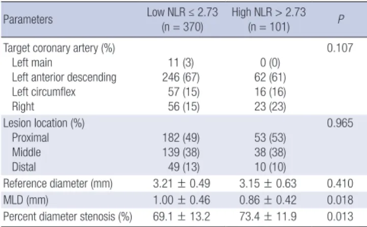
Coronary angiographic findings are summarized in Table 2. The minimal luminal diameter was significantly smaller and percent
diameter stenosis was significantly greater in high NLR group.
Grey-scale and VH- IVUS findings
Grey-scale IVUS findings are summarized in Table 3. At the pro- ximal reference, lumen cross-sectional area was significantly greater in high NLR group. At the minimum lumen area site, external elastic membrane cross-sectional area and plaque plus media cross-sectional area and plaque burden were significant- ly greater, and IVUS lesion was significantly longer in high NLR
Table 1. Baseline characteristics
Parameters Low NLR ≤ 2.73
(n = 315) High NLR > 2.73
(n = 84) P
Age (yr) 62.2 ± 10 62.2 ± 10 0.970
Male gender (%) 195 (62) 58 (69) 0.253
Clinical presentation (%) STEMI
NSTEMI UAP SAP
16 (4) 19 (5) 207 (56) 128 (35)
38 (37) 21 (21) 33 (33) 9 (9)
< 0.001
Diabetes mellitus (%) 87 (28) 32 (38) 0.080
Hypertension (%) 205 (65) 34 (41) < 0.001
Dyslipidemia (%) 128 (41) 25 (30) 0.077
Smoker (%) 119 (38) 45 (54) 0.024
Ejection fraction (%) 65.4 ± 7.3 60.4 ± 10.1 < 0.001 White blood cells (103/μL) 7.1 ± 2.1 10.2 ± 3.7 < 0.001 Neutrophil count (103/μL) 3.5 ± 1.2 7.6 ± 3.1 < 0.001 Lymphocyte count (103/μL) 2.5 ± 0.7 1.6 ± 0.6 < 0.001
NLR 1.5 ± 0.5 5.1 ± 2.5 < 0.001
Hemoglobin (g/dL) 13.5 ± 1.6 13.5 ± 1.8 0.938
Platelet count (103/μL) 227 ± 54 241 ± 90 0.178
Glucose (mg/dL) 133 ± 40 152 ± 50 0.004
Creatine kinase-MB (U/dL) 7.7 ± 21.2 38.0 ± 86.6 0.002
Troponin-I (ng/mL) 1.3 ± 6.9 21.0 ± 57.8 0.003
Creatinine (mg/dL) 0.87 ± 0.5 0.98 ± 0.7 0.096
Fibrinogen (mg/dL) 273 ± 67 295 ± 83 0.027
hs-CRP (mg/dL) 0.3 ± 1.0 1.2 ± 2.0 < 0.001
NT-pro-BNP (pg/mL) 176 ± 305 1206 ± 4231 0.044
Total cholesterol (mg/dL) 184 ± 41 185 ± 47 0.805
Triglyceride (mg/dL) 110 ± 69 122 ± 88 0.266
LDL cholesterol (mg/dL) 119 ± 36 117 ± 40 0.626
HDL cholesterol (mg/dL) 49 ± 20 50 ± 12 0.665
Data are presented as the No. (%) of patients or mean ± SD. NLR, Neutrophil-to- Lymphocyte ratio; STEMI, ST segment elevation myocardial infarction; NSTEMI, non- ST segment elevation myocardial infarction; UAP, unstable angina pectoris; SAP, sta- ble angina pectoris; hs-CRP, high-sensitivity C-reactive protein; NT-pro-BNP, N-termi- nal pro-B type natriuretic peptide; LDL, low-density lipoprotein; HDL, high-density li- poprotein.
Table 2. Coronary angiographic findings
Parameters Low NLR ≤ 2.73
(n = 370) High NLR > 2.73
(n = 101) P
Target coronary artery (%) Left main
Left anterior descending Left circumflex Right
11 (3) 246 (67)
57 (15) 56 (15)
0 (0) 62 (61) 16 (16) 23 (23)
0.107
Lesion location (%) Proximal Middle Distal
182 (49) 139 (38) 49 (13)
53 (53) 38 (38) 10 (10)
0.965
Reference diameter (mm) 3.21 ± 0.49 3.15 ± 0.63 0.410
MLD (mm) 1.00 ± 0.46 0.86 ± 0.42 0.018
Percent diameter stenosis (%) 69.1 ± 13.2 73.4 ± 11.9 0.013 Data are presented as the No. (%) of patients or mean ± SD. NLR, Neutrophil-to Lym- phocyte ratio; MLD, minimal luminal diameter.
Table 3. Grey-scale intravascular ultrasound findings
Parameters Low NLR ≤ 2.73
(n = 370)
High NLR > 2.73
(n = 101) P
Proximal reference EEM CSA (mm2) Lumen CSA (mm2) P&M CSA (mm2) Plaque burden (%)
18.2 ± 5.1 10.7 ± 2.9 7.5 ± 2.8 41 ± 7
19.5 ± 6.3 11.5 ± 3.4 8.0 ± 3.6 40 ± 8
0.064 0.032 0.188 0.509 Distal reference
EEM CSA (mm2) Lumen CSA (mm2) P&M CSA (mm2) Plaque burden (%)
13.0 ± 4.9 8.1 ± 2.9 5.0 ± 2.4 37 ± 7
14.0 ± 6.1 8.6 ± 3.7 5.4 ± 2.9 38 ± 7
0.164 0.225 0.129 0.319 Minimum lumen site
EEM CSA (mm2) Lumen CSA (mm2) P&M CSA (mm2) Plaque burden (%)
14.5 ± 4.6 4.3 ± 1.6 10.2 ± 3.9 69 ± 9
16.0 ± 6.0 4.1 ± 1.4 11.9 ± 5.3 73 ± 8
0.022 0.254 0.004
< 0.001 Largest necrotic core site
EEM CSA (mm2) Lumen CSA (mm2) P&M CSA (mm2) Plaque burden (%)
16.5 ± 5.3 5.7 ± 2.5 10.9 ± 3.9 65 ± 10
17.8 ± 6.0 5.5 ± 2.4 12.2 ± 4.7 68 ± 8
0.044 0.661 0.008 0.003 IVUS lesion length (mm) 23.2 ± 12.8 26.3 ± 12.0 0.026 Volumetric analysis
EEM volume (μL) Lumen volume (μL) TAV (μL) PAV (%)
343 ± 216 150 ± 95 193 ± 128 55.0 ± 6.8
506 ± 796 184 ± 106 250 ± 150 56.1 ± 8.7
0.044 0.002 0.001 0.178 Data are presented as the No. (%) of patients or mean ± SD. NLR, Neutrophil-to- Lymphocyte ratio; EEM, external elastic membrane; CSA, cross-sectional area; P&M, plaque plus media; IVUS, intravascular ultrasound; TAV, total atheroma volume; PAV, percent atheroma volume.
group. At the largest necrotic core site external elastic mem- brane cross-sectional area and plaque plus media cross-sec- tional area and plaque burden were significantly greater in high NLR group. By volumetric analysis, external elastic membrane volume and lumen volume and total atheroma volume were significantly greater in high NLR group.
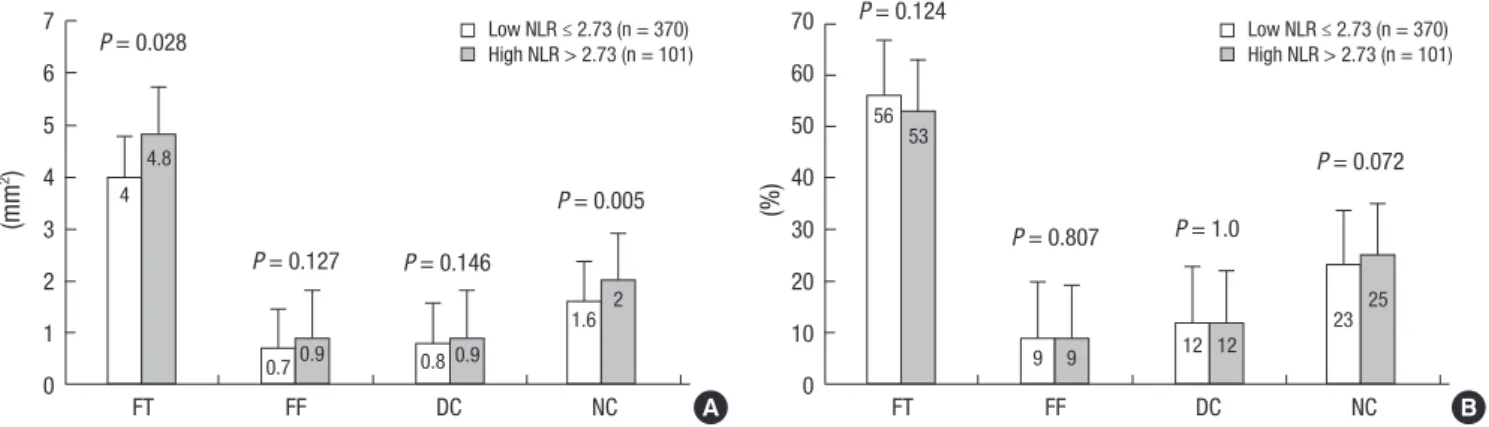
VH-IVUS findings are summarized in figures. At the minimum lumen area site, absolute fibrous and necrotic core areas were
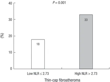
significantly greater in high NLR group (Fig. 1A). At the largest necrotic core site, absolute and relative necrotic core areas were significantly greater in high NLR group (Fig. 2). By volumetric analysis, absolute fibrous and fibro-fatty and dense calcium and necrotic core volumes were significantly greater in high NLR group (Fig. 3A). Also TCFA was observed more frequently in high NLR group compared with low NLR group (Fig. 4).
We performed separate analysis in the stable angina and ACS
Fig. 1. Absolute (A) and relative (B) plaque components at the minimum lumen area sites. Absolute fibrous and necrotic core areas are significantly greater in high NLR group.
FT, fibrous; FF, fibro-fatty; DC, dense calcium; NC, necrotic core.
P = 0.005
FT FF DC NC
7 6 5 4 3 2 1 0
Low NLR ≤ 2.73 (n = 370) High NLR > 2.73 (n = 101) P = 0.028
P = 0.127 4
4.8
0.70.9 0.8 0.9
1.6 2 P = 0.146
(mm2) P = 0.072
FT FF DC NC
70 60 50 40 30 20 10 0
Low NLR ≤ 2.73 (n = 370) High NLR > 2.73 (n = 101) P = 0.124
P = 0.807 56 53
9 9 12 12 23 25
P = 1.0
(%)
A B
Fig. 2. Absolute (A) and relative (B) plaque components at the largest necrotic core sites. Absolute necrotic core and relative necrotic core (%) areas are significantly greater in high NLR group.
P = 0.001
FT FF DC NC
6 5 4 3 2 1 0
Low NLR ≤ 2.73 (n = 370) High NLR > 2.73 (n = 101) P = 0.259
P = 0.315 3.9 4.3
0.6 0.7 1.1 1.1
2.1 P = 0.316 2.7
(mm2)
P = 0.050
FT FF DC NC
60 50 40 30 20 10 0
Low NLR ≤ 2.73 (n = 370) High NLR > 2.73 (n = 101) P = 0.313
P = 0.671 49 48
7 7
15 14
29 31 P = 0.627
(%)
A B
Fig. 3. Absolute (A) and relative (B) plaque components by volumetric analysis. Absolute fibrous and fibro-fatty and dense calcium and necrotic core volumes are significantly greater in high NLR group (A).
P = 0.001
FT FF DC NC
110 100 90 80 70 60 50 40 30 20 10 0
Low NLR ≤ 2.73 (n = 370) High NLR > 2.73 (n = 101) P = 0.003
P = 0.006 66
87
12 19 14 18
23 33 P = 0.013
(μL) P = 0.083
FT FF DC NC
70 60 50 40 30 20 10 0
Low NLR ≤ 2.73 (n = 370) High NLR > 2.73 (n = 101) P = 0.087
P = 0.676 57 55
11 11 12 12
20 22 P = 0.867
(%)
A B
patients. In a subgroup analysis, ACS patients had greater total atheroma volume and absolute necrotic core volume compared with stable angina patients (216.2 ± 141.9 μL vs. 177.2 ± 110.7 μL, P = 0.004, and 26.9 ± 23.2 μL vs. 20.0 ± 15.8 μL, P < 0.001, respectively), and TCFA was observed more frequently in ACS patients compared with stable angina patients (24% vs. 13%, P = 0.009).
We performed analysis in patients with ACS and stable angi- na using reference value of NLR (NLR: 2.7), respectively. We classified the lesions into two groups according to the NLR on admission in patients with ACS {low NLR group (NLR < 2.7 [n = 242]) vs. high NLR group (NLR ≥ 2.7 [n = 92]) and stable angina {low NLR group (NLR < 2.7 [n = 126]) vs. high NLR group (NLR ≥ 2.7 [n = 11]), respectively}. In ACS patients, at the mini- mum lumen area site and at the largest necrotic core site, high NLR group had greater absolute nectoric core area compared with low NLR group (1.58 ± 1.04 mm2 vs. 2.02 ± 1.38 mm2, P = 0.002, and 2.16 ± 1.11 mm2 vs. 2.72 ± 1.50 mm2, P < 0.001, re- spectively), and high NLR group had greater total atheroma volume and absolute necrotic core volume compared with low NLR group (201.7 ± 134.6 μL vs. 254.3 ± 153.8 μL, P = 0.002, and 24.3 ± 20.8 μL vs. 33.7 ± 27.7 μL, P = 0.001, respectively), and TCFA was observed more frequently in high NLR group com- pared with low NLR group (35% vs. 20%, P = 0.007). However, there were no significant differences in VH-IVUS parameters between high NLR group and low NLR group in stable angina patients using reference value (NLR: 2.7).
Independent predictors of TCFA
Independent predictors of TCFA are summarized in Table 4.
The following variables were tested to determine the indepen- dent predictors of TCFA (variables with P < 0.2 in univariate analysis): ST segment elevation myocardial infarction, NLR >
2.73, total atheroma volume, high-sensitivity C-reactive protein,
plaque burden at the minimum lumen area site, plaque burden at the largest necrotic core site, hypertension, creatine kinase- myocardial band, ejection fraction. ST segment elevation myo- cardial infarction (odds ratio [OR], 2.159; 95% CI, 1.000-4.660, P = 0.050) and NLR > 2.73 (OR, 1.848; 95% CI, 1.016-3.360, P = 0.044) and total atheroma volume (OR, 1.003; 95% CI, 1.001-1.004, P = 0.004) were independent predictors of TCFA.
DISCUSSION
The present VH-IVUS study demonstrated that 1) patients with high NLR had more acute coronary syndrome; 2) necrotic core components were significantly greater in high NLR group com- pared with low NLR group; 3) the TCFA was observed more fre- quently in high NLR group compared with low NLR group; and 4) ST segment elevation myocardial infarction, NLR > 2.73 and total atheroma volume were independent predictors of TCFA.
Atherosclerosis is known to be an inflammatory process and NLR is associated with enhanced inflammatory response (17).
Increased inflammatory response can cause atherosclerosis to destabilize and become clinical cardiovascular disease (18-21).
Muhmmed Suliman et al. (4) reported that high NLR increased mortality, thus providing an additional level of risk stratification in patients with acute coronary syndrome. In the present study, patients with high NLR had more incidence of acute coronary syndrome and had higher white blood cell counts and high-sen- sitivity C-reactive protein levels compared with those with low NLR.
Previous studies have reported that NLR was related to the development and progression of CAD (5, 22). In the present study, the percent diameter stenosis was significantly greater and total atheroma volume was significantly greater in patients with high NLR compared with those with low NLR. These re- sults suggest that inflammatory process indicated by high NLR are associated with the progression of atherosclerosis.
Vulnerable plaque in coronary artery can progress to plaque rupture and thrombosis, and have a strong potential to induce ACS (23, 24). Also inflammation and necrotic core size play a Table 4. Multivariate analysis for thin-cap fibroatheroma
Parameters OR 95% CI P
STEMI 2.159 1.000-4.660 0.050
NLR > 2.73 1.848 1.016-3.360 0.044
TAV 1.003 1.001-1.004 0.004
hs-CRP 1.106 0.914-1.340 0.301
Plaque burden at the MLA site 0.999 0.962-1.036 0.939 Plaque burden at the largest NC site 0.987 0.959-1.016 0.385
Hypertension 0.591 0.347-1.008 0.053
Creatine kinase-MB 0.996 0.990-1.003 0.248
Ejection fraction 0.992 0.962-1.023 0.629
STEMI, ST segment elevation myocardial infarction; NLR, Neutrophil to Lymphocyte ratio; TAV, total atheroma volume; hs-CRP, high-sensitivity C-reactive protein; MLA, minimum lumen area; NC, necrotic core.
Fig. 4. The incidence of thin-cap fibroatheroma. Thin-cap fibroatheroma is observed more frequently in high NLR group compared with low NLR group.
(%)
Thin-cap fibroatheroma Low NLR ≤ 2.73
40
30
20
10
0
P = 0.001
18
33
High NLR > 2.73
greater role in the progression of atherosclerosis in diabetic subjects who experienced sudden coronary death (25). In the present study, the necrotic core component was greater and TCFA within culprit lesions was observed more frequently in patients with high NLR compared with those with low NLR.
The results of the present study suggest that CAD patients with high NLR have a greater possibility having vulnerable plaque and higher inflammatory status, which can lead to acute coro- nary events. Therefore NLR can be used as a useful tool to de- tect not only significant atherosclerosis but also plaque vulner- ability in patients with CAD.
There are several limitations to be mentioned. First, the pres- ent study is retrospective single center study, so is subject to limitations inherent in this type of clinical investigation. Sec- ond, IVUS and VH-IVUS imaging were performed at the discre- tion of the individual operators leading to potential selection bias. Third, there is limitation using 20 MHz IVUS because this low frequency IVUS has a limitation to detect the plaque in de- tail, especially in the near field. Fourth, heavily calcified plaques may induce an artifact regarding the codification of plaques by VH-IVUS resulting in an increase in necrotic core content. Fifth, serial follow-up of serum neutrophil and lymphocyte levels were not performed. Therefore, we did not demonstrate the impact of sequential change of NLR levels on plaque components.
In conclusion, the present study demonstrates that CAD pa- tients with high NLR had more vulnerable plaque components (greater necrotic core containing plaques) than those with low NLR. Our results of the present study suggest that CAD patients with high NLR have a greater possibility having vulnerable plaque and higher inflammatory status, which can lead to acute coro- nary events. Therefore NLR can be used as a useful tool to de- tect not only significant atherosclerosis but also plaque vulner- ability in patients with CAD.
ORCID
Yun Ha Choi http://orcid.org/0000-0002-0224-0062 Young Joon Hong http://orcid.org/0000-0003-0192-8161 Youngkeun Ahn http://orcid.org/0000-0003-2022-9366 In Hyae Park http://orcid.org/0000-0001-5113-2095 Myung Ho Jeong http://orcid.org/0000-0003-4173-1494 REFERENCES
1. Libby P, Ridker PM, Maseri A. Inflammation and atherosclerosis. Circu- lation 2002; 105: 1135-43.
2. Margolis KL, Manson JE, Greenland P, Rodabough RJ, Bray PF, Safford M, Grimm RH Jr, Howard BV, Assaf AR, Prentice R. Leukocyte count as a predictor of cardiovascular events and mortality in postmenopausal women: the Women’s Health Initiative Observational Study. Arch Intern Med 2005; 165: 500-8.
3. Duffy BK, Gurm HS, Rajagopal V, Gupta R, Ellis SG, Bhatt DL. Usefulness of an elevated neutrophil to lymphocyte ratio in predicting long-term mortality after percutaneous coronary intervention. Am J Cardiol 2006;
97: 993-6.
4. Muhmmed Suliman MA, Bahnacy Juma AA, Ali Almadhani AA, Pathare AV, Alkindi SS, Uwe Werner F. Predictive value of neutrophil to lympho- cyte ratio in outcomes of patients with acute coronary syndrome. Arch Med Res 2010; 41: 618-22.
5. Arbel Y, Finkelstein A, Halkin A, Birati EY, Revivo M, Zuzut M, Shevach A, Berliner S, Herz I, Keren G, et al. Neutrophil/lymphocyte ratio is re- lated to the severity of coronary artery disease and clinical outcome in patients undergoing angiography. Atherosclerosis 2012; 225: 456-60.
6. Horne BD, Anderson JL, John JM, Weaver A, Bair TL, Jensen KR, Ren- lund DG, Muhlestein JB; Intermountain Heart Collaborative Study Group.
Which white blood cell subtypes predict increased cardiovascular risk? J Am Coll Cardiol 2005; 45: 1638-43.
7. Tamhane UU, Aneja S, Montgomery D, Rogers EK, Eagle KA, Gurm HS.
Association between admission neutrophil to lymphocyte ratio and out- comes in patients with acute coronary syndrome. Am J Cardiol 2008;
102: 653-7.
8. Núñez J, Núñez E, Bodí V, Sanchis J, Miñana G, Mainar L, Santas E, Mer- los P, Rumiz E, Darmofal H, et al. Usefulness of the neutrophil to lympho- cyte ratio in predicting long-term mortality in ST segment elevation myo- cardial infarction. Am J Cardiol 2008; 101: 747-52.
9. Cho KH, Jeong MH, Ahmed K, Hachinohe D, Choi HS, Chang SY, Kim MC, Hwang SH, Park KH, Lee MG, et al. Value of early risk stratification using hemoglobin level and neutrophil-to-lymphocyte ratio in patients with ST-elevation myocardial infarction undergoing primary percuta- neous coronary intervention. Am J Cardiol 2011; 107: 849-56.
10. Park JJ, Jang HJ, Oh IY, Yoon CH, Suh JW, Cho YS, Youn TJ, Cho GY, Chae IH, Choi DJ. Prognostic value of neutrophil to lymphocyte ratio in pa- tients presenting with ST-elevation myocardial infarction undergoing primary percutaneous coronary intervention. Am J Cardiol 2013; 111:
636-42.
11. Turak O, Ozcan F, Isleyen A, Tok D, Sokmen E, Buyukkaya E, Aydogdu S, Akpek M, Kaya MG. Usefulness of the neutrophil-to-lymphocyte ratio to predict bare-metal stent restenosis. Am J Cardiol 2012; 110: 1405-10.
12. Roberts WL, Moulton L, Law TC, Farrow G, Cooper-Anderson M, Sa- vory J, Rifai N. Evaluation of nine automated high-sensitivity C-reactive protein methods: implications for clinical and epidemiological applica- tions: part 2. Clin Chem 2001; 47: 418-25.
13. Reiber JH, van der Zwet PM, Koning G, von Land CD, van Meurs B, Ger- brands JJ, Buis B, van Voorthuisen AE. Accuracy and precision of quan- titative digital coronary arteriography: observer-, short-, and medium- term variabilities. Cathet Cardiovasc Diagn 1993; 28: 187-98.
14. Mintz GS, Nissen SE, Anderson WD, Bailey SR, Erbel R, Fitzgerald PJ, Pinto FJ, Rosenfield K, Siegel RJ, Tuzcu EM, et al. American College of Cardiology Clinical Expert Consensus Document on Standards for Ac- quisition, Measurement and Reporting of Intravascular Ultrasound Stu- dies (IVUS): a report of the American College of Cardiology Task Force on Clinical Expert Consensus Documents. J Am Coll Cardiol 2001; 37:
1478-92.
15. Nair A, Kuban BD, Tuzcu EM, Schoenhagen P, Nissen SE, Vince DG.
Coronary plaque classification with intravascular ultrasound radiofre- quency data analysis. Circulation 2002; 106: 2200-6.
16. Rodriguez-Granillo GA, García-García HM, Mc Fadden EP, Valgimigli M, Aoki J, de Feyter P, Serruys PW. In vivo intravascular ultrasound-de- rived thin-cap fibroatheroma detection using ultrasound radiofrequency data analysis. J Am Coll Cardiol 2005; 46: 2038-42.
17. Imtiaz F, Shafique K, Mirza SS, Ayoob Z, Vart P, Rao S. Neutrophil lym- phocyte ratio as a measure of systemic inflammation in prevalent chron- ic diseases in Asian population. Int Arch Med 2012; 5: 2.
18. Haim M, Boyko V, Goldbourt U, Battler A, Behar S. Predictive value of elevated white blood cell count in patients with preexisting coronary heart disease: the Bezafibrate Infarction Prevention Study. Arch Intern Med 2004; 164: 433-9.
19. Madjid M, Fatemi O. Components of the complete blood count as risk predictors for coronary heart disease: in-depth review and update. Tex Heart Inst J 2013; 40: 17-29.
20. Nakachi T, Kosuge M, Hibi K, Ebina T, Hashiba K, Mitsuhashi T, Endo M, Umemura S, Kimura K. C-reactive protein elevation and rapid angio- graphic progression of nonculprit lesion in patients with non-ST-segment elevation acute coronary syndrome. Circ J 2008; 72: 1953-9.
21. Burke AP, Tracy RP, Kolodgie F, Malcom GT, Zieske A, Kutys R, Pestaner J, Smialek J, Virmani R. Elevated C-reactive protein values and athero- sclerosis in sudden coronary death: association with different patholo- gies. Circulation 2002; 105: 2019-23.
22. Fowler AJ, Agha RA. Neutrophil/lymphocyte ratio is related to the sever- ity of coronary artery disease and clinical outcome in patients undergo- ing angiography: the growing versatility of NLR. Atherosclerosis 2013;
228: 44-5.
23. Virmani R, Burke AP, Farb A, Kolodgie FD. Pathology of the vulnerable plaque. J Am Coll Cardiol 2006; 47: C13-8.
24. Virmani R, Burke AP, Kolodgie FD, Farb A. Pathology of the thin-cap fi- broatheroma: a type of vulnerable plaque. J Interv Cardiol 2003; 16: 267- 72.
25. Burke AP, Kolodgie FD, Zieske A, Fowler DR, Weber DK, Varghese PJ, Farb A, Virmani R. Morphologic findings of coronary atherosclerotic pla- ques in diabetics: a postmortem study. Arterioscler Thromb Vasc Biol 2004; 24: 1266-71.