Clinical and Angiographic Outcomes of Drug-Eluting Stents in Patients With Large Vessel and Single Coronary Artery Lesion
Ung Kim, MD; Dong-Kie Kim, MD; Sang-Hoon Seol, MD; Tae-Hyun Yang, MD;
Dae-Kyung Kim, MD; Doo-Il Kim, MD; Dong-Soo Kim, MD; Sang-Hee Lee, MD;
Geu-Ru Hong, MD; Jong-Seon Park, MD; Dong-Gu Shin, MD; Young-Jo Kim, MD;
Yoon-Kyung Cho, MD; Hyung-Seop Kim, MD; Chang-Wook Nam, MD;
Seung-Ho Hur, MD; Kwon-Bae Kim, MD
Division of Cardiology, Department of Internal Medicine, (U. Kim, D.-K. Kim, S.-H. Seol, T.-H.
Yang, D.-K. Kim, D.-I. Kim, D.-S. Kim), Inje University Busan Baik Hospital; Yeungnam University Medical Center (S.-H. Lee, G.-R. Hong, J.-S Park, D.-G. Shin, Y.-J. Kim); Keimyung University Dongsan Hospital (Y.-K. Cho, H.-S. Kim, C.-W. Nam, S,-H. Hur, K.-B. Kim)
Address for correspondence:
Jong-Seon Park, MD Cardiovascular Division
Yeungnam University Medical Center 317-1, Daemyung-dong, Namgu Daegu, South Korea
[email protected]
Background: The aim of this study was to evaluate and compare the clinical and angiographic outcomes of 3 drug-eluting stents (DES) in patients with large vessel diameter and single coronary artery lesions.
Hypothesis: The efficacy of 3 DESs may be similar.
Methods: A total of 411 consecutive patients who visited 3 university hospitals from June 2004 to December 2007 and had a single coronary lesion which was treated with the use of a DES that was 3.5 mm in diameter were enrolled in this study. Patients were divided into 3 stent groups: Paclitaxel-eluting stent (PES, n = 105), Sirolimus-eluting stent (SES, n = 259), and Zotarolimus-eluting stent (ZES, n = 47). The study end point was a composite of major adverse cardiac events (MACE) including cardiac death, myocardial infarction (MI), and ischemia-driven target-vessel revascularization (TVR) for 12 months.
Results: Baseline characteristics were not different. Late loss was higher in the ZES group than the other stents (0.5 ± 0.4 mm in SES vs 0.3 ± 0.5 mm in PES, 0.7 ± 0.5 mm in ZES, P = 0.001). The total MACE-free survival rate was not significantly different between the SES group and the PES group (98.8% in SES vs 97.1%
in PES, P = 0.252) or the PES group and the ZES group (97.1% in PES vs 93.6% in ZES, P = 0.301). However, the SES group showed a significantly better MACE-free survival rate compared with the ZES group (98.8% in SES vs 93.6% in ZES, P = 0.018).
Conclusions: Clinical and angiographic outcomes of DES in a large vessel diameter and single coronary artery is excellent and SES appears to show better angiographic and clinical outcomes than ZES.
Introduction
Drug-eluting stents (DES) have shown great efficacy in the reduction of restenosis compared with bare-metal stents (BMS).
1However, the advent of safety and cost concerns shakes the firm position of DESs in a wide range of coro- nary lesions and patient subsets. The benefits of DESs were confined to lesions <3.0 mm.
2Although there were several randomized studies and other large registries comparing DES and BMS,
2 – 5no data was reported comparing the different DESs.
We seek to evaluate and compare the clinical and angiographic outcomes of 3 different DESs in patients with large vessel diameter and single coronary artery lesion.
Methods
Study Population and Grouping
A total of 411 consecutive patients who visited 3 qualified centers in South Korea (Yeungnam University Medical
Center, Keimyung University Dongsan Hospital, and Inje University Busan Paik Hospital) from June 2004 to December 2007 and who underwent single vessel coronary intervention in a large vessel and were treated with a 3.5 mm DES were studied. Patients were divided into 3 groups according to DES; Paclitaxel-eluting stent (PES; Taxus, Boston Scientific Corp, Natick, MA; n = 105), Sirolimus- eluting stent (SES; Cypher, Cordis, Johnson & Johnson, Roden, The Netherlands; n = 259), and Zotarolimus-eluting stent (ZES; Endeavor, Medtronic, Minneapolis, MN;
n = 47).
Intervention
Percutaneous coronary intervention (PCI) was performed
with standard techniques. Use of an intravascular ultrasound
to identify optimal stent expansion and apposition and DES
selection and stenting techniques for bifurcationlesion were
left to the discretion of the operator. All patients received
aspirin (325 mg orally) and a loading dose of 300 mg of
maintain life-long aspirin therapy. Prior to October 2006, patients who received SES were prescribed clopidogrel for 3 or 6 months depending on the complexity of the procedure, whereas patients treated with PES were given a 6-month prescription. After that time, all patients were prescribed clopidogrel for 1 year.
Coronary angiography was performed after administra- tion of 0.2 mg of intracoronary nitroglycerin. During the procedure, heparin was given at a bolus dose of 100 U/kg with an additional bolus to maintain activated clotting time
>250 seconds. The use of glycoprotein IIb/IIIa inhibitors was left to the operator’s discretion.
Quantitative Coronary Analysis
Intracoronary nitroglycerin (0.1–0.2 mg) was given before and after each intervention to achieve maximal dilation.
Quantitatively, CAG was performed immediately before and after stenting by an experiencedtechnician who was blinded to the type of stent deployed. Angiographic measurements included proximal, distal reference, minimum lumen diameter (MLD), percentage of lesion stenosis, and lesion length. Acute gain was measured and defined as the difference between the MLD after stent deployment and baseline MLD.
Study End Points and Definitions
Large vessels were defined as those coronary arteries that received a stent ≥3.5 mm in diameter by operator’s visual assessment. The end points of this study were a composite of major adverse cardiac events (MACE) including cardiac death, myocardial infarction (MI), and ischemia-driven target-vessel revascularization (TVR). Procedural success was defined as residual diameter stenosis ≤30% and the absence of in-hospital MACE such as cardiac death, MI, or TVR. Clinical success was defined as procedural success without in-hospital complications such as cardiac death, MI, or coronary artery bypass graft (CABG) within 24 hours of the index procedure. Myocardial infarction was defined as typical ischemic chest pain and/or ST-segment and/or T wave abnormalities with creatine kinase-MB increase ≥2 times the reference values without any new pathologic Q waves.
Ischemia-driven TVR was defined as emergency or elective CABG or repeat PCI in the target vessel for chest pain or a positive test for ischemia (exercise stress test, stress echocardiogram, 24-h Holter monitor, resting echocardiogram evidence of ST-segment depression or elevation in >1 lead, or radionuclide study showing reversible defect).
acute coronary syndrome with angiographic documentation of either vessel occlusion or thrombuswithin or adjacent to a previously successfully stented vessel or autopsy evidence of stent thrombosis; (2) Probable: acute MI in the distribution of the treated vessel or unexplained death <30 days; and (3) Possible: unexplained death >30 days.
6Major adverse cardiac events, including clinical follow- up, was done in 30 days, 3 months, 6 months, and 1 year after PCI. Angiographic follow-up was recommended in all living patients at 6 to 8 months after PCI. The 1-year clinical follow-up data were collected by physician’s appointment or by telephone interview.
Statistical Analysis
Data are expressed as means ± SD for continuous variables and as frequencies for categorical variables. Categorical data were analyzed with an χ
2test and continuous variables were evaluated with a Student t test or 1-way analysis of variance (ANOVA) test. The cumulative incidences of adverse cardiac events were estimated according to the Kaplan-Meier method. Differences between the event-free survival curves for the 3 groups were compared using a log-rank test. Probability values <0.05 were considered significant. Data were analyzed with SPSS 12.0 for Windows (SPSS, Chicago, IL).
Results
Baseline characteristics are shown in Table 1 and there were no significantdifferences among groups. Most patients were men (72% in PES, 74% in SES, and 78% in ZES, P = 0.710) and patients with MI, ST-elevation, or non–ST- elevation, had considerable portion in diagnosis (43.8%
in group 1, 41.4% in group 2, and 55.3% in group 3,
P = 0.286). Angiographic and procedural outcomes are
represented in Table 2. Although it was not statistically
significant, the left anterior descending (LAD) artery was
a prominent intervention site for SES and ZES (56.8% vs
63.8%) and the LAD and right coronary artery (RCA)
were prominent intervention sites for PES (42.9% each,
P = 0.054). Most lesions were B1 or B2 according
to the American College of Cardiology/American Heart
Association classification (73.3% in PES, 73.7% in SES,
76.6% in ZES, P = 0.972). Total stent length was different
among the groups (23.6 mm in PES, 24.2 mm in SES,
21.1 mm in ZES, P = 0.047). Medications such as aspirin,
clopidogrel, cilostazol, and statin were not different among
the groups (Table 2). The data of quantitative coronary
analysis are shown in Table 3. A bifurcation lesion was
detected in 3 patients in the PES group, 9 patients in
the SES group, and 3 patients in the ZES group. In
Clinical Investigations continued
Table 1. Baseline Characteristics of Patients PES
(n = 105) SES
(n = 259) ZES
(n = 47) P Value
Age (yrs) 63 ± 9 61 ± 9 59 ± 12 0.078
Gender (male) 76 (72.4%) 192 (74.1%) 37 (78.7%) 0.710 Diabetes mellitus 23 (21.9%) 64 (24.7%) 12 (25.5%) 0.826 Hypertension 50 (47.6%) 111 (42.9%) 20 (43.5%) 0.706 Dyslipidemia 43 (41.3%) 101 (39.8%) 18 (39.1%) 0.953 Smoking 45 (42.9%) 102 (61.1%) 20 (42.6%) 0.813 Previous PCI 12 (11.4%) 21 (8.1%) 2 (4.3%) 0.318 Previous CVA 1 (1.0%) 6 (2.3%) 1 (2.1%) 0.692 Ejection fraction, % 55 ± 10 56 ± 11 52 ± 11 0.060
Diagnosis 0.286
Silent ischemia 2 (1.9%) 1 (0.4%) 0 Stable angina 34 (32.4%) 85 (32.8%) 11 (23.4%) Unstable angina 23 (21.9%) 66 (25.5%) 10 (21.3%) NSTEMI 14 (13.3%) 39 (15.1%) 5 (10.6%) STEMI 32 (30.5%) 68 (26.3%) 21 (44.7%) Abbreviations: CVA, cerebrovascular accident; NSTEMI, non– ST- elevation myocardial infarction; PCI, percutaneous coronary interven- tion; PES, Paclitaxel-eluting stent; SES, Sirolimus-eluting stent; STEMI, ST-elevation myocardial infarction; ZES, Zotarolimus-eluting stent.
all cases, a provisional stent technique was used, but another stent was not implanted in the daughter side branch.
At follow-up, minimal lumen diameter (2.7 mm in PES, 3.0 mm in SES, 2.5 in ZES, P = 0.001), diameter stenosis (19.2% in PES, 11.6% in SES, 27.5% in ZES, P = 0.001), and late loss (0 .5 ± 0.4 mm in PES, 0.3 ± 0.5 mm in SES, 0.7 ± 0.5 mm in ZES, P = 0.001) were significantly different among the groups. Angiographic follow-up was performed in 55% (227/411) of patients. Restenosis was found in a total of 7 patients (3.1%, 7/227); 4 patients (2.7%) in the SES group, 1 patient in the PES group (1.8%), and 2 patients in the ZES group (7.7%, P = 0.333). Their patterns, according to type of DES, were represented as 1 edge type in the PES group; 1 body, 2 edges, 1 total occlusion type in the SES group; and 1 focal diffuse type in the ZES group, and this was not significantly different (P = 0.072).
Clinical follow-up was done in all patients (100%).
In-hospital outcomes showed 1 death in the SES and ZES groups due to cardiogenic shock after PCI presented as ST- segment elevation myocardial infarction (STEMI) and 1 MI
Table 2. Angiographic and Procedural Characteristics of Patients PES
(n = 105) SES
(n = 259) ZES
(n = 47) P Value
Site of PCI 0.054
LAD 45 (42.9%) 147 (56.8%) 30 (63.8%)
LCX 15 (14.2%) 30 (11.6%) 2 (4.3%)
RCA 45 (42.9%) 82 (31.7%) 15 (31.9%)
Type of lesion
a0.972
A 8 (7.6%) 20 (7.7%) 3 (6.4%)
B1/B2 77 (73.3%) 191 (73.8%) 36 (76.6%)
C 20 (19.1%) 48 (18.5%) 8 (17.0%)
Infarct-related artery 46 (25.6%) 108 (60.0%) 26 (55.3%) 0.223 Bifurcation lesion
(>2.5 mm)
3 (2.9%) 9 (3.5%) 3 (6.4%) 0.564
Stent used
Length (mm) 23.6 ± 8.6 24.2 ± 8.0 21.1 ± 5.5 0.047 Number of stents 1.0 ± 0.2 1.0 ± 0.2 1.0 ± 0.0 0.417 Postdilatation 13 (15.1%) 59 (22.8%) 8 (17.0%) 0.258 Postprocedural
medication
Aspirin 105 (100%) 259 (100%) 47 (100%) 1.000 Clopidogrel 105 (100%) 259 (100%) 47 (100%) 1.000 Cilostazol 17 (16.0%) 28 (10.5%) 7 (14.9%) 0.295 Statin 62 (58.5%) 183 (68.6%) 34 (73.9%) 0.091
Abbreviations: LAD, left anterior descending artery; LCX, left circumflex artery; PCI, percutaneous coronary intervention; PES, Paclitaxel-eluting stent; RCA, right coronary artery; SES, Sirolimus-eluting stent; ZES, Zotarolimus-eluting stent.
a
According to the American College of Cardiology/American Heart Association classification.
in the PES group. However, at 30 days after PCI, no MACE
was detected in any groups. At 12 months, 1 death due to
probable stent thrombosis at 5 months after PCI, 1 MI, and 1
TVR were observed in the SES group. At 12 months, 2 TVRs
were found in the ZES group and 2 TVRs were detected
in the PES group (Table 4). Cumulative total MACE was
detected in 9 cases, which was shown as 3 in the PES group
Baseline
RVD (mm) 3.4 ± 0.2 3.4 ± 0.1 3.4 ± 0.2 0.508 MLD (mm) 0.3 ± 0.2 0.3 ± 0.2 0.3 ± 0.3 0.316 DS (%) 90.3 ± 7.4 89.1 ± 8.3 90.3 ± 8.7 0.390 LL (mm) 19.5 ± 7.0 20.0 ± 7.6 17.6 ± 5.2 0.122 Postprocedure
RVD (mm) 3.4 ± 0.1 3.4 ± 0.1 3.4 ± 0.1 0.251 MLD (mm) 3.3 ± 0.1 3.3 ± 0.2 3.3 ± 0.2 0.684 DS (%) 5.4 ± 3.8 4.4 ± 3.2 5.0 ± 3.7 0.057 Acute gain 2.9 ± 0.3 2.9 ± 0.3 2.9 ± 0.3 0.347 Follow-up
RVD (mm) 3.4 ± 0.2 3.4 ± 0.3 3.4 ± 0.1 0.371 MLD (mm) 2.7 ± 0.5 3.0 ± 0.5 2.5 ± 0.6 0.001 DS (%) 19.2 ± 12.8 11.6 ± 14.8 27.5 ± 17.0 0.001 Late loss (mm) 0.5 ± 0.4 0.3 ± 0.5 0.7 ± 0.5 0.001 Abbreviations: DS, diameter stenosis; LL, lesion length; MLD, minimal lumen diameter; PES, Paclitaxel-eluting stent; RVD, reference vessel diameter; SES, Sirolimus-eluting stent; ZES, Zotarolimus-eluting stent.
(2.9%), 3 in the SES group (1.2%), and 3 in the ZES group (6.4%) and this was not statistically significant (P = 0.068;
Table 4).
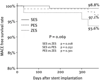
MACE-free survival was represented in Figure 1. The MACE-free survival rate was not significantly different between the SES and PES groups (98.8% in SES vs 97.1%
in PES, P = 0.252) or the PES and ZES groups (97.1%
in PES vs 93.6% in ZES, P = 0.301). However, the SES group showed a significantly better MACE-free survival rate compared with the ZES group (98.8% in SES vs 93.6% in ZES, P = 0.018).
Discussion
The major findings of this study show that the efficacy of DES in large vessel diameter and single coronary artery lesion is associated with low incidences of adverse cardiac events for 1 year and compared with ZES, SES had better clinical outcomes, although there were no significant differences between SES and PES or PES and ZES.
Reference vessel diameter is of importance in restenosis in patients undergoing PCI.
7Restenosis rates are quite low in large arteries after bare-metal stent (BMS) implantation.
3,8 – 11Steinberg et al
12reported that patients
In-hospital MACE 1 (1.0%) 1 (0.4%) 1 (2.1%) 0.414
Death 0 1 (0.4%) 1 (2.1%) 0.204
Myocardial infarction 1 (1.0%) 0 0 0.232
30-day MACE 0 0 0 1.000
Death 0 0 0 1.000
Myocardial infarction 0 0 0 1.000
TVR 0 0 0 1.000
12-month MACE 2 (1.9%) 3 (1.2%) 2 (4.3%) 0.315
Death 0 1 (0.4%) 0 0.745
Myocardial infarction 0 1 (0.4%) 0 0.745
TVR 2 (1.9%) 1 (0.4%) 2 (4.3%) 0.064
Total MACE 3 (2.9%) 3 (1.2%) 3 (6.4%) 0.068
Death 0 2 (0.8%) 1 (2.1%) 0.360
Myocardial infarction 1 (1.0%) 1 (0.4%) 0 0.232
TVR 2 (1.9%) 1 (0.4%) 2 (4.3%) 0.064
Stent thrombosis
Acute 0 0 0 1.000
Subacute 0 0 0 1.000
Late 0 1 (0.4%) 0 0.745
Abbreviations: MACE, major adverse cardiac event; PES, Paclitaxel- eluting stent; SES, Sirolimus-eluting stent; TVR, target-vessel revascu- larization; ZES, Zotarolimus-eluting stent.
treated with ≥3.5 mm DES and BMS had similar low incidence of MACE and target-lesion revascularization (TLR) and TVR in both groups, with no superiority of DES over BMS in this lesion. Quizhpe et al
13showed excellent 1-year clinical outcomes after large vessel (>3 mm) PCI between DES and BMS. However, the efficacy of different DESs in large vessel diameter with single lesion has not been reported. Our study showed that all DESs had good MACE-free survival rates in this lesion subset, especially SES.
We compared 3 different DESs, but the clinical outcomes
of ZES were different. This may be due to the different
tendency of late luminal loss among DES, although the
number of enrolled patients was small. Mean late luminal
loss of SES and PES was reported as 0.17–0.29 mm,
however, that of ZES was 0 .61 ± 0.49 mm and 0.65 ±
0 .49 mm in larger caliber (>2.9 mm).
14Our study also
Clinical Investigations continued
100%
95%
90%
85%
80%
0 100 200 300
Days after stent implantation
MACE free survival rate
P = 0.069
SES vs ZES PES vs ZES SES vs PES
p = 0.018 p = 0.301 p = 0.252