Coronary Flow Reserve in Non-Infarcted Myocardium Predicts Long-Term Clinical Outcomes in Patients
Undergoing Percutaneous Coronary Intervention
Rongchao Cheng
1, Xiaoming Zhu
2, Yunling Li
1, Xiuping Bai
1, Li Xue
1, and Li Wei
11Department of Cardiology, the Forth Affiliated Hospital of Harbin Medical University, Harbin;
2Department of Economic Management, Heilongjiang Nongken Vocational College, Harbin, China.
Purpose: Coronary flow reserve (CFR) is recognized as an indicator of myocardial perfusion. The aim of this study was to assess the relationship between CFR in the non-infarcted myocardium and the incidence of major adverse cardiac events (MACEs).
Materials and Methods: 100 consecutive patients with acute myocardial infarction (AMI) undergoing percutaneous coronary in- tervention (PCI) were enrolled in the present study, and divided into MACE and non-MACE groups according to the incidence of 12-month MACEs. Left ventricular function and CFR were analyzed using two-dimensional echocardiography and myocardial contrast echocardiography at one week after PCI. Cardiac troponin I levels were assayed to estimate peak concentrations thereof.
Results: The MACE group was associated with lower CFR, compared to the non-MACE group (2.41 vs. 2.77, p<0.001). In the mul- tivariable model, CFR in the non-infarcted myocardium was an independent predictor of 12-month MACE (hazard ratio: 0.093, 95% confidence interval: 0.020–0.426, p=0.002) after adjustment for baseline demographic and clinical characteristics.
Conclusion: CFR in the non-infarcted myocardium is a useful marker for predicting 12-month MACEs in patients with AMI un- dergoing primary PCI.
Key Words: Myocardial infarction, microvascular dysfunction, coronary flow reserve, myocardial contrast echocardiography
INTRODUCTION
Coronary flow reserve (CFR) is defined as the magnitude of increased coronary flow from the basal state to that achieved following maximal coronary vasodilation, and is recognized as an indicator of myocardial perfusion.1 Poor clinical out- comes have been found to be closely related with dysfunction of microcirculation perfusion, both in patients with satisfactory and those with unsatisfactory revascularization of the infarct- related artery.2-4 A large number of experiments have shown
that myocardial infarction (MI) area and left ventricular func- tion are largely affected by microcirculation function, which is closely related to recovery of left ventricular function and long- term left ventricular remodeling.5-7 About 25% of patients who have successfully undergone angioplasty still suffer from mi- crovascular dysfunction.8-10 It has been shown that microvas- cular dysfunction is not only in the infarcted myocardium but also in the non-infarcted myocardium. However, the clinical importance thereof has not been fully elucidated.11-12
The purpose of this study was to evaluate the relationship be- tween CFR in the non-infarcted myocardium and long-term clinical outcome in patients with acute myocardial infarction (AMI) and to analyze factors influencing the value of CFR in the non-infarcted myocardium.
MATERIALS AND METHODS
Study population
A total of 100 patients (74 males and 26 females) with first AMI Received: August 1, 2017 Revised: November 20, 2017
Accepted: November 26, 2017
Corresponding author: Dr. Li Wei, Department of Cardiology, the Fourth Affiliated Hospital of Harbin Medical University, No. 37, YiYuan Street, NanGang District, Harbin 150001, China.
Tel: 86-451-8593-9357, Fax: 86-451-8593-9357, E-mail: [email protected]
•The authors have no financial conflicts of interest.
© Copyright: Yonsei University College of Medicine 2018
This is an Open Access article distributed under the terms of the Creative Com- mons Attribution Non-Commercial License (http://creativecommons.org/licenses/
by-nc/4.0) which permits unrestricted non-commercial use, distribution, and repro- duction in any medium, provided the original work is properly cited.
pISSN: 0513-5796 · eISSN: 1976-2437 Yonsei Med J 2018 Mar;59(2):252-257
https://doi.org/10.3349/ymj.2018.59.2.252
and single-vessel disease who were undergoing primary per- cutaneous coronary intervention (PCI) within 12 hours after symptom onset were enrolled in the study and followed up for 12 months. The study protocol was approved by the Institu- tional Review Board of the Fourth Hospital of Harbin Medical University. Patients under unstable condition, severe extra- cardiac disease, or severe valve disease were excluded. Informed consent was obtained from all patients.
Serum parameter
As the levels of creatinine kinase or cardiac troponin I (cTnI) in serum are important parameters to diagnose AMI, cTnI levels were chosen for assay in this study, and were measured on ad- mission and every six hours during the first 24 hours.
Percutaneous coronary intervention
PCI was performed with standard techniques and the stent was placed in all patients. All patients received aspirin (300 mg loading dose) and ticagrelor (180 mg loading dose) prior to PCI. A regimen of 180 mg ticagrelor per day was continued for at least 12 months after PCI, and 100 mg aspirin daily was pre- scribed indefinitely. Successful primary angioplasty was de- fined as a final thrombolysis in MI flow grade 3 in the infarct- related artery and residual stenosis of <20%.
Two-dimensional echocardiography
Two-dimensional echocardiography (Philips iE33, Philips Healthcare, Andover, MA, USA) was performed one week af- ter PCI to measure left ventricular end diastolic volume, left ventricular end systolic volume, and calculated left ventricular ejection fraction (LVEF).
Myocardial contrast echocardiography
Myocardial contrast echocardiography (MCE) was performed one week after PCI, using intravenous injection of sonovue at baseline and during hyperemia. Meanwhile, dobutamine was injected by intravenous infusion during hyperemia (at a start- ing dose of 5 μg/kg/min, followed by an increasing dose of 10 μg/kg/min up to a dose of 20 μg/kg/min in 3-minute stages).
MCE images were acquired both at baseline and hyperemia.
Sonovue (Bracco, Milan, Italy) was used as the intravenous con- trast agent and administered by an infusion syringe-pump.13
CFR measurement
The left ventricular was divided into 17 segments. The non-in- farcted myocardium far away from the infarcted myocardium was selected to ensure that there was no interaction between these two. Regions of interest (ROIs) were placed and tracked manually within the myocardium at baseline and after hyper- emia. The software package automatically calculated the mean acoustic intensity of each ROI and generated time-in- tensity curves that were subsequently fitted to a monoexpo- nentially function: y=A(1-e-βt)(q). Myocardial blood flow (MBF)
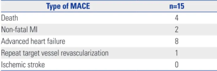
Table 1. Distribution of Patients with MACE
Type of MACE n=15
Death 4
Non-fatal MI 2
Advanced heart failure 8
Repeat target vessel revascularization 1
Ischemic stroke 0
MACE, major adverse cardiac event; MI, myocardial infarction.
was calculated as the product of A×β. Basal and hyperemic MBF was derived, and CFR (MBF at hyperemia/MBF at base- line) was calculated for each patient.14-16 All images were inde- pendently analyzed by two experienced observers, who were blinded to the clinical data, angiographic results, and other respective imaging.
Clinical outcomes and definitions
The primary endpoint of this study was the 12-month com- mutative incidence of major adverse cardiac event (MACE).
MACE comprised all causes of death, non-fatal MI, advanced heart failure, repeat target vessel revascularization, and isch- emic stroke. All patients were divided into MACE and non- MACE groups according to the incidence of 12-month MACE.
Statistical analysis
Statistical analysis was performed using SPSS 20.0 software (IBM Corp., Armonk, NY, USA). Continuous variables are ex- pressed as means±standard deviations, and were analyzed by Student’s t-tests. Categorical variables are expressed as per- centages and counts, and were analyzed by the chi square test.
p-values less than 0.05 were considered statistically significant.
Variables with a p value <0.05 in univariate analysis were in- cluded in the multivariate cox regression model, and results are presented as hazard ratios (HRs) with 95% confidence in- tervals (CIs). Multivariate cox regression analysis was per- formed to assess the independent predictors of MACE. A re- ceiver-operating characteristic (ROC) curve was drawn to evaluate the diagnostic value of CFR in the non-infarcted myo- cardium and peak cTnI in MACE and to determine the opti- mum cutoff level for CFR. Survival analysis was conducted using Kaplan-Meier survival curves, and the differences were compared using the log-rank test.
RESULTS
Patients
Among all patients in our study, MACEs occurred in 15 patients (15%) (all causes of death, n=4; non-fatal MI, n=2; advanced heart failure, n=8; repeat target vessel revascularization, n=1;
and ischemic stroke, n=0) (Table 1).
Clinical variables in the MACE and non-MACE groups
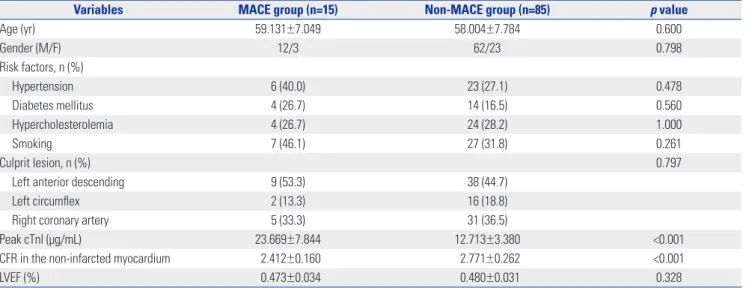
Mean peak cTnI in the MACE group was significantly higher than that in the non-MACE group (23.67 vs. 12.71 μg/mL, p<0.001). Mean CFR in the non-infarcted myocardium in the MACE group was significantly lower than that in the non- MACE group (2.412 vs. 2.771, p<0.001) (Table 2). There were no differences in the other variables between these two groups.
ROC curve and cut-off values
When predicting MACE by ROC curves, the sensitivity and specificity of CFR <2.305 were 91% and 86%, respectively [area
under the curve (AUC)=0.958], and the sensitivity and speci- ficity of peak cTnI >18.50 were 80% and 94%, respectively (AUC=
0.935) (Fig. 1).
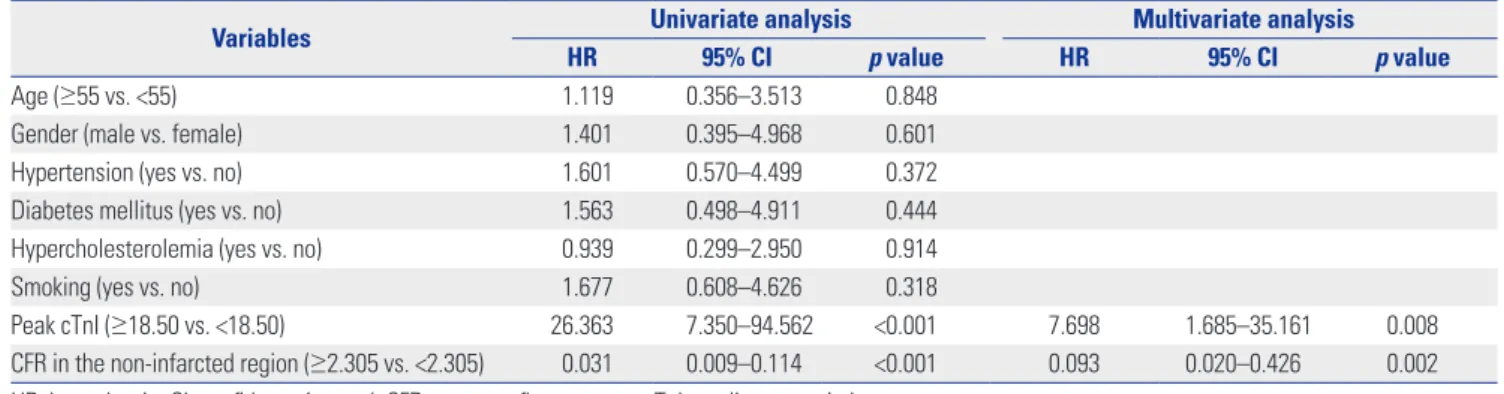
Predictive value of CFR in the non-infarcted myocardium on 12-month MACE
In univariate analysis, CFR in the non-infarcted myocardium and peak cTnI showed a significant association with 12-month MACE (p<0.001). These two factors with a p<0.05 were includ- ed in a multivariate cox regression analysis, and the interac- tion between the two factors was corrected. Therein, the risk Table 2. Comparison of the Two Study Groups according to Clinical Variables (n=100)
Variables MACE group (n=15) Non-MACE group (n=85) p value
Age (yr) 59.131±7.049 58.004±7.784 0.600
Gender (M/F) 12/3 62/23 0.798
Risk factors, n (%)
Hypertension 6 (40.0) 23 (27.1) 0.478
Diabetes mellitus 4 (26.7) 14 (16.5) 0.560
Hypercholesterolemia 4 (26.7) 24 (28.2) 1.000
Smoking 7 (46.1) 27 (31.8) 0.261
Culprit lesion, n (%) 0.797
Left anterior descending 9 (53.3) 38 (44.7)
Left circumflex 2 (13.3) 16 (18.8)
Right coronary artery 5 (33.3) 31 (36.5)
Peak cTnI (µg/mL) 23.669±7.844 12.713±3.380 <0.001
CFR in the non-infarcted myocardium 2.412±0.160 2.771±0.262 <0.001
LVEF (%) 0.473±0.034 0.480±0.031 0.328
MACE, major adverse cardiac event; cTnI, cardiac troponin I; CFR, coronary flow reserve; LVEF, left ventricular ejection fraction.
Values are presented as number (%) or mean±standard deviation.
1.0
0.8
0.6
0.4
0.2
0.0
1.0
0.8
0.6
0.4
0.2
0.0
Sensitivity Sensitivity
0.0 0.2 0.4 0.6 0.8 1.0 0.0 0.2 0.4 0.6 0.8 1.0
1-specificity CFR 1-specificity peak cTnI
ROC curve ROC curve
Fig. 1. The best cut-off values of CFR in the non-infarcted myocardium and peak cTnI were analyzed using ROC curves. CFR in the non-infarcted re- gion (AUC=0.958), cut-off=2.305, sensitivity=0.91, specificity=0.86). Peak cTnI (AUC=0.935), cut-off=18.50, senstivity=0.80, specificity=0.94. CFR, coronary flow reserve; cTnI, cardiac troponin I; ROC, receiver-operating characteristic; AUC, area under the curve.
of MACE in patients with high CFR (CFR≥2.305) was lower than that in patients with low CFR (CFR<2.305) (HR=0.093, 95% CI: 0.020–0.426, p=0.002) (Table 3).
Kaplan-Meier curves of cumulative survival rate (one-year survival)
Kaplan-Meier curves revealed median survival times of 7.353
±0.802 months in the CFR <2.305 group and 11.795±0.133 months in the CFR ≥2.305 group (p<0.001) (Fig. 2).
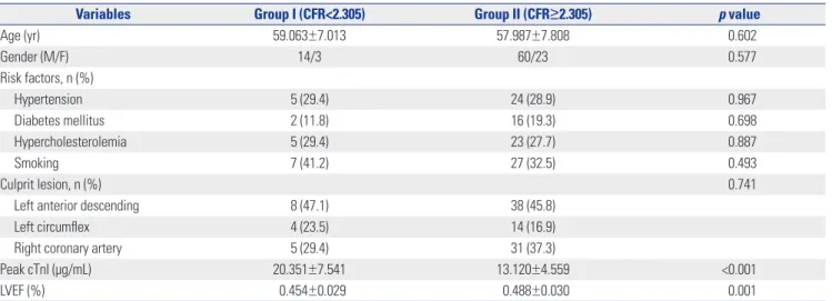
The impact factors of CFR
According to the CFR cut-off value of 2.305 in the non-infarct- ed myocardium, all patients were divided into two subgroups:
Group I (CFR<2.305) and Group II (CFR≥2.305). Peak cTnI was higher in Group I than in Group II (p<0.001), while LVEF was lower in Group I than in Group II (p=0.001) (Table 4).
Table 3. Factors Associated with Major Adverse Cardiac Event in Univariate and Multivariate Cox Regression Analysis
Variables Univariate analysis Multivariate analysis
HR 95% CI p value HR 95% CI p value
Age (≥55 vs. <55) 1.119 0.356–3.513 0.848
Gender (male vs. female) 1.401 0.395–4.968 0.601
Hypertension (yes vs. no) 1.601 0.570–4.499 0.372
Diabetes mellitus (yes vs. no) 1.563 0.498–4.911 0.444
Hypercholesterolemia (yes vs. no) 0.939 0.299–2.950 0.914
Smoking (yes vs. no) 1.677 0.608–4.626 0.318
Peak cTnI (≥18.50 vs. <18.50) 26.363 7.350–94.562 <0.001 7.698 1.685–35.161 0.008
CFR in the non-infarcted region (≥2.305 vs. <2.305) 0.031 0.009–0.114 <0.001 0.093 0.020–0.426 0.002 HR, hazard ratio; CI, confidence interval; CFR, coronary flow reserve; cTnI, cardiac troponin I.
1.0
0.8
0.6
0.4
0.2
0.0
Cum survival
MACE time (months) Overall survival
0 2 4 6 8 10 12
CFR
<2.305
≥2.305
<2.305-censored
≥2.305-censored
Fig. 2. Kaplan-Meier curves of cumulative survival (one-year survival).
The results showed median survival times of 7.353±0.802 months in the CFR <2.305 group and 11.795±0.133 months in the CFR ≥2.305 group (p<
0.001). MACE, major adverse cardiac event; CFR, coronary flow reserve.
DISCUSSION
To the best of our knowledge, this is the first study to investigate the association between CFR in the non-infarcted myocardium and 12-month adverse clinical outcomes in patients with AMI undergoing PCI. The non-MACE group was more likely to have lower peak cTnI and higher CFR in the non-infarcted myocardium than the MACE group. Multivariate analysis iden- tified CFR in the non-infarcted myocardium as an indepen- dent predictor of 12-month MACE after adjusting for multiple cardiovascular risk factors. Additionally, we found the mean survival time of the low CFR group (CFR<2.305) to be signifi- cantly shorter than that of the high CFR group (CFR≥2.305).
Moreover, we further analyzed factors related with CFR value, among which only peak cTnI and LVEF were statistically sig- nificant. Thus, we inferred that CFR value is closely associated with the MI size and left ventricular function.
MCE is the gold standard method to assess myocardial mi- crocirculation.17 The predictive value of CFR measured by MCE for no reflow phenomenon and left ventricular remodeling af- ter PCI has been revealed by several clinical studies.6,18 De- crease in CFR in the non-infarcted myocardium was correlated with an increase in the extent of ischemia. Alterations in CFR in the non-infarcted myocardium had important clinical im- plications after revascularization.19 Furthermore, some rele- vant biomarkers and other easily available clinical parameters have also been studied to predict the incidence of MACE, such as mean platelet volume and neutrophil counts.20
Microvascular dysfunction is found not only in the infarcted region but also in the remote region, which has been discov- ered long before. In MI animal models, CFR and coronary re- sistance changes were found in the non-infarcted areas, which were independent of changes in hemodynamic parameters.21 Similar studies in humans also verified that coronary vasodi- lator responses decreased in patients with non-infarcted myo- cardium after PCI.22,23 However, the etiology and clinical impor- tance of microvascular dysfunction in the non-infarcted myo- cardium are not fully understood.
Several mechanisms for CFR in the non-infarcted myocar- dium seem to have a correlation with MACE. The decrease of
CFR in the non-infarcted myocardium is representative of more extensive necrosis and deterioration of cardiac function, which was also proved in our study.24 Microvascular dysfunction in the non-infarct area is an indicator of enlargement of the isch- emic range, which can reduce myocardial collateral circula- tion to the infarction area and increase the area of MI. Also, the cardiospecific overexpression of angiotensin II type I receptor and decrease in microvessel density in the non-infarcted myo- cardium may lead to some cardiovascular events, mainly cardi- ac remodeling and heart failure.25 Moreover, investigators have also suggested that alterations in CFR in the non-infarcted myocardium also could be attributed to increased sympathetic nervous system activity, leading to coronary vasoconstriction and decreased vasodilator reserve.26 Furthermore, some schol- ars indicate that microvascular dysfunction after MI is associ- ated with impaired sympathetic innervation and function even in the non-infarcted myocardial tissues. Impaired sym- pathetic innervation might be associated with electrical insta- bility. Adverse structural and electrophysiological remodeling at non-infarcted regions after MI are responsible for clinically ventricular arrhythmias, leading to sudden cardiac death even- tually. Ventricular tachyarrhythmias are life threatening cardiac arrhythmias and the most common causes of sudden cardiac death.27
In our study, the incidence of MACE after PCI was 15%, slight- ly higher than that in other studies, which may be related to the small sample size of patients selected. Moreover, CFR was measured only once after PCI, and we could not evaluate chang- es in microvascular perfusion. Also, some problems in the ex- amination of MCE are still difficult to figure out at present, such as a number of segments appearing as acoustic shadow and artifacts.28 In addition, the relevant mechanism between CFR in the non-infarcted myocardium and MACE is planned to be explored in future study. We should also draw more attention to apparently “normal” non-infarction regions for further un-
derstanding of the mechanism of sudden cardiac death.
In conclusion, our results clearly show that CFR in the non- infarcted myocardium is associated with prognostic signifi- cance for 12-month MACE in patients with AMI undergoing primary PCI. The use of CFR is advised in contemporary clini- cal practice.
ACKNOWLEDGEMENTS
This work was supported by the Department of Medical Ultra- sonics of the Second Affiliated Hospital of Harbin Medical University. We also thank Dr. Xueqi Li (the Forth Affiliated Hos- pital of Harbin Medical University) for providing amendment opinions.
ORCID
Rongchao Cheng https://orcid.org/0000-0001-6683-8613 Li Wei https://orcid.org/0000-0002-3139-4247
REFERENCES
1. Bowers TR, O’Neill WW. Coronary rotablation and reserve: can they occur together? Cathet Cardiovasc Diagn 1995;36:277.
2. Bierig SM, Mikolajczak P, Herrmann SC, Elmore N, Kern M, Labo- vitz AJ. Comparison of myocardial contrast echocardiography de- rived myocardial perfusion reserve with invasive determination of coronary flow reserve. Eur J Echocardiogr 2009;10:250-5.
3. Kisanuki A, Yuasa T, Kuwahara E, Takasaki K, Yoshifuku S, Otsuji Y, et al. Reproducibility of intravenous intermittent triggered myo- cardial contrast echocardiography in healthy subjects. Jpn Heart J 2004;45:461-73.
4. Kaul S. Myocardial contrast echocardiography: basic principles.
Prog Cardiovasc Dis 2001;44:1-11.
5. Wita K, Filipecki A, Lelek M, Bochenek T, Elžbieciak M, Wróbel W, et al. Prediction of left ventricular remodeling in patients with STEMI treated with primary PCI: use of quantitative myocardial contrast echocardiography. Coron Artery Dis 2011;22:171-8.
Table 4. Comparison of Clinical Variables (n=100)
Variables Group I (CFR<2.305) Group II (CFR≥2.305) p value
Age (yr) 59.063±7.013 57.987±7.808 0.602
Gender (M/F) 14/3 60/23 0.577
Risk factors, n (%)
Hypertension 5 (29.4) 24 (28.9) 0.967
Diabetes mellitus 2 (11.8) 16 (19.3) 0.698
Hypercholesterolemia 5 (29.4) 23 (27.7) 0.887
Smoking 7 (41.2) 27 (32.5) 0.493
Culprit lesion, n (%) 0.741
Left anterior descending 8 (47.1) 38 (45.8)
Left circumflex 4 (23.5) 14 (16.9)
Right coronary artery 5 (29.4) 31 (37.3)
Peak cTnI (µg/mL) 20.351±7.541 13.120±4.559 <0.001
LVEF (%) 0.454±0.029 0.488±0.030 0.001
CFR, coronary flow reserve; cTnI, cardiac troponin I; LVEF, left ventricular ejection fraction.
Values are presented as number (%) or mean±standard deviation.
6. Porter TR, D’Sa A, Turner C, Jones LA, Minisi AJ, Mohanty PK, et al. Myocardial contrast echocardiography for the assessment of coronary blood flow reserve: validation in humans. J Am Coll Cardiol 1993;21:349-55.
7. Swinburn JM, Lahiri A, Senior R. Intravenous myocardial contrast echocardiography predicts recovery of dysynergic myocardium early after acute myocardial infarction. J Am Coll Cardiol 2001;38:
19-25.
8. Mengozzi G, Rossini R, Palagi C, Musumeci G, Petronio A, Lim- bruno U, et al. Usefulness of intravenous myocardial contrast echo- cardiography in the early left ventricular remodeling in acute myo- cardial infarction. Am J Cardiol 2002;90:713-9.
9. Yamamuro A, Akasaka T, Tamita K, Yamabe K, Katayama M, Takagi T, et al. Coronary flow velocity pattern immediately after percuta- neous coronary intervention as a predictor of complications and in-hospital survival after acute myocardial infarction. Circulation 2002;106:3051-6.
10. Camici PG, Crea F. Coronary microvascular dysfunction. N Engl J Med 2007;356:830-40.
11. Wu JC, Yun JJ, Dione DP, Heller EN, Deckelbaum LI, Sinusas AJ. Se- vere regional ischemia alters coronary flow reserve in the remote perfusion area. J Nucl Cardiol 2000;7:43-52.
12. Kern MJ, Bach RG, Mechem CJ, Caracciolo EA, Aguirre FV, Miller LW, et al. Variations in normal coronary vasodilatory reserve strati- fied by artery, gender, heart transplantation and coronary artery disease. J Am Coll Cardiol 1996;28:1154-60.
13. Bokor D. Diagnostic efficacy of SonoVue. Am J Cardiol 2000;86:
19-24.
14. Eliasen P, Amtorp O. Effect of intracoronary adenosine upon re- gional blood flow, microvascular blood volume and hematocrit in canine myocardium. Int J Microcirc Clin Exp 1984;3:3-12.
15. Wei K, Jayaweera AR, Firoozan S, Linka A, Skyba DM, Kaul S. Quan- tification of myocardial blood flow with ultrasound-induced de- struction of microbubbles administered as a constant venous infu- sion. Circulation 1998;97:473-83.
16. Tiemann K, Pohl C, Schlosser T, Goenechea J, Bruce M, Veltmann C, et al. Stimulated acoustic emission: pseudo-Doppler shifts seen during the destruction of nonmoving microbubbles. Ultrasound Med Biol 2000;26:1161-7.
17. Kaul S. Myocardial contrast echocardiography: 15 years of research and development. Circulation 1997;96:3745-60.
18. Galiuto L, Garramone B, Scarà A, Rebuzzi AG, Crea F, La Torre G,
et al. The extent of microvascular damage during myocardial con- trast echocardiography is superior to other known indexes of post- infarct reperfusion in predicting left ventricular remodeling: re- sults of the multicenter AMICI study. J Am Coll Cardiol 2008;51:
552-9.
19. Yang L, Xia C, Mu Y, Guan L, Wang C, Tang Q, et al. Prognostic val- ue of real time myocardial contrast echocardiography after per- cutaneous coronary intervention. Echocardiography 2016;33:
421-30.
20. Duffy BK, Gurm HS, Rajagopal V, Gupta R, Ellis SG, Bhatt DL.
Usefulness of an elevated neutrophil to lymphocyte ratio in pre- dicting long-term mortality after percutaneous coronary inter- vention. Am J Cardiol 2006;97:993-6.
21. Aoki H, Matsunari I, Nomura Y, Fujita W, Komatsu R, Miyazaki Y, et al. Myocardial sympathetic innervation, function, and oxida- tive metabolism in non-infarcted myocardium in patients with prior myocardial infarction. Ann Nucl Med 2013;27:523-31.
22. Feldman RL, Macdonald RG, Nichols WW, Conti CR, Pepine CJ.
Effects of acute coronary occlusion on hemodynamics in an adja- cent coronary artery in dogs. Am J Cardiol 1984;54:1103-7.
23. Uren NG, Crake T, Lefroy DC, de Silva R, Davies GJ, Maseri A. Re- duced coronary vasodilator function in infarcted and normal myo- cardium after myocardial infarction. N Engl J Med 1994;331:222-7.
24. Neizel M, Futterer S, Steen H, Giannitsis E, Reinhardt L, Loss- nitzer D, et al. Predicting microvascular obstruction with cardiac troponin T after acute myocardial infarction: a correlative study with contrast-enhanced magnetic resonance imaging. Clin Res Cardiol 2009;98:555-62.
25. de Boer RA, Pinto YM, Suurmeijer AJ, Pokharel S, Scholtens E, Humler M, et al. Increased expression of cardiac angiotensin II type 1 (AT(1)) receptors decreases myocardial microvessel densi- ty after experimental myocardial infarction. Cardiovasc Res 2003;
57:434-42.
26. Tsai CF, Ueng KC, Wu DJ, Tsai TP, Lin CS. Remodeled left ventric- ular myocardium remote to infarction sites is the arrhythmogenic substrate for sudden cardiac death. Med Hypotheses 2010;75:368- 71.
27. Bekkers SC, Yazdani SK, Virmani R, Waltenberger J. Microvascu- lar obstruction: underlying pathophysiology and clinical diagno- sis. J Am Coll Cardiol 2010;55:1649-60.
28. Mayer S, Grayburn PA. Myocardial contrast agents: recent ad- vances and future directions. Prog Cardiovasc Dis 2001;44:33-44.