• 검색 결과가 없습니다.

Comparison of Plaque Composition in Diabetic and Non-Diabetic Patients With Coronary Artery Disease Using Multislice CT Angiography

N/A
N/A
Protected

Academic year: 2022

Share "Comparison of Plaque Composition in Diabetic and Non-Diabetic Patients With Coronary Artery Disease Using Multislice CT Angiography"

Copied!
6
0
0

로드 중.... (전체 텍스트 보기)

전체 글

(1)

581

Open Access

Comparison of Plaque Composition in Diabetic and Non-Diabetic Patients With Coronary Artery Disease Using Multislice CT Angiography

Yong-Seop Kwon, MD

1

, Jae-Sik Jang, MD

3

, Chang-Won Lee, MD

1

, Dong-Kie Kim, MD

2

, Ung Kim, MD

2

, Sang-Hoon Seol, MD

2

, Doo-Il Kim, MD

2

, Young-Wan Jo, MD

3

, Han Young Jin, MD

3

, Jeong-Sook Seo, MD

3

, Tae-Hyun Yang, MD

3

, Dae-Kyeong Kim, MD

3

and Dong-Soo Kim, MD

3

1

Department of Internal Medicine, Busan St. Mary’s Hospital, Busan,

2

Department of Internal Medicine, Inje University College of Medicine, Haeundae Paik Hospital, Busan,

3

Department of Internal Medicine, Inje University College of Medicine, Busan Paik Hospital, Busan, Korea

ABSTRACT

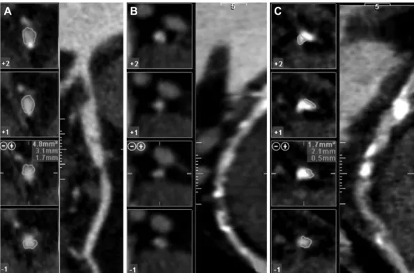
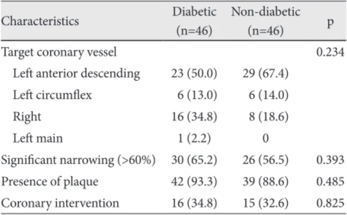
Background and Objectives: Plaque composition rather than degree of luminal narrowing may be predictive of future cor- onary events in high risk patients. The purpose of this study was to compare degree of plaque burden and composition with mul- tislice computed tomography (MSCT) angiography between diabetic and non-diabetic patients. Subjects and Methods: A total of 452 consecutive MSCT angiography examinations were performed between July 2007 and June 2009. Of these, the pa- tients who underwent invasive coronary angiography were evaluated for the presence and type of atherosclerotic plaque and se- verity of luminal narrowing. Results: Ninety two (46 in the diabetic group and 46 in the non-diabetic group) patients under- went both MSCT angiography and invasive coronary angiography. Among them, 30 patients (65.2%) in the diabetic group and 26 patients (56.5%) in the non-diabetic group had significant coronary narrowing on MSCT angiography. Sixteen patients (34.8%) in the diabetic group and 15 patients (32.6%) in non-diabetic group underwent coronary angioplasty and stenting. Forty-two patients (93.3%) in the diabetic group and 39 patients (88.6%) in the non-diabetic group had multiple types of coronary plaque (p=0.485). MSCT angiography was similar to conventional coronary angiography in its ability to predict significant coronary ar- tery disease in that the area under the curve was 0.88 (95% confidence interval, 0.81 to 0.95). Diabetic patients had more mixed plaque compared with non-diabetic patients. Conclusion: Differences in coronary plaque composition between diabetic and non-diabetic patients can be determined noninvasively by MSCT angiography. In patients with diabetes, mixed plaque types contribute to the total plaque burden to a higher degree than in non-diabetic patients. (Korean Circ J 2010;40:581-586) KEY WORDS: Coronary artery diseases; Diabetes.

Received: March 12, 2010 Revision Received: May 3, 2010 Accepted: May 11, 2010

Correspondence: Jae-Sik Jang, MD, Department of Medicine, Inje University College of Medicine, Busan Paik Hospital, 633-165 Gaegeum- dong, Jin-gu, Busan 614-735, Korea

Tel: 82-51-890-6947, Fax: 82-51-892-0273 E-mail: [email protected]

cc

This is an Open Access article distributed under the terms of the Cre- ative Commons Attribution Non-Commercial License (http://creativecom- mons.org/licenses/by-nc/3.0) which permits unrestricted non-commer- cial use, distribution, and reproduction in any medium, provided the origi- nal work is properly cited.

Introduction

Patients with diabetes have a significant risk for atheroscle- rotic vascular disease, and aggressive management of risk fac-

tors is recommended in these patients, irrespective of symp- toms or known coronary artery disease (CAD). Therefore, the role of coronary imaging in diabetic patients is not only to pro- ve the presence of coronary narrowing but to detect those pa- tients with a higher risk in whom further testing may be war- ranted in order to identify those with significant inducible myo- cardial ischemia.

Multislice computed tomography (MSCT) angiography can

accurately visualize the coronary arteries and permits detec-

tion of significant coronary artery stenosis when compared

with conventional coronary angiography.

1-3)

Previous reports

on detection and characterization coronary atherosclerotic pla-

que has revealed that MSCT angiography enables the meas-

urement of the degree of stenosis and well correlated with meas-

urement by intravascular ultrasound (IVUS)

4)5)

and conven-

(2)

tional coronary angiography.

Although IVUS is the gold standard for characterizing mor- phology plaque or degree of stenosis in patients undergoing coronary angiography, noninvasive detection and characteri- zation of coronary plaque would be an attractive alternative mo- dality. Noninvasive evaluation of coronary atherosclerotic pla- ques would improve risk stratification of both symptomatic and asymptomatic patients and also of high risk diabetic pa- tients.

7)

The purpose of this study was to compare the coronary plaque burden and composition in diabetic and non-diabetic patients with angina pectoris undergoing both MSCT angiog- raphy with conventional coronary angiography and evaluate any differences that might explain the poorer clinical outcomes in diabetic patients with CAD.

Subjects and Methods

Subjects

A total 452 consecutive MSCT angiography examinations were performed between July 2007 and June 2009. The study group consisted of the patients who underwent both MSCT angiography and conventional coronary angiography. Refer- ral for coronary angiography was based on the presence of symptoms, abnormal or inconclusive previous treadmill exer- cise electrocardiography (ECG), and/or nuclear testing. Pa- tients in sinus rhythm without contraindications to MSCT angiography were included. Exclusion criteria were (supra-) ventricular arrhythmia, renal insufficiency, known allergy to contrast media and pregnancy. An institutional review board approved the study and all participants provided written in- formed consent.

Data acquisition

MSCT angiography data were acquired with a 64 multi-de- tector CT scanner (Toshiba Medical Systems, Japan). Accord- ing to a previously published protocol,

8)

images were acquired with 16×0.75 mm slice collimation, a gantry rotation time of 420 ms, table feed of 2.8 mm/rotation, tube energy of 120 kV, and an effective tube current of 500 mAs. Eighty milliliters of contrast agent (Iodhexodol 320 g/cm

3

, Visipaque, GE-He- althcare Amersham, Piscataway, NJ, USA) were injected intra- venously at a rate of 4 mL/s, followed by a 30 mL saline chaser.

All patients with a heart rate > 65 beats/min received 5 mg me- toprolol intravenously before the MSCT scan.

Overlapping trans-axial images were reconstructed using a medium sharp convolution kernel (B35f) with an image ma- trix of 512×512 pixels, slice thickness of 1 mm, and an incre- ment of 0.5 mm using an ECG-gated half-scan algorithm with a resulting temporal resolution of 210 ms in the center of rotation. Image reconstruction was retrospectively gated to the ECG. The position of the reconstruction window within the cardiac cycle was individually optimized to minimize mo-

tion artifacts.

Data evaluation

Data were evaluated on a remote workstation using dedi- cated software (Vitrea, Vital Images, USA). For each patient, the entire coronary arterial tree was inspected for the presence of coronary plaques. Observers graded image quality of cor- onary segments as interpretable or not interpretable. Only pa- tients with interpretable image quality in all coronary seg- ments with lesions >50% were included in the analysis. To evaluate lesions, original axial images, multi-planar recon- structions perpendicular to the vessel centerline and cross-sec- tional reconstructions (1-mm thickness) were rendered at the site of maximal lumen narrowing and at reference sites prox- imal and distal to the lesion. Segments with motion artifacts or poor contrast-to-noise ratio were excluded from the anal- ysis and patients in whom all of the following segments (the left main, the proximal and middle segment of the left ante- rior descending artery and the right coronary artery and the proximal segment of the circumflex artery) could be assessed were included. Interpretable lesions identified on MSCT an- giography subsequently were visually classified as non-calci- fied, mixed plaques, or calcified plaques. Examples of each type of lesion are provided in Fig. 1.

Coronary plaques were defined as structures >1 mm

2

within the coronary artery whose density differed from that of the con- trast-enhanced vessel lumen. Plaques were divided into calci- fied {density >130 Hounsfield Unit (HU) in native scans}, non- calcified, or mixed lesions (Fig. 1). Non-calcified lesions were identified on the basis of their lower density compared with the contrast-enhanced vessel lumen (density <130 HU) as des- cribed by Schroder et al.

9)

Mixed lesions were defined as a plaque area consisting of >50% of non-calcified plaque tissue.

Statistical analysis

To test differences in demographics and risk factors be- tween diabetic and non-diabetic patients, we performed an unpaired t test (for continuous variables) or a chi-square test (for categorical variables). For assessment of the independent relationship of different plaques subtypes with diabetes mel- litus, logistic regression analysis (plaque subtype as the depen- dent variable) was used. All statistical analyses were perform- ed using Statistical Package for the Social Sciences (SPSS) soft- ware (version 15.0, SPSS Inc, Chicago, IL, USA). A p<0.05 was considered to indicate statistical significance.

Results

Patient characteristics

Of the 452 patients, 92 (46 in the diabetic group and 46 in the

non-diabetic group) patients underwent both MSCT angiogra-

phy and invasive coronary angiography. Demographic and cli-

(3)

nical characteristics of the patients are summarized in Table 1.

No significant differences in risk factors were present between the two groups. Patients with diabetes were more likely to be hy- pertensive compared with those without diabetes. Smoking history, hypercholesterolemia, previous history of percutane- ous coronary intervention and coronary artery bypass graft surgery were not different between the two groups. Twenty- two (47.8%) patients in the diabetic group and 17 (37.0%) in the non-diabetic group had multi-vessel disease (p=0.291).

Diagnostic performance of multislice computed tomography angiography

Fig. 2 shows a receiver-operating-characteristic curve de-

scribing the diagnostic performance of MSCT angiography to identify coronary stenosis of 60% or more in the culprit ves- sel. The area under the curve (AUC) was 0.88 {95% confidence interval (CI), 0.81 to 0.95}. The sensitivity and specificity for the quantitative analysis were 95% (95% CI, 92 to 96) and 71%

(95% CI, 69 to 72), respectively, with positive and negative predic- tive value of 75% (95% CI, 71 to 78) and 97% (95% CI, 95 to 98).

CT angiographic characteristics

The characteristics of coronary stenosis according to dia- betes status are presented in Table 2. The right coronary artery tended to be more frequently involved in diabetic patients, whereas left anterior descending artery involvement was more prevalent in non-diabetic patients. There was no statistically

A B C

Fig. 1. Example of different plaque types as visually assigned on the multislice computed tomography angiography. A: non-calcified plaque.

B: mixed plaque. C: calcified plaque.

Table 1. Baseline clinical characteristics of the study population*

Characteristics Diabetic

(n=46) Non-diabetic

(n=46) p

Age 65.0±9.6 62.8±8.8 0.258

Male sex 23 (50.0) 24 (52.2) 0.835

Smoking 13 (29.5) 13 (30.2) 0.944

Hypertension 38 (84.4) 29 (63.0) 0.021

Hypercholesterolemia 24 (53.3) 19 (43.2) 0.338

Previous PCI 3 (6.5) 2 (4.3) 0.646

Previous CABG 0 2 (4.3) 0.153

Multivessel disease 22 (47.8) 17 (37.0) 0.291

LV ejection fraction (%) 61.3 62.8 0.241

*Data shown as numbers (%) or mean±SD. PCI: percutaneous cor- onary intervention, CABG: coronary artery bypass graft, LV: left ventricular

Fig. 2. Diagnostic performance of MSCT angiography. Receiver-op- erating-characteristic curve describing the diagnostic performance of MSCT angiography to identify coronary stenosis of 60% or more in culprit vessel. The area under the curve was 0.88 (95% confidence interval, 0.81 to 0.95). MSCT: multislice computed tomography.

1.0 0.8 0.6 0.4 0.2 0.0

1-specificity

Se ns iti vi ty

0.0 0.2 0.4 0.6 0.8 1.0

(4)

significant difference in the prevalence of significant coronary narrowing (>60%) between the two groups (65.2 vs. 56.5%, p=0.393). Atherosclerotic plaque was present in 42 (93.3%) diabetic patients and 39 (88.6%) non-diabetics, a difference which was not statistically significant. Coronary interven- tion was done in 16 (34.8%) diabetic patients and 15 (32.6%) non-diabetics. The remaining patients were treated conser- vatively with optimal medical treatment.

Coronary CT angiographic characteristics of the study pop- ulation are shown in Table 3. More than half the patients in both groups had single vessel disease. Triple vessel disease was found in 8 (17.4%) patients with diabetes and 3 (6.8%) in pa- tients without diabetes. Also, the severity of luminal narrow- ing of target vessels was not significantly different between the two groups (p=0.677). When assessed according to types of coronary plaque, patients with diabetes had a higher preva- lence of mixed plaques compared with non-diabetic patients (50% vs. 25.6%, p=0.024), whereas the other kinds of plaque did not show such differences (non-calcified plaques: 16.7%

vs. 28.2%, p=0.212, calcified plaques: 33.3% vs. 46.2%, p=

0.238) (Table 4). Fig. 3 shows the composition of different kinds of plaque according to the presence of diabetes mellitus.

The results of multivariate logistic regression analysis are shown in Table 5. After adjustment for age and sex, diabetics were shown to be more likely to have mixed plaques {odds ra- tio (OR): 2.91, 95% CI 1.11-0.62, p=0.03}; whereas, after ad- justment for age, sex, history of hypertension, smoking, and hypercholesterolemia, patients with diabetes showed a trend toward more mixed plaque but the difference was not statis-

Table 2. Multislice CT angiographic characteristics of the study po- pulation*

Characteristics Diabetic

(n=46) Non-diabetic

(n=46) p

Target coronary vessel 0.234

Left anterior descending 23 (50.0) 29 (67.4) Left circumflex 06 (13.0) 06 (14.0)

Right 16 (34.8) 08 (18.6)

Left main 1 (2.2) 0

Significant narrowing (>60%) 30 (65.2) 26 (56.5) 0.393 Presence of plaque 42 (93.3) 39 (88.6) 0.485 Coronary intervention 16 (34.8) 15 (32.6) 0.825

*Data shown as numbers (%)

Table 3. Multislice CT angiographic characteristics of the study pop- ulation*

Characteristics Diabetic

(n=46) Non-diabetic

(n=46) p

CT diagnosis 0.240

1 vessel disease 24 (52.2) 29 (65.9) 2 vessel disease 14 (30.4) 12 (27.3) 3 vessel disease 08 (17.4) 3 (6.8)

CT severity 0.677

Mild <20% 2 (4.3) 1 (2.2)

Mild to moderate 20-40% 3 (6.5) 4 (8.9) Moderate 40-60% 11 (23.9) 15 (33.3) Moderate to severe 60-70% 11 (23.9) 11 (24.4) Severe 70-90% 12 (26.1) 06 (13.3) Critical >90% 07 (15.2) 08 (17.8)

*Data shown as numbers (%)

Table 4. Comparison of plaque composition based on multislice CT angiography

Characteristics Diabetic (n=42)

Non-diabetic

(n=39) p

Types of plaque

Non-calcified plaque 07 (16.7) 11 (28.2) 0.212 Mixed plaque 21 (50.0) 10 (25.6) 0.024 Calcified plaque 14 (33.3) 18 (46.2) 0.238 Data shown as numbers (%)

Fig. 3. Bar graph demonstrating the relative distribution of the differ- ent lesion types in diabetic and non-diabetic patients (p=0.035).

100 80 60 40 20

0 Diabetic Non-diabetic Non-calcified plaque Mixed plaque Calcified plaque

Table 5. Multivariable adjusted analysis demonstrating the asso- ciation between diabetes mellitus and the presence of plaques ac- cording to subtypes

Non diabetic (n=42) OR (95% CI)

Diabetic (n=39)

OR (95% CI) p

Mixed

Model 1 1 (ref) 2.91 (1.11-7.62) 0.03

Model 2 1 (ref) 2.87 (0.95-8.70) 0.06

Non calcified

Model 1 1 (ref) 0.52 (0.17-1.58) 0.25

Model 2 1 (ref) 0.88 (0.21-3.74) 0.86

Calcified

Model 1 1 (ref) 0.56 (0.22-1.40) 0.21

Model 2 1 (ref) 0.53 (0.19-1.50) 0.24

Model 1: adjusted for age and gender. Model 2: adjusted for age, gen- der, hypertension, smoking and hypercholesterolemia. OR: odds ra- tio, CI: confidence interval, ref: reference

(%)

(5)

tically significant (OR: 2.87, 95% CI 0.95-8.70, p=0.062).

Discussion

A major finding of this study was that MSCT angiography can accurately identify burden of atherosclerotic plaque and differences in its composition and that plaque features were closely related to the presence of diabetes (Fig. 3). The extent of coronary plaques was similar in diabetic and non-diabetic patients when assessed by MSCT angiography. The propor- tion of mixed plaques seen using MSCT angiography was lar- ger in patients with diabetes when compared with patients without diabetes.

A similar finding was reported recently by Ibebuogu et al.

10)

showing that diabetic patients had a higher proportion of co- ronary segments with mixed plaques compared with non-di- abetic patients (1.67±2.01 vs. 1.23±1.61, p=0.05) using MSCT angiography. In our study, a similar extent of coronary athero- sclerotic narrowing was observed in diabetic patients on MS- CT angiography and although statistically not significant, the presence of plaques was higher in diabetic patients. This find- ing is in line with a previous study

11)

exploring invasive coro- nary plaque characterization on gray-scale IVUS in 654 pa- tients, of which 128 had diabetes. They reported a significantly larger plaque burden in diabetic patients. Increased plaque ex- tent on MSCT angiography, as well as gray-scale IVUS, might explain a higher event rate in diabetic patients compared with non-diabetics.

Previous studies using MSCT angiography have suggested that coronary calcium content is associated with total plaque burden and therefore increases severity of luminal narrowing.

Therefore, a high calcium score level was regarded as a surro- gate marker for increased cardiovascular risk and events.

12)13)

However, it is unclear how well the presence and amount of calcium can predict coronary events in various patient pop- ulations with different cardiovascular risk factors, such as diabetes. In our study, diabetic patients had an increased num- ber of mixed plaques compared with patients without diabe- tes. Differences in plaque composition may in part explain the increased risk of CAD and mortality in diabetic pati- ents.

14)

Indeed, previous studies support the hypothesis that plaque composition, as well as stenosis severity, is predictive of future coronary events. As shown in Mann and Davies’s st- udy,

15)

lipid core size and minimal cap thickness have been known as major determinants of plaque vulnerability, while absolute plaque size and degree of coronary narrowing were not. So, vulnerable plaques may occur across the full spectrum of coronary disease, suggesting that relatively mild coronary lesions may contribute to coronary events.

16)17)

IVUS is the gold standard for plaque detection and char- acterization and for the assessment of plaque progression in follow-up studies.

18)

It has the advantage of a higher spatial

resolution but is invasive and limited due to its low penetra- tion depth.

19)

Although current MSCT angiography technol- ogy is not as good when compared to IVUS in terms of spa- tial resolution, it allows non-invasive evaluation of the vessel wall.

20)

Additionally, a major advantage of non-invasive im- aging modality is that it is less time consuming for both the pa- tient and the operator. MSCT angiography also has fewer acute risks, such as coronary spasm related to IVUS catheterization, acute vessel closure, dissection and distal embolization.

21)

A recent study using virtual histology IVUS demonstrated that mixed plaques were more highly associated with high risk features when compared with calcified or non-calcified plaques.

22)

Also, thin-cap fibroatheromas (TCFA) were more frequently present in mixed plaques and the increased am- ount of necrotic core was shown to be a feature of increased risk to plaque rupture.

23)

Nevertheless, the proportion of TC- FAs was identical in diabetic and non-diabetic patients. An- other study demonstrated that TCFA lesions were known to be more prevalent in patients with acute coronary syndromes than in patients with stable CAD.

24)

The higher proportion of mixed plaques in diabetic patients in our study can be re- garded as having high risk lesions because vulnerable plaque presented with TCFA; less extensive calcification and high pro- portion of necrotic core tissue are frequently observed in mix- ed plaques on MSCT angiography. As previously mentioned, Pundziute et al.

25)

demonstrated that mixed plaques on MSCT angiography were related more with adverse cardiovascular ev- ents on one-year follow-up. Among the predictors of coronary cardiac events in that study, the number of segments with mix- ed plaques were one of the significant independent predic- tors (hazard ratio 1.6, 95% CI 1.2-2.0, p=0.0003). Therefore, the predominance of mixed plaques in symptomatic diabetic patients with CAD might explain the higher incidence of coro- nary events, including unstable acute coronary syndrome, and thus hospital admission.

On the contrary, in a recent study, 49% of plaques in dia- betic patients were classified as completely calcified.

26)

This finding could be explained by a higher risk profile of the pa- tients in the study when compared with the population of in the previous study. Further studies are necessary to better understand the mechanisms that lead to plaque instability and cardiovascular events in diabetic patients. Although ear- lier studies have shown that diabetics are more likely to have multi-vessel disease compared with non-diabetics,

27)

in our sel- ected symptomatic patients, using a 60% stenosis cut-off did not show a significant difference between the two groups (47.8%, 37.0%, p=0.291).

There were several limitations in this study which should be

considered. This study was a retrospective and non-randomiz-

ed study. Also, there might be many confounding factors that

could not be properly accounted for in the analysis and there

were no available validated algorithms that allow quantifica-

(6)

tion of plaque stenosis, volume, or composition. Because most patients who underwent MSCT angiography were asympto- matic and didn’t have any obstructive findings on their exami- nation, we selected only patients who had a high likelihood of CAD by performing both MSCT angiography and conven- tional angiography. As a consequence, many patients were ex- cluded and selection bias may have affected the results. Thus, we should interpret the results very carefully and not extend them to most asymptomatic diabetic patients.

Another limitation of this study is the lack of IVUS as a gold standard for plaque characterization, although previous stud- ies demonstrated a strong correlation between MSCT and IV- US examinations for assessing plaque characteristics, as well as for the measurement of the plaque volumes.

22)28)

Also, the prognostic value of MSCT angiography in diabetic and non- diabetic patients was not determined. Finally, the study pop- ulation was small. Further studies should address whether certain plaque characteristics in diabetic patients may indeed be related to adverse outcomes.

In conclusion, MSCT angiography may be useful for the identification of CAD in diabetic and non-diabetic patients.

There were significant differences in coronary atherosclerotic plaque burden and composition between diabetic and non- diabetic patients, with a higher proportion of mixed plaques, a more vulnerable form of atherosclerotic plaque, in diabet- ics. Furthermore, MSCT angiography may give accurate in- formation about plaque characteristics according to different coronary risk factors, thereby identifying high risk features warranting a more intensive anti-atherosclerotic treatment.

REFERENCES

1) Achenbach S, Ulzheimer S, Baum U, et al. Noninvasive coronary an- giography by retrospectively ECG-gated multi-slice spiral CT. Cir- culation 2000;102:2823-8.

2) Hoffmann U, Moselewski F, Cury RC, et al. Predictive value of 16-slice multi-detector spiral computed tomography to detect significant obst- ructive coronary artery disease in patients at high risk for coronary ar- tery disease: patient-versus segment-based analysis. Circulation 2004;

110:2638-43.

3) Oh HJ, Kwon K, Park SH, et al. CT coronary angiography using mul- tidetector computed tomography in coronary artery disease : a compa- rative study to quantitative coronary angiography. Korean Circ J 2004;

34:1167-73.

4) Achenbach S, Moselewski F, Ropers D, et al. Detection of calcified and non-calcified coronary atherosclerotic plaque by contrast-enhanced, submillimeter multi-detector spiral computed tomography: a segment based comparison with intravascular ultrasound. Circulation 2004;

109:14-7.

5) Kim SY, Kim KS, Lee YS, et al. Assessment of non-calcified coronary plaques using 64-slice computed tomography: comparison with intra- vascular ultrasound. Korean Circ J 2009;39:95-9.

6) Cury RC, Pomerantsev EV, Ferencik M, et al. Comparison of the de- gree of coronary stenoses by multi-detector computed tomography ver- sus by quantitative coronary angiography. Am J Cardiol 2005;96:

784-7.

7) Naghavi M, Libby P, Falk E, et al. From vulnerable plaque to vul- nerable patient: a call for new definitions and risk assessment strate- gies: part I. Circulation 2003;108:1664-72.

8) Hoffmann U, Moselewski F, Nieman K, et al. Noninvasive assess-

ment of plaque morphology and composition in culprit and stable le- sions in acute coronary syndrome and stable lesions in stable angina by multi-detector computed tomography. J Am Coll Cardiol 2006;47:

1655-62.

9) Schroeder S, Kopp AF, Baumbach A, et al. Noninvasive detection and evaluation of atherosclerotic coronary plaques with multi-slice computed tomography. J Am Coll Cardiol 2001;37:1430-5.

10) Ibebuogu UN, Nasir K, Gopal A, et al. Comparison of atherosclerotic plaque burden and composition between diabetic and non diabetic pa- tients by non invasive CT angiography. Int J Cardiovasc Imaging 2009;

25:717-23.

11) Nicholls SJ, Tuzcu EM, Crowe T, et al. Relationship between cardio- vascular risk factors and atherosclerotic disease burden measured by intravascular ultrasound. J Am Coll Cardiol 2006;47:1967-75.

12) Wayhs R, Zelinger A, Raggi P. High coronary artery calcium scores pose an extremely elevated risk for hard events. J Am Coll Cardiol 2002;

39:225-30.

13) Kim D, Choi SY, Choi EK, et al. Distribution of coronary artery calci- fication in an asymptomatic Korean population: association with risk factors of cardiovascular disease and metabolic syndrome. Korean Circ J 2008;38:29-35.

14) Herlitz J, Karlson BW, Lindqvist J, Sjolin M. Rate and mode of death during five years of follow-up among patients with acute chest pain with and without a history of diabetes mellitus. Diabet Med 1998;

15:308-14.

15) Mann JM, Davies MJ. Vulnerable plaque: relation of characteristics to degree of stenosis in human coronary arteries. Circulation 1996;

94:928-31.

16) Giroud D, Li JM, Urban P, Meier B, Rutishauer W. Relation of the site of acute myocardial infarction to the most severe coronary arterial st- enosis at prior angiography. Am J Cardiol 1992;69:729-32.

17) Davies MJ, Thomas AC. Plaque fissuring: the cause of acute myocar- dial infarction, sudden ischaemic death, and crescendo angina. Br He- art J 1985;53:363-73.

18) Kopp AF. Angio-CT: heart and coronary arteries. Eur J Radiol 2003;

45(Suppl 1):S32-6.

19) Escolar E, Weigold G, Fuisz A, Weissman NJ. New imaging techniques for diagnosing coronary artery disease. CMAJ 2006;174:487-95.

20) Carrascosa PM, Capunay CM, Garcia-Merletti P, Carrascosa J, Garcia MF. Characterization of coronary atherosclerotic plaques by multi-de- tector computed tomography. Am J Cardiol 2006;97:598-602.

21) Hausmann D, Erbel R, Alibelli-Chemarin MJ, et al. The safety of intra- coronary ultrasound: a multicenter survey of 2207 examinations. Cir- culation 1995;91:623-30.

22) Pundziute G, Schuijf JD, Jukema JW, et al. Head-to-head comparison of coronary plaque evaluation between multislice computed tomogra- phy and intravascular ultrasound radiofrequency data analysis. JACC Cardiovasc Interv 2008;1:176-82.

23) Kolodgie FD, Virmani R, Burke AP, et al. Pathologic assessment of the vulnerable human coronary plaque. Heart 2004;90:1385-91.

24) Rodriguez-Granillo GA, Garcia-Garcia HM, McFadden EP, et al. In vivo intravascular ultrasound-derived thin-cap fibroatheroma detec- tion using ultrasound radiofrequency data analysis. J Am Coll Cardi- ol 2005;46:2038-42.

25) Pundziute G, Schuijf JD, Jukema JW, et al. Prognostic value of mul- tislice computed tomography coronary angiography in patients with known or suspected coronary artery disease. J Am Coll Cardiol 2007;

49:62-70.

26) Pundziute G, Schuijf JD, Jukema JW, et al. Noninvasive assessment of plaque characteristics with multi-slice computed tomography cor- onary angiography in symptomatic diabetic patients. Diabetes Care 2007;30:1113-9.

27) Hambly RI, Sherman L, Mehta J, et al. Reappraisal of the role of the diabetic state in coronary artery disease. Chest 1976;70:251-7.

28) Springer I, Dewey M. Comparison of multislice computed tomogra- phy with intravascular ultrasound for detection and characterization of coronary artery plaques: a systematic review. Eur J Radiol 2009;71:

275-82.

수치

Fig. 2 shows a receiver-operating-characteristic curve de-
Table 3. Multislice CT angiographic characteristics of the study pop- pop-ulation* Characteristics Diabetic (n=46) Non-diabetic(n=46) p CT diagnosis 0.240     1 vessel disease 24 (52.2) 29 (65.9)     2 vessel disease 14 (30.4) 12 (27.3)     3 vessel diseas

참조

관련 문서

All the previous stud- ies regarding the measurement of plaque area with CT adopted the coronary artery angiography protocol, which targeted peak enhancement of the arterial lumen

Objective: To compare multi-detector CT (MDCT) using 128-slice coronary CT angiography (Definition AS+, Siemens Medical Solution, Forchheim, Germany) with ECG-based maximum

Diagnostic performance of noninvasive fractional flow reserve derived from coronary computed tomography angiography in suspected coronary artery disease: the NXT trial

Abbreviations: BMI = body mass index, CABG = coronary artery bypass graft surgery, CAD = coronary artery disease, CCTA = coronary computed tomography angiography, ICA =

The aim of this study was to investigate the diagnostic accuracy and usefulness of 64-slice MDCT coronary angiography for detecting coronary artery disease in Korean patients with