Review Articles 순환기:순환기:제순환기:순환기:제제제 2 7 권권권 제권 제제 7 호제 호호 1997호
심혈관계통 장기에서의 Apoptosis
고려대학교 의과대학 순환기내과학교실
서 홍 석
Apoptosis in Cardiovascular System
Hong Seog Seo, M.D., Ph.D.
Department of Internal Medicine, Korea University Guro Hospital, College, of Medicine, Korea University, Seoul, Korea
서 론
척추동물 조직에서 두가지 세포 죽음이 있다. 1885 년부터 토끼의 난소세포의 maturation 관찰을 통한 세포 죽음의 또다른 형태가 있음을 보고하였다고 Majno와 Joris등은 기술하고 있으며1), 1972년에 Kerr가 이를 apoptosis라 명명하였다2). Majno와 Joris등은 세포의 죽음을 두가지로 명백히 구별하여
“cell death by suicide(apoptosis)”와“cell death by muder (accidental cell death)” 로 나누었으며1),
“oncosis”는 ischemic cell death의 형태학적 모양 을 기술한 것이고, necrosis는 두가지 세포죽음에 의 해 초래되는 세포 죽음의 형태학적 변화를 지칭하고 자 하였다1). 그러나 흔히 임상에서 apotosis의 상대 되는 죽음의 형태인 accidental cell death를 괴사 (necrosis)로 총칭하여 사용하고 있으며, 이는 허혈, 지속적 고열, 물리적 또는 화학적 충격 등의 강하고 급격한 injury가 주어졌을 때 세포가 죽는 가장 흔한 형태의 죽음이다3). 세포의 괴사는 초기에 mitochon- dria의 모양과 기능에 변화가 오고 단 시간 내에 세포 의 homeostasis가 파괴된다. 따라서 세포막에 심한 damage를 주어 삼투압 유지가 깨어짐으로써, 세포가 붓고 파괴되고, 그 내용물이 주위 조직으로 스며들어 염증반응을 일으키게 된다3). 이러한 염증 반응은 효과 적 세포잔해물의 제거역활을 함으로써 repair 과정을
돕게 된다3). Apoptosis는 또 하나의 세포사의 형태로 써 형태학적, 종말론적 의미로 programmed cell death 와 같은 의미로 사용되기도 한다. 척추동물에서는 ㅜ.체 내의 일부 세포의 죽음이 일상적인 것으로, 또 심지어 정확히 예측할수 있는 일로써, 받아들여지고 있으나, 무 척추 동물에서는 특별한 경우를 제외하고는, 개개의 세 포의 죽음이 program되지는 않는다. Apoptosis는 세 포가 죽은 동안, 괴사와는 다른 일연의 구조적 변화가 동반되고, 조직 통제의 정상적 과정으로 일어나는 것을 nematode인 Caenorhabditis elegans의 연구4)를 통해 밝혀짐으로 최근 많은 주목을 받게 되었다. 그예로써, 수명이 짧은 세포(백혈구)의 죽음, self-reactive T cell의 제거, growth factor제거로 인한 세포의 퇴화, 태생기의 morphogenetic cell death, 일부 면역체계 세 포의 죽음 등을 들 수 있다5).
Apoptosis의 형태학적 특징(Fig. 1)
Apoptosis의 세포변화는 수없이 다양하지만 실제 로 세포의 죽음과 직접 관련이 있는지는 확실치 않다.
세포막은 주름이 지며 bleb이 형성되어(zeiosis), 결 국 세포가 여러조각으로 나누어져 apoptotic body를 형성한, 세포막은 끝까지 유지가 되고 삼투압도 조절 된다3). 따라서 세포 내용물의 유출은 없으며 염증반 응도 동반되지 않는다. 전체적 세포의 크기가 줄어들 며, 전자현미경상 cytoplasm은 농축이 되나 orga-
nelle들은 정상 모양을 유지한다3). 이는 정확한 원인 은 아직 모르나, 세포가 짧은시간내에 물을 소실하게 되고 ion들도 균형있게 잃어 버림으로 삼투압이 정상 으로 유지가 되기 때문이다3). 생화학적으로 이때 세 포가 RNA와 단백질의 합성이 급격히 줄고, 분해가 늘어난다. 세포핵 변화는 세포에 따라 약간씩은 틀리 지만 대개 핵의 크기가 줄고, chromatin이 dense해지 고 patch 형태로 collapse되어 나중에 nuclear enve- lop 주위에 crescent를 형성하였다가 한 개 또는 여 러개의 dense sphere를 형성하게 된다(Fig. 1). 이러 한 변화는 endonuclease의 활성화에 의해 nucleos- omal core들 간의 수백만 linker 지역에 double stra- nded break가 일어나 결국 약 185base pair 간격의 DNA fragmentation이 일어나 세포가 회복 불가능 상태를 만들고 DNA 전사가 중단된다3). 물론 일부 세 포의 apoptosis에서 이러한 nucleosomal DNA cle- avage가 동반되지 않는 경우도 있으며, double-str- anded breaks대신 extensive single-stranded ni- cking이 주로 관찰되는 경우도 있다. 또한 DNA의 전 사가 중단된다고 해도 apoptosis에서 처럼 그 자체가 바로 빠른 시간내 세포의 죽음을 야기하는 것이 아니
므로 apoptosis에 또다른 과정이 관여되어 있으리라 여겨진다. 더불어 몇몇보고에 따르면, nucleases를 억 제하는 aurin tricarboxylic acid가 apoptosis에 동반 된 모든 변화를 억제하나, aurin이 다른 여러 세포대 사반응을 같이 억제하기 때문에 DNA파괴가 역시 apoptosis의 결정적 과정이라고 단언하기가 어려운 실정이다6-8). 결론적으로 Apoptosis의 특징을 정리하 면1) 형태학적, 생화학적 특성을 가지고 있으며, mi- tosis의 반대개념으로 이해되고 있다.2) 형태학적으로 세포가 줄고 농축되며 세포핵의 chromatin이 핵막주 위로 이동하면서 pyknotic해지고 부드러운 덩어리를 형성하다가 반달이나 배모양을 형성하게 된다. 또한 핵이 부서질수도 있으며(karyorhexis) 세포막의 융 기가 돌출되기도 한다(budding phenomenon).3) 이 러한 과정이 진행이되면 apoptosis body을 형성하여 탐식세포가 먹거나, 주위세포가 먹든지 또는 그대로 방치되어 있기도 한다.4) 그러나 mitochondria를 비롯 한 세포소체들의 부종이 없으며,5) 생화학적으로 DNA 가 185bp 간격으로 일정하게 잘려지는 특성이 유전자 에 의해 조절이 되는데6) 이 apoptosis는 개체내의 생 물학적 시계나 호르몬, cytokines, killer cells 및 여 Fig. 1. The stages of apoptosis in a lymphocyte. These stages are best seen in isolated culture;in vivo,
phagocytosis will intervene:(a) The normal cell has a sparse cytoplasm and heterogeneous nuclear chromatin. Cell volume is about 90 fL. (b) The cell has lost some volume, and its cytoplasmic organelles are new tightly packed. There is clumping of chromatin. At this stage, membrane changes that can lead to phagocytosis are present. (c) The cell exhibits zeiosis. (d) The chormatic has collapsed down into crescents along the nuclear envelope. This is readily observed using cell- permeant DNA dyes and a loght microscope. Cell volumen is now about 70 fL. (e) The nucleus has collapsed into a black hole. (f) The collapsed nucleus frequently breaks up into spheres. Some DNA has probably been lost from the cell by now, as apoptotic bodies are blebbing off it. (g) The cell fragments into apoptotic bodies. Each of these continues to exclude vital dyes for some time.
러 화학적, 물리적, virus등의 외적인 인자에 의해 시 작되고 매우 빨리 진행이되어 34분이내에 모든 과정 이 끝나기도 한다.
Apoptosis의 유전자적 제어(Fig. 2)
Apoptosis 연구의 잘 design된 model에서보면, 형 태학적 변화와 세포가 죽기위해 새로운 유전자의 발 현이 있다3). 따라서 세포가 apoptosis 형태로 죽음은 어떤 신호에 의해 죽는 것이지 억지로 타의적으로 죽 임을 당하는 것이 아니라는 것을 알 수 있다3). 이러한 사실은 유전자의 같은 complement를 가지고 있는 thymocyte와 T-cell을 비교해 봄으로써 명백히 알 수 있다. 즉 대부분의 thymocyte는 glucocorticoids 에 노출시 죽으나, 거의 같은 수의 glucocorticoid receptor를 가지고 있는 T-cell은 거의 죽지 않는다.
따라서 같은 자극에 thymocyte는‘자살 유전자’를 발현시킨 반면, T-cell은 그렇지 않은 것이다3). 최근 이 자살유전자가 무엇인지는 계속 연구가 수행되어 상당한 진전이 있다. 즉 apoptosis가 일어나는 경우 그러한 자극에 노출된 후 새로운 유전자의 발현이 있 으며, mRNA나 단백질 합성이 억제되는 경우 apop-
tosis가 억제 되는 몇예가 있어 총칭하여 유도(indu- ction) 기전에 해당한다3). 또하나의 모델은 HL-60 세포 계열에서는 mRNA나 단백질 합성이 억제되는 경우 apoptosis가 일어나 마치 suicide program이 체절적으로 수행이 되나 반감기가 짧은 여러 인자들 에 의해 억제되는 소위 release 기전에 의해 apop- tosis가 일어나기도 한다3). 이 두가지 기전은 생화학 적으로 매우 밀접한 관련성이 있는 것으로 알려져 있 다. 세째로 transduction 기전에 의한 경우로써, cyt- otoxic T cell에 의한 taret cell의 apoptosis이며, 이 경우 macromolecular synthesis의 억제가 apoptosis 에 아무런 효과가 없던지, 양성적 또는 음성적 효과 를 다양하게 나타낸다. 따라서 apoptosis가 일어나 게하는 모든 분자물질이 언제든 세포내에 존재함을 시사한다3).
Apoptosis와 관련된 각각의 유전자들은 apoptosis 와 두가지 방법으로 연결되어 apoptosis가 일어나게 세포내에서 발현이되던가, apoptosis과정을 조절되게 한다. 예로써 c-myc protooncogene은 세포가 pr- oliferation과 apoptosis의 선택을 조절하는데 일익을 담당하고 있다. c-myc을 발현하는 섬유아세포(fibr- oblast)는 세포배양액의 혈청 농도를 낮추면 wild- Fig. 2. A partial list of the agents that have been reported to induce or inhibit apoptosis.
type 세포에서 처럼 성장정지가 오는 대신 곧바로 apoptosis가 일어난다9). c-myc이 세포성장을 pro- gram하고 있으며, 이 program이 혈청내의 성장요소 결핍 또는 이차적 oncogene등으로 파괴가 되면 세포 가 자살을 수행하게 된다고 볼 수 있다. Antigen receptor crosslinking으로 세포가 죽게되는 T-cell hybridoma의 경우 c-myc antisense oligonucle- otide으로 그세포들의 apoptosis를 막을수 있음이 보 고된 바 있다. 따라서 c-myc 발현이 apoptosis에 필 요하며, 다른 mitogenic stimuli가 없는 상태에서 발 현된다는 것은 비정상적이며, 그것은 곧바로 apopt- osis를 일으키는 program으로 연결된다. adenovirus 의 E1A는 이 c-myc과 같은 유전자에 해당한다11).
Anti-oncogene p53 역시 apoptosis와 유관하다.
이유전자의 산물은 세포증식을 억제시키며, 대신 세포 가 분화하게끔 방향전환을 시킨다12). 많은 세포계대배 양에서 분화는 세포사와 같은 결과를 초래하며, my- eloid 또는 epithelial cell 계통은 apoptosis를 일으킨
다13,14). p-53의 진정한 역할은 damaged cell이 회복
되는 동안 그세포를 G1 phase로 묶어두는 것이며, G1 phase로의 정지가 불가능한 세포는 자살 program를 작동시키게 하여 없애버린다15).
Fas는 TNF(tumor necrosis factor)와 NGFR (ne- rve growth factor receptor)에 해당하는 membra- nspanning protein을 생성하는 유전자이다16,17) Fas 를 발현하는 세포가 fas 항체와 결합하면 apoptosis 가 일어난다. Fas는 human cell surface molecule인 APO-1과 같다. 임파구 증식을 일으키는 lpr 돌연변 이를 일으키는 쥐의 경우 fas 유전자의 결함에 의한
것으로 정상보다 잘 죽지않는 임파구가 형성되어 결 국 lymphoaccumulation이 초래되는 것이다18). 이러 한 Fas/Apo-1 system은 정상조직의 turnover 뿐아 니라, 악성종양을 비롯한 여러조건에서 치료의 일환으 로 apoptosis를 가동시키는데 응용의 가능성이 높이 제시되고 있다3).
Putative oncogene인 bcl-2는 종종 human follicular B-cell lymphomas에 과발현이 된다. IL- 3-dependent B-lymphoblastoid cell 계통에 bcl-2 를 형질변환시켜 발현되면, IL-3에 세포가 indep- endent하게 되어 IL-3를 없애도 세포가 죽지않게
된다19,20). 따라서 이러한 특성상 bcl-2는 anti-
apoptosis gene의 하나임을 시사하는 바이다3). 그러 나 bcl-2가 모든 상황에서 apoptosis를 막지는 않아, cytotoxic T cell에 의한 target cell의 apoptosis는 예방하지 못하며, 모든 계통의 세포에 다 작용하지는 않는다21).
아울러 apoptosis 세포에 발현이 증가된 몇 개의 유 전자가 있으나 아직 그역할이 정확히 규명되지는 않았다.
clusterin과 SGP-2 단백 등을 생성하는 TRPM-2가 비뇨기계통 세포의 apoptosis에 발현되나22) 태생기 발 생학적 세포사에서는 발현되지는 않는다23). 이 유전자 발현 산물은 분비와 지질 운반에 어떤역할을 하리라 알 려져 있으며, damage에 대한 반응에 관여되어 있으리 라 여겨진다. RP-2와 RP-8은 흉선세포의 apoptosis 유도후에 많이 발현되는 유전자 family이다24). RP-8 은 적어도 임파조직에서 뿐 아니라 태생기 사지와 중 추신경계의 형성시 일어나는 apoptosis에서 발현되는 유전자로써 많은 관심이 기울여지는 바이다25).
Fig. 3. A hypothetical model for the regulation of apoptotic cell death. As diagrammed, the major end point of apoptotic cell death is the removal of the dying cell by phagocytosis. One of the difficulties in determining the contribution of apoptosis to the pathogenesis of disease is the rapidity with which the phagocytosis of apoptotic cells occurs in vivo. Both the death repressor BCL2 and ICE are members of larger gene families.
Apoptosis가 수행되기 위해 세포내 여러 signal이 나 효소들 등의 변화가 일어나지만 아직 명백한 기전 은 밝혀지지 않았다. 이제까지 알려진 세포내 signal transductin을 포유동물에서의 경우를 중심으로 볼 때, 우선 세포막에 존재하는 Fas나 TNF receptor-1 등이 activation되면26), 이들 수용체의 내포내 dea- th domain과 FADD 등의 death domain이 서로 작 용하여 interleukin-1β converting enzyme(ICE) 를 activation시키며27,28), 이는 yama(어원:Hindu 의 죽음의 신에서 유래)/CPP32β로 하여금 핵내의 protein poly(ADP-ribose) polymerase(PARP)를 활성분해시켜 결국 apoptosis의 endonuclease ac- tivation으로 연결되어 DNA가 잘려지는 것으로 알 고 있다(Fig. 3)29).
검 사 방 법
Apoptosis의 발현시간은 매우 빠르게 진행되어 대 개 2~4시간 내에 이러한 현상이 일어나기 때문에 검 사시간대가 발현 확인에 결정적인 역할을 함로 이를 꼭 고려해야 한다. 우선 앞에서 설명한 여러 형태학적 변화를 전자현미경(transmission electron microsc- opy)을 통해 확인할 수 있으며, time-lapse video- microscopy)로 녹화기록하면 가장 확실히 확인할 수 있다30). 또한 세포핵의 변화를 이용한 검사 방법으로 acridine orange, propidium iodide나 Hoechst 33342를 이용한 형광 세포-영구 DNA 염색을 하거 나3), terminal deoxynucleotidyl transferase을 이용 한 terminal deoxynucleotidyl transferase-med-
iated dUTP-biotin nick end labeling(TUNEL)32)과 Klenow fragment of DNA polymerase I 효소를 이 용한 in situ end-labeling detection(ISEL)33)을 이 용하건, 전기영동후 일정한 간격(약 185bp)으로 끊어 진 nucleotides를 사다리 형태로 확인하는 방법 등이 흔히 사용되고 있다.
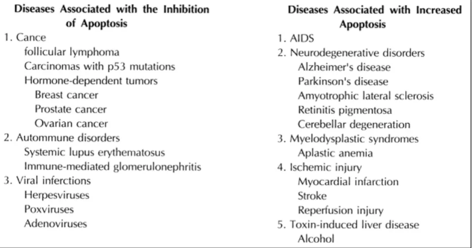
순환기질환에서 Apoptosis의 임상적의의(Fig. 4)
Apoptosis는 여러 생물학적, 병리적 현상에서 결정 적인 역할을 담당하는 것으로 여겨진다. 예로서 발생 기 장기형성을 위해서 뿐아니라, tissue remodeling, cancer, ischemia와 infarction, 면역계통질환 그리고 Alzheimer질환, Parkinson 질환 등의 신경퇴화질환 등을 들수 있다34). 즉 apoptosis가 일어나는데 어떤 결함이 있어 잘 일어나지 않음으로써 cancer patho- genesis나 자가면역질환의 발생을 초래할수 있으며, 지 나친 apoptosis등의 발현이 신경퇴화질환이나 AIDS 와같은 면역결핍 질환등을 초래한다34,36). 현재까지 순환기계통에서의 apoptosis 또한 몇몇 보고가 있다.
Bennett등은 정상적 동맥층의 smooth muscle cell에 서도 2.7~3.25%가 관찰되어 이는 cell mass 조절과 유관하리라 여기고 있으며30), 동맥경화의 죽상편내의 smooth muscle cell은 8.7~16.8%의 높은 비율을 보고하였으며30), Han등의 죽상편내 smooth muscle cell과 macrophage의 apoptosis의 보고37)와 더불어 Schwartz는 동맥경화질환에서 macrophage들의ap-
Fig. 4. Diseases associated with the induction or inhibition of apoptotic cell death.
optosis에 의한 acellular lipid core 형성과 plaque instability, 또한 smooth muscle cell의 apoptosis에 의한 vascular remodeling과의 유관성을 제시하였 다38). Isner등은 원발성 동맥경화병변위(43%)보다 매우높은 재협착부위의 apoptosis율(93%)을 보고하 였다39). Gottlieb등은 apoptosis와 허혈후 재관류 손 상이 심근세포의 apoptosis에 의한 것을 제시한 바 있다40). 최근 심근증에서도 virus 등의 침범과 심근허 혈 및 심근경색등으로 인한 failing heart의 심근세포 apoptosis가 심근증 발생의 병태생리학적 설명을 시 도하고 있으며, 선천성 완전 방실 차단의 경우 His- purkinje system의 세포가 선택적으로 apoptosis가 일어난 것이 보고 된 바도 있다41).
결 론
Apoptosis가 처음 기술된 후 20여년이 지나서 여 러 세포생물학 분야에서 hot topic으로 관심이 집중되 고 있다. 때로 왜 이러한 현상이 일어나는지 알수 없 으며, 어떤 조건에서 세포가 죽는다는 것을 알더라도, apoptosis을 이해하기는 많은 어려움이 있다. 결국 apoptosis는 척추동물세포의 타고난 특성의 하나로 생체내에서 흔히 일어나는 현상으로 여겨지며, 그에 반해 괴사를 통한 세포사는 실제로 극한 상황을 제 외하고는 거의 일어나지 않는 것으로 볼수 있다. 이 러한 apoptosis가 치료에 응용될 수 있는 여러 가능 성을 고려해 볼 때, apoptosis에 관여하는 기전들과 물질들을 정확히 찾아낼 필요가 있다. 생명의 진화는 불필요한 세포를 없애기 위한 고도의 system을 수 백만년에 걸쳐 발전 시켜왔다. 이런 현상을 통해 우 리는 우리 신체의 어떤 과정을 생명현상에 유리하게 끔 의지대로 작용시키고 있으며, 이것이 어느 한쪽으 로 치우져 균형이 파괴될 때 신채에 질병으로 발현 됨을 알수 있다. 따라서 순환기 계통에서도 예외없이 이러한 현상이 정상조직에서도 일어남이 관찰되고 있으며, 병과 관련이 된 apoptosis와 proliferation 등을 잘 관찰하고 이해함으로써 새로운 개념으로서 의 병인론에 접근과 더불어 그것을 통해 보다 근본 적인 질병 치유에 이르는 길을 열 수 있으리라 생각 하는 바이다.
References
1) Manjo G, Joris I:Apoptosis, incosis, and necrosis. Am J Pathol 14:3, 1995
2) Kerr JFR, Wyllie AH, Currie AR:Apoptosis:A basic biological phenomenon with wide-ranging implications in tissue kinetics. Br J Cancer 26:239, 1972
3) Cohen JJ:Apoptosis. Immuology Today, 14:126, 1993 4) Ellis HM, Horvitz HR:Genetic control of programmed
cell death in thenematode Caenorhabditis elegans. Cell 44:817, 1986
5) Ellis RE, Yuan JY, Horvitz HR:Mechnisms and functi- ons of cell death. Annu Rev Cell Biol 7:663, 1991 6) McConkey DJ, Hartzell P, Nicotera P, Orenius S:Calci-
um-activated DNA fragmenation kills immature thymoc- ytes. FASEB J 3:1843, 1989
7) Shi YF, Szalay MG, Paskar L, Boyer M, Singh B:
Activation-induced cell death in T cell hybridomas is due to apoptosis. Morphologic aspects and DNA fragment- ation. I Immunol 144:3326, 1990
8) Crompton T:IL-3-dependent cells die by apoptosis on re- moval of their growth factor. Growth Factors 4:109. 1991 9) Evan GI, Wyllie AH, Gilbert CS, Littlewood TD, Land H,
Brooks M, Waters CM, Penn L, Hancock DC:Induction of apoptosis in fibroblasts by c-myc protein. Cell 69: 119, 1992
10) Shi Y, Glynn JM, Guuilbert LJ, Cotter TG, Bissonnette RP, Green DR:Science 257:212, 1992
11) Rao L, Debbas M, Sabbatini P, Hockenbery D, Korsm- eyer S, White E:The adenovirus E1A proteins induce apoptosis, which is inhibited by the E18 19-kDa and Bcl- 2 proteins. Proc Natl Acad Sci USA 89:7742, 1992 12) Ginsberg D, Michael-Michalovitz D, Ginsberg D, Oren
M:Induction of growth arrest by a temperature-sensitive p53 mutant is correlated with increased nuclear localiz- ation and decreased stability of the protein. Mol Cell Biol 11:582, 1991
13) Yonish-Rouach E, Resnitzky D, Lotem J, Sachs L, Kimchi A, Oren M:Wild-type p53 induces apoptosis ofmyeloid leukemic cells that is inhibited by interleukin-6. Nature 352:345, 1991
14) Shaw P, Bovey R, Tardy S, Sahli R, Sordat B, Costa J:
Induction of apoptosis by wild type p53 in a human colon tumor-derived cell line. Proc Natl Acad Sci USA 89: 4495, 1992
15) Lane DP:Cancer. p53, guardian of the genome. 358:15, 1992
16) Itoh N, Yonehara S, Ishii A, Yonehara M, Mizushima S, Sameshima M, Hase A, Seto Y, Nagata S:The polypep- tide encoded by the cDNA for human cell surface anti- gen Fas can mediate apoptosis. 66:233, 1991 17) Oehm A, Behrmann I, Falk W, Pawlita M, Maier G, Klas
C, Li-Weber M, Richards S, Dhein J, Trauth BC:
Purification nd molecular cloning of the APO-1 cell surface antigen, a member of the tumor necrosis factor- nerve growth factor receptor superfamily. Sequenceiden- tify with the Fas antigen. J Biol Chem 267:10709 18) Watanabe-Fukunaga R, Brannan CT, Copeland NG,
Jenkins NA, Nagata S:Lymphoprolifertive disorder in mice explained by defects in Fas antigen that mediates apoptosis. Nature 356:314, 1992
19) Vaux DL, Cory S, Adams JM:Bcl-2 gene promotes ha- emopoietic cell survival and cooperates with c-myc to immortalize pre-B cells. Nature 335:440, 1988 20) Nunez G, London L, Hockenbery D, Alexander M, Mc-
Kearn JP, Korsmeyer SJ:Deregulated Bcl-2 gene expre- ssion selectively prolongs survival of growth factor-deprived hemopolietic cell lines. J Immunol 144:3602, 1990 21) Vaux DL, Aquila HL, Weissman IL:Bcl-2 prevents death
of factor-deprived cells but fails to prevent apoptosis in targets of cell mediated killing. Int Immunol 4:821, 1992 22) Buttyan R, Olsson CA, Rintar J, Chang C, Bandyk M,
Ng PY, Sawczuk IS:Induction of the TRPM-2 gene in cells undergoing porgrammed death. Mol Cell Biol 9: 3473, 1989
23) Garden GA, Bothwell M, Rubel EW:Lack of corre- spondence between mRNA expression for a putatue cell death molecule(SGP-2) and neural cell death in the ce- ntral nervous system. J Neurobiol 22:590, 1991 24) Owens GP, Hahn WE, Cohen JJ:Identification of mRANs
associated with programmed cell death in immature thy- mocytes. Mol Cell Biol 11:4177, 1991
25) Owen GP, Cohen JJ:Identication of genes involved in programmed cell death. Cancer Metastasis Rev 11:149, 1992
26) Nagata S, Golstein P:The Fas death factor. Science 267: 1149, 1995
27) Chinnaiyan AM, O'Rourke K, Kewari M, Dixit VM:
FADD, a novel death domain-containing protein, inter- acts with the death domain of Fas and initiates apoptosis.
Cell 81:505, 1995
28) Hsu H, Xiong J, Goeddel DV:The TNF receptor 1-ass- ociated protein TRADD signals cell death and NF-kB ac- tivation. Cell 81:495, 1995
29) Tewari M, Quan LT, O’Rourke K, Desnoyers eng S, Beidler DR, Poirier GG, Salvesen GS, Dixit VM:Yama /CPP32β, a mammalian homolog of CED-3, is a CrmA- inhibitable protease the cleaves the death substrate poly(ADP-ribose) polymerase. Cell 81:801, 1995 30) Bennett MR, Evan GI, Schwartz SM:Apoptosis of
human vascular smooth muscle cells derived from nor- mal vessels and coronary ahterosclerotic plaques. J Clin Invest 95:2266, 1995
31) Crompton T, Peitsch MC, McDonald HR, Tschopp J:
Propidium iodide staining correlates with the extent of DNA degradation in isolated nuclei. Biochem Biophys Res Commun 183:532, 1992
32) Gavrieli Y, Sherman Y, Ben-Sasson SA:Identification of programmed cell death in situ via specific labeling of nuclear DNA fragmentation. J Cell Biol 119:493, 1992 33) Ansari B, Coates PJ, Greenstein BD, Hall PA:In situ
end-labelling detects DNA strand Breaks in apoptosis and other physiological and pathological states. J Pathol 170:1, 1993
34) Thompson CB:Apoptosis in the pathogenesis and tre- atment of disease. Science 267:1456, 1995
35) Ameisen JC, Capron A:Cell dysfunction and depletion in AIDS:The prormmed cell death hypothesis. Immunol Today 12:102, 1991
36) Ameisen JC:Programmed cell death and AIDS:From hypothesis to experiment. Immunol Today 13:388, 1992 37) Han DK, Haudenschild CC, Hong MK, Tinkel BT, Leon MB, Liau G:Evidence for apoptosis in human atherog- enesis and in a rat vascualr injury model. Am J Pathol 147:267
38) Schwartz SM, Bennett MR:Death by any other name.
Am J Pathol 147:229, 1995
39) Isner JM, Kearney M, Bortman S, Passeri J:Apoptosis in human atherosclerosis and restenosis. Circulation 91: 2703, 1995
40) Olivetti G, Abbi R, Quaini F, Kajstura J, Cheng W, Nita- hara J, Quaini E, Loreto CD, Beltrami CA, Drajewski S, Reed JC, Anversa P:Apoptosis in the failing human he- art. New Engl J Med 336:1131, 1997
41) James TN:Congenital disorders of cardiac rhythm and conduction. J Cardiogasc Electrophysiol 4:702, 1993