Multiple Molecular Targets of Sensitizers in Tumor Necrosis Factor (TNF)-Related Apoptosis-Inducing Ligand (TRAIL/Apo2L)-Mediated Apoptosis
Kyoung-jin Min and Taeg Kyu Kwon*
Department of Immunology, School of Medicine, Keimyung University, Daegu 704-701, Korea Received October 13, 2011 /Revised October 31, 2011 /Accepted October 31, 2011
Tumor necrosis factor (TNF)-related apoptosis-inducing ligand (TRAIL/Apo2L) is a recently identified member of the TNF ligand family that can initiate apoptosis through the activation of their death receptors. TRAIL has been paid attention as a potential anti-cancer drug, because it selectively induces apoptosis in tumor cells in vitro and in vivo but not in most normal cells. However, recent studies have shown that some cancer cells including malignant renal cell carcinoma and hepatocellular carci- noma, are resistant to the apoptotic effects of TRAIL. Therefore, single treatment with TRAIL may not be sufficient for the treatment of various malignant tumor cells. Understanding the molecular mecha- nisms of TRAIL resistance and identification of sensitizers capable of overcoming TRAIL resistance in cancer cells is needed for the establishment of more effective TRAIL-based cancer therapies.
Chemotherapeutic drugs induce apoptosis and the upregulation of death receptors or activation of in- tracellular signaling pathways of TRAIL. Numerous chemotherapeutic drugs have been shown to sen- sitize tumor cells to TRAIL-mediated apoptosis. In this study, we summarize biological agents and drugs that sensitize tumors to TRAIL-mediated apoptosis and discuss the potential molecular basis for their sensitization.
Key words : TRAIL, death receptor, sensitization, cancer therapy
*Corresponding author
*Tel:+82-53-580-3882, Fax:+82-53-580-3795
*E-mail : [email protected]
Introduction
Apoptosis is an important regulatory mechanism on de- velopment, tissue homeostasis, immune response, and elimi- nation of damaged cells and non-necessary cells. Apoptosis is tightly controlled and demand energy. Balance between cell death and cell survival maintain healthy tissue, organ, and body. Unfortunately, deregulation of apoptosis leads to cancer, and then contributes to malignant tumor.
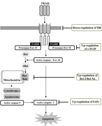
In mammals, apoptosis was induced via distinct two pathways. One is an intrinsic pathway that is activated by intracellular signals, such as DNA damage, excessive re- active oxygen species, and hypoxia. Intracellular signals, which stimulate intrinsic pathway, result in opening of the mitochondrial transition pore. Successively, mitochondrial transmembrane potential is lost, and pro-apoptotic proteins, such as cytochrome c, apoptosis-inducing factors, Omi/HtrA2, EndoG and Smac/DIABLO, released from in- termembrane space to cytoplasm. Released cytochrome c forms apoptosome, which is composed of apoptotic pro- tease-activating factor (Apaf), cytochrome c, and caspase-9.
Caspase-9 induces activation of caspase-3, 6, and 7.
Omi/HtrA2 and Smac/DIABLO bind to inhibitors of apop- tosis, thus induce to disrupting complex of inhibitors of apoptosis protein (IAP) and caspase-3 or 9. The other is an extrinsic pathway. Activation of death receptor is involved in this pathway. Tumor necrosis factor (TNF) family of cyto- kines, such as TNF, Fas ligand, and TNF-related apopto- sis-inducing ligand (TRAIL), bind to their receptor, and re- cruit caspase-8 into a death inducing signalling complex (DISC) at the plasma membrane. Caspase-8 activation leads to cleave caspase-3 (active form) or induce outer mitochon- drial membrane permeabilization, and then release cyto- chrome c. Among of them, TRAIL-mediated apoptosis is promising tumor therapeutic. TRAIL selectively induces apoptosis in tumor cells, but does not cause toxicity to most normal cells, which is supported by the presence of large numbers of decoy receptors on normal cells [29].
However, although TRAIL is a charming agent to treat-
ment against tumor cells, many papers reported that a lot
of tumor cells have resistance or change into TRAIL-induced
apoptosis resistant cells. TRAIL-resistant cancer cells can be
sensitized by chemotherapeutic drugs in vitro and in vivo,
indicating that combination therapy could induce sensitivity
of tumor cells against TRAIL. Therefore, understanding the
- Review -
molecular mechanisms of TRAIL resistance and ways to sen- sitize tumor cells to undergo apoptosis by TRAIL are im- portant points for effective cancer therapy.
Multiple molecular targets of sensitizers in the TRAIL-mediated apoptosis
TRAIL
Wiley et al. and Pitti et al., first discovered TRAIL [31,49].
TRAIL is composed of five exons of 222, 138, 42, 106, and 1245 nucleotides and four introns of 8.2, 3.2, 2.3, and 2.3 kb, and located on chromosome 3. While TRAIL genes lack TATA and CAAT boxes, the promoter of TRAIL include pu- tative response elements for GATA, activator protein a (AP1), CCAAT/enhancer binding protein (C/EBP), specific protein-1 (SP-1), octamer-binding transcription factor 1 (OCT-1), AP3, polyomavirus enhancer activator 3 homolog (PEA3), cleavage stimulation factor (CF-1), NF-κB, and inter- feron-sensitive response element (ISRE) [17,46].
Most tissue is detected TRAIL expression in human. For examples, TRAIL was detected by immunohistochemistry in hepatocytes and bile duct epithelium of liver, neurons of brain, tubuli contorti of kidney, myocytes of heart, luminal epithelium and crypt cells of colon, bronchial epithelium, alveolar septa, and vascular endothelium of lung, germ cells and leydig cells of testis, and immune cells, such as natural killer cells, macrophages, T cells and dendritic cells [35].
Furthermore, spleen and prostate also expressed TRAIL [49].
TRAIL, which is a member of TNF superfamilies, anch- ored to the cell membrane, carboxy terminus is located in extracellular space, and has the receptor binding domain as a type II transmembrane polypeptide of 281 amino acids.
Cleavage of C-terminal domain of TRAIL by cysteine pro- tease makes soluble TRAIL. TRAIL is composed of two anti- pareallel β-pleated sheets, which form a β sandwich, and then binds to the adjacent TRAIL monomer to form a homotrimer. TRAIL acts as a homotrimer and this homo- trimer has stronger biological activity than monomer of TRAIL. Cysteine residue at position 230 of TRAIL is im- portant for stability and activity as zinc ion bind to cysteine residue [2].
TRAIL receptors
TRAIL could bind to five different membrane receptors;
Death receptor 4 (DR4; TRAIL-R1), DR5 (TRAIL-R2), decoy receptor (DcR1; TRAIL-R3), DcR2 (TRAIL-R4), and
Osteoprotegerin (OPG) [8]. There is a different functionally between death receptors and decoy receptors. Death re- ceptors have a cytoplasmic death domain, which could re- cruit apoptosis signalling molecules, and trigger apoptosis.
On the other hands, decoy receptors lack cytoplasmic death domain, and inhibit apoptosis by death receptor as sequester TRAIL in extracellular space. OPG, as soluble protein, could bind to TRAIL with low affinity.
Apoptosis signalling by TRAIL receptor
TRAIL homotrimer binds to death receptors, DR4 and DR5, and then recruit FAS-associated protein with death do- main (FADD) via interaction of death domain in the carboxyl terminus of receptor. FADD, as an adaptor protein, recruits caspase 8 or 10 via death effector domain (DED), and then forms the death inducing stimulation complex (DISC). DISC could trigger activation of initiating caspase via inducing au- tocatalytic processing, and caspase is released into cytoplasm.
Activated initiating caspase induces activation of effector caspase-3, 6, and 7. In type I cells, activation of caspase-8 via formation of DISC is sufficient to induce apoptosis.
However, in type II cells, mitochondria pathway is involved in induction of apoptosis. Caspase-8 or 10 cleaves Bid into truncated Bid (tBid), which in turn translocates to the mi- tochondria and then induces the activation of Bcl-2-antago- nist/killer (Bak) and Bcl2-associated X protein (Bax), pro-apoptotic protein. Activated Bak and Bax change the mi- tochondrial membrane potential, and release cytochrome c into cytoplasm.
Non-apoptosis signalling by TRAIL receptor TRAIL could also activate non-apoptotic signalling de- pending on the cell types, duration and strength of signal, and recruiting down stream signalling molecules. TRAIL in- duces activation of nuclear factor-κB (NF-κB), mi- togen-activated protein kinase (MAPKs), protein kinase B/Akt via various combination of signalling proteins, such as FADD, TNFR type I- associated death domain protein (TRADD), caspase-8, 10, cellular FLICE-like inhibitory pro- tein (c-FLIP), TNF receptor associated factor 2 (TRAF2), and RPA-interacting protein (RIP) [5,33,40].
Physiological signalling by TRAIL receptor TRAIL signalling is associated with immune system.
Monocytes, T cells, dendritic cells, and natural killer cells
increased TRAIL (soluble and membrane bound form) ex-
pression by several stimuli, such as interferon, and ex- pression of TRAIL in monocytes and dendritic cells is corre- lated with cytotoxicity against tumor cells [1]. Therefore, TRAIL signalling is involved in immune response. In addi- tion, TRAIL is regarded as tumorigenesis and metastasis. In TRAIL knock out mice or mice treated with antibody block- ade of TRAIL, tumor growth and metastasis are promoted [4,36]. In contrast, a few papers were reported that TRAIL is not associated with tumorigenesis in intestinal and skin tumor [9,51].
TRAIL as therapeutic agent
TRAIL, is a potent anti-cancer agent, promotes apoptosis in tumor cells, while has no or minimal effects on death of normal cells, because level of decoy receptors is higher in normal cells than in tumor cells. Soluble TRAIL and mono- clonal antibodies, anti-DR4 and anti-DR5, are selectively in- duced apoptosis in tumor cells. TRAIL is a promising ther- apeutic agent for the treatment of cancer. However, therapy using TRAIL is limited to TRAIL-sensitive tumor. In re- cently, many researchers were reported that a number of cancer cells are resistant to TRAIL, such as pancreatic cancer, neuroblastoma, chronic lymphocytic leukaemia (CLL), as- trocytoma, meningioma, medulloblastoma and malignant melanoma. Therefore, we need more efficient therapeutic strategy.
Mechanism of resistance to TRAIL
Apoptosis by TRAIL is modulated at several stages in the apoptotic signalling pathways. Tumor cells could escape from apoptosis via activation of survival mechanisms.
Therefore, we need to study for TRAIL-resistant mechanisms.
Death receptor expression
TRAIL resistant is associated with 8p chromosome deletion. Death receptors are located on this sites, which is a hot-spot for deletions and frequently appear allelic loss [18]. Down-regulation of death receptor was detected in sev- eral cancer types, such as non-Hodgkin’s lymphoma and non-small cell lung cancers, head and neck cancers, gastric cancers, and breast cancers [7]. Hypermethylation of TRAIL gene decreases TRAIL receptor gene expression, and down-regulation of death receptor is one of TRAIL-resistant mechanism [43]. Furthermore, post-translational mod-
ification of the death receptor changes signal transition by TRAIL. O-glycosylation of death receptor promotes TRAIL-induced clustering of DR4 and DR5. O-glycosylation by GALNT14, a O-glycosyltransferase, increases response against TRAIL in pancreatic carcinoma, non-small-cell lung carcinoma and melanoma cell lines [44]. S-palmitoylation of DR4 is also involved in oligomerization of DR4, and then promotes TRAIL-induced signalling [44].
c-FLIP and caspase-8 expression
TRAIL-resistance is correlated with c-FLIP expression in lung and breast cancers, colon and hepatocellular carcinoma, malignant melanoma, leukemia, and glioblastoma. c-FLIP competes with caspase-8 for DISC binding sites, thus high levels of c-FLIP could block cleavage of caspase-8. Thus, c-FLIP inhibits TRAIL-induced death signal. Furthermore, some cells, such as lung carcinoma, neuroblastoma, leuke- mia, and colon cells, are expressed low levels of caspase-8.
Methylation and point mutation of genes results in low lev- els of caspase-8 expression [39].
Modulation of components in mitochondria
Bcl-2 family is divided into two groups, pro-apoptotic
Fig. 1. Activation of extrinsic and intrinsic apoptosis pathway by TRAIL. Extrinsic (death receptor) and intrinsic path- ways (mitochondria) were involved in TRAIL-induced apoptosis.
Fig. 2. Mechanisms of resistance to TRAIL.
(Bax and Bak) and anti-apoptotic (Bcl-2 and Bcl-xL).
Overexpression of Bcl-2 or Bcl-xL blocked TRAIL-induced apoptosis in adenocarcinoma and pancreatic carcinoma, and inactivation of Bax and Bak also failed to induce apoptosis by TRAIL in some cells, such as solid tumor and colon carci- nomas and colorectal cancer cells [34,42,52]. Balance of Bcl-2 family between pro-apoptotic protein and anti-apoptotic protein decides sensitivity against TRAIL. Therefore, al- though TRAIL induces extrinsic apoptosis, crosstalk between death receptor signalling and mitochondrial signalling is as- sociated with TRAIL-induced apoptosis.
IAP family contains baculovirus IAP repeat (BIR domain), which could bind caspases, and C-terminal RING domain, which ubiquitinate. When IAP make complex with caspase, they ubiquitinate and then degrade. Member of IAP contain X-linked IAP (XIAP), c-IAP1, c-IAP2, survivin, livin, Ts-IAP, ILP-2 and Bruce. Among of them, XIAP has the most potent function and bind caspase 3, 7, and 9. Overexpression of IAPs is associated with TRAIL resistance [28].
Modulation of signalling pathways
PI3K/Akt signalling is involved in expression of c-FLIP, XIAP, and Bcl-2 [48, 30]. Increased expression of these pro- teins induces inactivation of Bax, Bad, and caspase.
Therefore, activation of PI3K/Akt increases resistance against TRAIL. In fact, increased activation of PI3K/Akt sig- nalling is already reported in TRAIL-resistant cells, such as colon cancer, gastric cancers, and leukemia [23,32,47].
MAPKs is identified three subfamilies; ERK, JNK, and p38 MAPK. The role of MAPK is controversial on TRAIL-in- duced apoptosis. ERK has been known as survival signalling. In breast cancer cells, activation of ERK inhibits TRAIL-induced apoptosis [20], while inhibition of JNK sig- nalling induces TRAIL-sensitize in hepatocellular carcinoma cells [27]. In contrast, activation of JNK and p38 MAPK en- hance TRAIL-induced apoptosis in hepatocellular carcinoma [45]. Therefore, understanding about role of MAPKs on TRAIL-induced apoptosis needs more information and fur- ther studies.
The role of NF-κB signalling on TRAIL-induced apoptosis is also controversial. TRAIL homotrimer binds DR4 or DR5, and could activate NF-κB signalling [13]. XIAP could en- hance of IκB degradation, and then increase NF-κB activity [10, 22]. Thus, activation of NF-κB desensitizes cells to TRAIL. However, in the TRAIL-resistant cells, NF-κB has an apposite function, that is, NF-κB induced proliferation and survival [6]. There is one of possibility to determine death or life. The component of NF-κB has a different function. Rel A (p65) acts as anti-apoptotic signalling.
Overexpression of RelA induced c-IAP and decreased DR expression. On the other hands, c-Rel acts as a pro-apoptotic signalling, which increase DR expression and decrease IAPs expression [3].
Strategies to induce TRAIL sensitivity
As mentioned earlier, tumor cells have exhibit resistance
to TRAIL-induced apoptosis. Therefore, to induce cellular
apoptosis need a new therapeutic strategy. Previous studies
reported that TRAIL induced tumor cells apoptosis in the
presence of other chemopreventive drugs, such as curcumin,
withaferin A, luteolin, quercetin, resveratrol, and silibinin,
which restore TRAIL sensitivity. The sensitizing mechanisms
of these drugs are diverse; 1) up-regulation of death receptor
(DR) expression levels [14], 2) decrease of c-FLIP expression
[21], 3) up-regulation of pro-apoptotic components and
down-regulation of anti-apoptotic components in Bcl-2 fam-
ily [41], 4) reduction of IAPs family [19], 5) modulation of
signalling molecules.
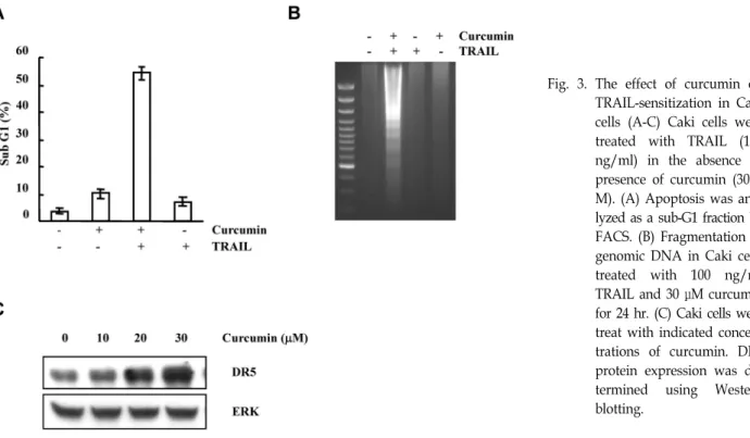
Fig. 3. The effect of curcumin on TRAIL-sensitization in Caki cells (A-C) Caki cells were treated with TRAIL (100 ng/ml) in the absence or presence of curcumin (30 μ M). (A) Apoptosis was ana- lyzed as a sub-G1 fraction by FACS. (B) Fragmentation of genomic DNA in Caki cells treated with 100 ng/ml TRAIL and 30 μM curcumin for 24 hr. (C) Caki cells were treat with indicated concen- trations of curcumin. DR5 protein expression was de- termined using Western blotting.
Novel strategy via modulation of TRAIL-related components
Up-regulation of death receptor; Curcumin and Calyculin A
Curcumin is a very famous flavoring agent in foods, and is a major component of the Curcuma species. Previous stud- ies reported that curcumin have a antiproliferative activity and acticarcinogenic activity in vitro and in vivo [11,24].
However, the mechasnisms underlying the effects of curcu- min on cancer cells are not understood. Curcumin enhanced TRAIL-induced cell death in Caki cells, renal cancer cell line (Fig. 3A). Furthermore, Treatment with curcumin and TRAIL in Caki cells increased typical ladder pattern of inter- nucleosomal fragmentation, which is a hallmark of apoptosis (Fig. 3B). Molecular mechanisms underlying the synergistic induction of apoptosis by curcumin and TRAIL in Caki cells is an up-regulation of DR5 (Fig. 3C). Treatment with curcu- min induced the expression of DR5 protein in a dose-de- pendent manner. These results sugget that curcumin enhan- ces TRAIL-induced apoptosis by DR5 upregulation in Caki cells.
State of phosphorylation and dephosphorylation is im- portant on transition cellular signal. Phosphorylaion is mod- ulate by protein kinases, such as MAPK, protein kinase A, protein kinase C, and PI3K/Akt. On the other hands, protein
phosphatases (PPs) restore protein to dephosphorylated state. The balance between kinase and phosphatase is key regulator for the cellular signalling cascade. Phosphatase is divided into four groups as their substrates; 1) serine/threo- nine phosphatase, 2) protein histidine phosphatase, 3) pro- tein tyrosine phosphatase, and 4) dual-specific phosphatase.
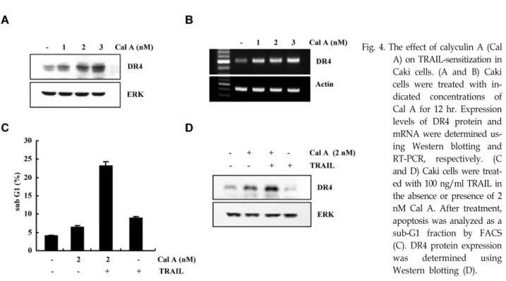
Among them, we have an interesting on serine/threonine phosphatase. Calyculin A has known as a potent ser- ine/threonine phosphatase inhibitor, and it inhibits phos- phatase 1 and 2.
In previous studies, inhibitors of phosphatase induced tu- mor cell apoptosis. Interestingly, caliculin A increased apop- tosis in human osteoblstic osteosarcoma MG63 cells [37]. As shown in Fig.4A and B, calyculin A significantly induced DR4 protein and mRNA expression in a dose-dependent manner. This data indicate that calyculin A may regulate expression of TRAIL sensitizing components. Caki cells are not induced apoptosis by TRAIL alone. However, when Caki cells were co-treated with calyculin A and TRAIL, sub-G1 phase cells increased (Fig. 4C). In addition, DR4 expression was dramatically increased in calyculin A and TRAIL co-treated- Caki cells, compared with calyculin A alone (Fig.
4D). This result suggest that calyculin A-mediated TRAIL
sensitization is an attractive strategy for treatment of
TRAIL-resistant cancer cells.
Fig. 5. The effect of Withaferin A (Wit A) on TRAIL-sensitiza- tion in Caki cells. (A) Caki cells were treated with the indicated concentrations of Wit A for 24 hr. c-FLIP pro- tein expression was de- termined using Western blotting. (B and C) Caki/
vector and Caki/c-FLIP cells were treated with 100 ng/ml TRAIL in the absence or presence of 1.2 μM Wit A for 24 hr. Apoptosis was ana- lyzed as a sub-G1 fraction by FACS (B). PARP and c-FLIP protein expression were de- termined using Western blotting (C).
Fig. 4. The effect of calyculin A (Cal A) on TRAIL-sensitization in Caki cells. (A and B) Caki cells were treated with in- dicated concentrations of Cal A for 12 hr. Expression levels of DR4 protein and mRNA were determined us- ing Western blotting and RT-PCR, respectively. (C and D) Caki cells were treat- ed with 100 ng/ml TRAIL in the absence or presence of 2 nM Cal A. After treatment, apoptosis was analyzed as a sub-G1 fraction by FACS (C). DR4 protein expression was determined using Western blotting (D).
Down-regulation of cFLIP; Withaferin A
Withaferin A is a steroidal lactone isolated from the me- dicinal plant Withania somnifera. It has been known as an important prototype of the withanolide class of natural products. Previous studies reported that withaferin A in- hibited tumor cell growth, metastasis and angiogenesis [26, 50]. In addition, withaferin A significantly inhibited c-FLIP
expression in a dose-dependent manner in Caki cells (Fig.
5A). Furthermore, overexpression of c-FLIP in Caki cells
(Caki/c-FLIP) inhibited withaferin A–facilitated TRAIL-in-
duced apoptosis, compared with that of Caki/vector cells
(Fig. 5B). Cleavage of PARP was also significantly inhibited
by overexpression of c-FLIP (Fig. 5C). These results indicate
that down-regulation of c-FLIP is associated with withaferin
Fig. 6. The effect of Mithramycin A (Mi A) on TRAIL-sensitiza- tion in Caki cells (A) Caki cells were treated with the indicated concentrations of Mi A in the absence or pres- ence of 100 ng/ml TRAIL for 24 hr. (B and C) Caki/vector and Caki/XIAP cells were treated with 200 nM Mi A and 100 ng/ml TRAIL for 24 hr. Apoptosis was analyzed as a sub-G1 by FACS (B). XIAP and PARP protein expression were de- termined using Western blotting (C).
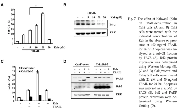
Fig. 7. The effect of Kahweol (Kah) on TRAIL-sensitization in Caki cells (A and B) Caki cells were treated with the indicated concentrations of Kah in the absence or pres- ence of 100 ng/ml TRAIL for 24 hr. Apoptosis was an- alyzed as a sub-G1 fraction by FACS (A). Bcl2 protein expression was determined using Western blotting (B).
(C and D) Caki/vector and Caki/Bcl2 cells were treated with 20 μM and 50 ng/ml TRAIL for 24 hr. Apoptosis was analyzed as a sub-G1 by FACS (B). Bcl2 and PARP protein expression were de- termined using Western blotting (D).
A-enhanced TRAIL-induced apoptosis.
Down-regulation of XIAP; Mithramycin A
Mithramycin A (also known as Plicamycin), which is an anticancer and antibiotic agent, was isolated from strepto-
myces griseus. Mithramycin A have an anticancer effects viainteraction with double-stranded DNA with GC base specif- icity in genes promoter [25]. Furthermore, mithramycin A could regulate expression of TRAIL sensitized components.
Caki cells treated with mithramycin A was significantly in-
hibited XIAP expression (Fig. 6A). Overexpression of XIAP
(Caki/XIAP) was inhibited apoptosis and PARP protein ex-
pression in mithramycin A and TRAIL-treatd cells, compare with Caki/vector cells (Fig. 6B and C).
Down-regulation of Bcl-2; Kahweol
Kahweol is a diterpene molecule, that is found in coffee beans. The compound have a multiple functions, such as an- ti-caner, anti-tumor, and anti-inflammation [12,16,38].
Kahweol acts as anti-carcinogenic drugs through induction of phase II detoxifying enzymes and reduction of signal transducer and activator of transcription 3 (STAT 3) [15,38].
In addition, kahweol sensitized TRAIL-induced apoptosis in Caki cells. As shown in Fig. 7A, kahweol induced TRAIL-mediated cell death in a dose-dependent manner.
Bcl-2 expression also blocked in kahweol and TRAIL-treated cells, compare with kahweol or TRAIL alone (Fig. 7B).
Overexpression of Bcl-2 (Caki/Bcl-2) significantly decreased TRAIL-induced cell death in the presence of kahweol, com- pare with Caki/vector (Fig. 7C). Cleavage of PARP also blocked in Caki/Bcl2 cells (Fig. 7D). Therefore, kahweol en- hanced TRAIL-sensitization via down-regulation of Bcl2.
Conclusion
TRAIL has been known as a potent anti-cancer drug, be- cause TRAIL specifically induces apoptosis in tumor cells, not normal cells. However, some cancer cells have a resist-
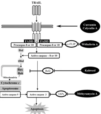
Fig. 8. Schematic diagram of TRAIL sensitization.
ance against TRAIL, through induction of c-FLIP, IAPs, and anti-apoptotic Bcl2 family expression, and reduction of DR4, DR5, and pro-apoptotic Bcl2 family expression. Single treat- ment with TRAIL is not sufficient strategy to induce apopto- sis in tumor cells. Several agents, such as curcumin, calyculin A, withaferin A, mithramycin A, and kahweol, could en- hance sensitivity against TRAIL through modulation of TRAIL-related signal components (Fig. 8). Therefore, we need to understand the mechanism of TRAIL-resistance and find a novel TRAIL-sensitizing drug for cancer therapy.
Acknowledgment
This work was supported by Mid-career Researcher pro- gram through NRF grant funded by the MEST (No.
2011-0016239).
References
1. Almasan, A. and A. Ashkenazi. 2003. Apo2L/TRAIL: apop- tosis signaling, biology, and potential for cancer therapy.
Cytokine Growth Factor Rev. 14, 337-348.
2. Bodmer, J. L., P. Meier, J. Tschopp, and P. Schneider. 2000.
Cysteine 230 is essential for the structure and activity of the cytotoxic ligand TRAIL. J. Biol. Chem. 275, 20632-20637.
3. Chen, X., K. Kandasamy, and R. K. Srivastava. 2003.
Differential roles of RelA (p65) and c-Rel subunits of nuclear factor kappa B in tumor necrosis factor-related apoptosis-in- ducing ligand signaling. Cancer Res. 63, 1059-1066.
4. Cretney, E., K. Takeda, H. Yagita, M. Glaccum, J. J. Peschon, and M. J. Smyth. 2002. Increased susceptibility to tumor ini- tiation and metastasis in TNF-related apoptosis-inducing li- gand-deficient mice. J. Immunol. 168, 1356-1361.
5. Di, P. R. and G. Zauli. 2004. Emerging non-apoptotic func- tions of tumor necrosis factor-related apoptosis-inducing li- gand (TRAIL)/Apo2L. J. Cell Physiol 201, 331-340.
6. Ehrhardt, H., S. Fulda, I. Schmid, J. Hiscott, K. M. Debatin, and I. Jeremias. 2003. TRAIL induced survival and pro- liferation in cancer cells resistant towards TRAIL-induced apoptosis mediated by NF-kappaB. Oncogene 22, 3842-3852.
7. Fulda, S. and K. M. Debatin. 2004. Modulation of TRAIL signaling for cancer therapy. Vitam. Horm. 67, 275-290.
8. Gli-Esposti, M. 1999. To die or not to die--the quest of the TRAIL receptors. J. Leukoc. Biol. 65, 535-542.
9. Grosse-Wilde, A., O. Voloshanenko, S. L. Bailey, G. M.
Longton, U. Schaefer, A. I. Csernok, G. Schutz, E. F. Greiner, C. J. Kemp, and H. Walczak. 2008. TRAIL-R deficiency in mice enhances lymph node metastasis without affecting pri- mary tumor development. J. Clin. Invest 118, 100-110.
10. Hofer-Warbinek, R., J. A. Schmid, C. Stehlik, B. R. Binder, J. Lipp, and M. R. de. 2000. Activation of NF-kappa B by XIAP, the X chromosome-linked inhibitor of apoptosis, in
endothelial cells involves TAK1. J. Biol. Chem. 275, 22064-22068.
11. Huang, M. T., R. C. Smart, C. Q. Wong, and A. H. Conney.
1988. Inhibitory effect of curcumin, chlorogenic acid, caffeic acid, and ferulic acid on tumor promotion in mouse skin by 12-O-tetradecanoylphorbol-13-acetate. Cancer Res. 48, 5941-5946.
12. Huber, W. W., S. Prustomersky, E. Delbanco, M. Uhl, G.
Scharf, R. J. Turesky, R. Thier, and R. Schulte-Hermann.
2002. Enhancement of the chemoprotective enzymes glucur- onosyl transferase and glutathione transferase in specific or- gans of the rat by the coffee components kahweol and cafestol. Arch. Toxicol. 76, 209-217.
13. Jeremias, I. and K. M. Debatin. 1998. TRAIL induces apopto- sis and activation of NFkappaB. Eur. Cytokine Netw. 9, 687-688.
14. Jung, E. M., J. W. Park, K. S. Choi, J. W. Park, H. I. Lee, K. S. Lee, and T. K. Kwon. 2006. Curcumin sensitizes tumor necrosis factor-related apoptosis-inducing ligand (TRAIL)-mediated apoptosis through CHOP-independent DR5 upregulation. Carcinogenesis 27, 2008-2017.
15. Kim, H. G., Y. P. Hwang, and H. G. Jeong. 2009. Kahweol blocks STAT3 phosphorylation and induces apoptosis in hu- man lung adenocarcinoma A549 cells. Toxicol. Lett. 187, 28-34.
16. Kim, J.Y., K. S. Jung, K. J. Lee, H. K. Na, H. K. Chun, Y.
H. Kho, and H. G. Jeong. 2004. The coffee diterpene kahweol suppress the inducible nitric oxide synthase expression in macrophages. Cancer Lett. 213, 147-154.
17. Kirshner, J. R., A. Y. Karpova, M. Kops, and P. M. Howley.
2005. Identification of TRAIL as an interferon regulatory fac- tor 3 transcriptional target. J. Virol. 79, 9320-9324.
18. LeBlanc, H. N. and A. Ashkenazi. 2003. Apo2L/TRAIL and its death and decoy receptors. Cell Death. Differ. 10, 66-75.
19. Lee, T. J., E. M. Jung, J. T. Lee, S. Kim, J. W. Park, K. S.
Choi, and T. K. Kwon. 2006. Mithramycin A sensitizes can- cer cells to TRAIL-mediated apoptosis by down-regulation of XIAP gene promoter through Sp1 sites. Mol. Cancer Ther.
5, 2737-2746.
20. Lee, T. J., J. T. Lee, J. W. Park, and T. K. Kwon. 2006.
Acquired TRAIL resistance in human breast cancer cells are caused by the sustained cFLIP(L) and XIAP protein levels and ERK activation. Biochem. Biophys. Res. Commun. 351, 1024-1030.
21. Lee, T. J., H. J. Um, D. S. Min, J. W. Park, K. S. Choi, and T. K. Kwon. 2009. Withaferin A sensitizes TRAIL-induced apoptosis through reactive oxygen species-mediated up-reg- ulation of death receptor 5 and down-regulation of c-FLIP.
Free Radic. Biol. Med. 46, 1639-1649.
22. Levkau, B., K. J. Garton, N. Ferri, K. Kloke, J. R. Nofer, H.
A. Baba, E. W. Raines, and G. Breithardt. 2001. xIAP induces cell-cycle arrest and activates nuclear factor-kappaB : new survival pathways disabled by caspase-mediated cleavage during apoptosis of human endothelial cells. Circ. Res. 88, 282-290.
23. Martelli, A. M., P. L. Tazzari, G. Tabellini, R. Bortul, A. M.
Billi, L. Manzoli, A. Ruggeri, R. Conte, and L. Cocco. 2003.
A new selective AKT pharmacological inhibitor reduces re- sistance to chemotherapeutic drugs, TRAIL, all-trans-reti- noic acid, and ionizing radiation of human leukemia cells.
Leukemia 17, 1794-1805.
24. Mehta, K., P. Pantazis, T. McQueen, and B. B. Aggarwal.
1997. Antiproliferative effect of curcumin (diferuloylmethane) against human breast tumor cell lines. Anticancer Drugs 8, 470-481.
25. Miller, D. M., D. A. Polansky, S. D. Thomas, R. Ray, V. W.
Campbell, J. Sanchez, and C. A. Koller. 1987. Mithramycin selectively inhibits transcription of G-C containing DNA.
Am. J. Med. Sci. 294, 388-394.
26. Mohan, R., H. J. Hammers, P. Bargagna-Mohan, X. H. Zhan, C. J. Herbstritt, A. Ruiz, L. Zhang, A. D. Hanson, B. P.
Conner, J. Rougas, and V. S. Pribluda. 2004. Withaferin A is a potent inhibitor of angiogenesis. Angiogenesis. 7, 115-122.
27. Mucha, S. R., A. Rizzani, A. L. Gerbes, P. Camaj, W. E.
Thasler, C. J. Bruns, S. T. Eichhorst, E. Gallmeier, F. T.
Kolligs, B. Goke, and E. N. De Toni. 2009. JNK inhibition sensitises hepatocellular carcinoma cells but not normal hepatocytes to the TNF-related apoptosis-inducing ligand.
Gut. 58, 688-698.
28. Ng, C. P., A. Zisman, and B. Bonavida. 2002. Synergy is achieved by complementation with Apo2L/TRAIL and acti- nomycin D in Apo2L/TRAIL-mediated apoptosis of pros- tate cancer cells: role of XIAP in resistance. Prostate 53, 286-299.
29. Pan, G., J. Ni, Y. F. Wei, G. Yu, R. Gentz, and V. M. Dixit.
1997. An antagonist decoy receptor and a death do- main-containing receptor for TRAIL. Science 277, 815-818.
30. Perianayagam, M. C., N. E. Madias, B. J. Pereira, and B.
L. Jaber. 2006. CREB transcription factor modulates Bcl2 transcription in response to C5a in HL-60-derived neutrophils. Eur. J. Clin. Invest 36, 353-361.
31. Pitti, R. M., S. A. Marsters, S. Ruppert, C. J. Donahue, A.
Moore, and A. Ashkenazi. 1996. Induction of apoptosis by Apo-2 ligand, a new member of the tumor necrosis factor cytokine family. J. Biol. Chem. 271, 12687-12690.
32. Rychahou, P. G., C. A. Murillo, and B. M. Evers. 2005.
Targeted RNA interference of PI3K pathway components sensitizes colon cancer cells to TNF-related apoptosis-induc- ing ligand (TRAIL). Surgery 138, 391-397.
33. Secchiero, P., A. Gonelli, E. Carnevale, D. Milani, A.
Pandolfi, D. Zella, and G. Zauli. 2003. TRAIL promotes the survival and proliferation of primary human vascular endo- thelial cells by activating the Akt and ERK pathways.
Circulation 107, 2250-2256.
34. Song, J. J., J. Y. An, Y. T. Kwon, and Y. J. Lee. 2007. Evidence for two modes of development of acquired tumor necrosis factor-related apoptosis-inducing ligand resistance.
Involvement of Bcl-xL. J. Biol. Chem. 282, 319-328.
35. Spierings, D. C., E. G. de Vries, E. Vellenga, F. A. van den Heuvel, J. J. Koornstra, J. Wesseling, H. Hollema, and J. S.
de. 2004. Tissue distribution of the death ligand TRAIL and its receptors. J. Histochem. Cytochem. 52, 821-831.
36. Takeda, K., Y. Hayakawa, M. J. Smyth, N. Kayagaki, N.
Yamaguchi, S. Kakuta, Y. Iwakura, H. Yagita, and K.
Okumura. 2001. Involvement of tumor necrosis factor-re- lated apoptosis-inducing ligand in surveillance of tumor metastasis by liver natural killer cells. Nat. Med. 7, 94-100.
37. Tanaka, H., K. Yoshida, H. Okamura, H. Morimoto, T.
Nagata, and T. Haneji. 2007. Calyculin A induces apoptosis and stimulates phosphorylation of p65NF-kappaB in human osteoblastic osteosarcoma MG63 cells. Int. J. Oncol. 31, 389-396.
38. Tao, K. S., W. Wang, L. Wang, D. Y. Cao, Y. Q. Li, S. X.
Wu, and K. F. Dou. 2008. The multifaceted mechanisms for coffee's anti-tumorigenic effect on liver. Med. Hypotheses 71, 730-736.
39. Teitz, T., T. Wei, M. B. Valentine, E. F. Vanin, J. Grenet, V. A. Valentine, F. G. Behm, A. T. Look, J. M. Lahti, and V. J. Kidd. 2000. Caspase 8 is deleted or silenced preferen- tially in childhood neuroblastomas with amplification of MYCN. Nat. Med. 6, 529-535.
40. Tran, S. E., T. H. Holmstrom, M. Ahonen, V. M. Kahari, and J. E. Eriksson. 2001. MAPK/ERK overrides the apop- totic signaling from Fas, TNF, and TRAIL receptors. J. Biol.
Chem. 276, 16484-16490.
41. Um, H. J., J. H. Oh, Y. N. Kim, Y. H. Choi, S. H. Kim, J.
W. Park, and T. K. Kwon. 2010. The coffee diterpene kah- weol sensitizes TRAIL-induced apoptosis in renal carcinoma Caki cells through down-regulation of Bcl-2 and c-FLIP.
Chem. Biol. Interact. 186, 36-42.
42. Van Geelen, C.M., E. G. de Vries, and J. S. de. 2004. Lessons from TRAIL-resistance mechanisms in colorectal cancer cells: paving the road to patient-tailored therapy. Drug Resist. Updat. 7, 345-358.
43. van Noesel, M. M., B. S. van, P. A. Voute, J. G. Herman, R. Pieters, and R. Versteeg. 2003. Clustering of hyper- methylated genes in neuroblastoma. Genes Chromosomes Cancer 38, 226-233.
44. Wagner, K. W., E. A. Punnoose, T. Januario, D. A. Lawrence, R. M. Pitti, K. Lancaster, D. Lee, G. M. von, S. F. Yee, K.
Totpal, L. Huw, V. Katta, G. Cavet, S. G. Hymowitz, L.
Amler, and A. Ashkenazi. 2007. Death-receptor O-glyco-
sylation controls tumor-cell sensitivity to the proapoptotic ligand Apo2L/TRAIL. Nat. Med. 13, 1070-1077.
45. Wang, C., T. Chen, N. Zhang, M. Yang, B. Li, X. Lu, X.
Cao, and C. Ling. 2009. Melittin, a major component of bee venom, sensitizes human hepatocellular carcinoma cells to tumor necrosis factor-related apoptosis-inducing ligand (TRAIL)-induced apoptosis by activating CaMKII-TAK1- JNK/p38 and inhibiting IkappaBalpha kinase-NFkappaB. J.
Biol. Chem. 284, 3804-3813.
46. Wang, Q., Y. Ji, X. Wang, and B. M. Evers. 2000. Isolation and molecular characterization of the 5'-upstream region of the human TRAIL gene. Biochem. Biophys. Res. Commun. 276, 466-471.
47. Wang, W. Q., H. Zhang, H. B. Wang, Y. G. Sun, Z. H. Peng, G. Zhou, S. M. Yang, R. Q. Wang, and D. C. Fang. 2010.
Programmed cell death 4 (PDCD4) enhances the sensitivity of gastric cancer cells to TRAIL-induced apoptosis by in- hibiting the PI3K/Akt signaling pathway. Mol. Diagn. Ther.
14, 155-161.
48. Wang, X., W. Chen, W. Zeng, L. Bai, Y. Tesfaigzi, S. A.
Belinsky, and Y. Lin. 2008. Akt-mediated eminent ex- pression of c-FLIP and Mcl-1 confers acquired resistance to TRAIL-induced cytotoxicity to lung cancer cells. Mol. Cancer Ther. 7, 1156-1163.
49. Wiley, S. R., K. Schooley, P. J. Smolak, W. S. Din, C. P.
Huang, J. K. Nicholl, G. R. Sutherland, T. D. Smith, C.
Rauch, and C. A. Smith. 1995. Identification and character- ization of a new member of the TNF family that induces apoptosis. Immunity 3, 673-682.
50. Yokota, Y., P. Bargagna-Mohan, P. P. Ravindranath, K. B.
Kim, and R. Mohan. 2006. Development of withaferin A an- alogs as probes of angiogenesis. Bioorg. Med. Chem. Lett. 16, 2603-2607.
51. Yue, H. H., G. E. Diehl, and A. Winoto. 2005. Loss of TRAIL-R does not affect thymic or intestinal tumor develop- ment in p53 and adenomatous polyposis coli mutant mice.
Cell Death Differ. 12, 94-97.
52. Zhang, L. and B. Fang. 2005. Mechanisms of resistance to TRAIL-induced apoptosis in cancer. Cancer Gene Ther. 12, 228-237.