REVIEW ARTICLE
간, 췌장 질환에서 Necroptosis
윤재훈, 전대원
1, 최호순
1한림대학교 의과대학, 한양대학교 의과대학1 내과학교실
Necroptosis in Liver and Pancreatic Diseases
Jai Hoon Yoon, Dae Won Jun1 and Ho Soon Choi1
Departments of Internal Medicine, Hallym University College of Medicine, Chuncheon, Hanyang University College of Medicine, Seoul1, Korea
Cell death is an integral part of life of an organism that is necessary to maintain organs and tissues. Apoptosis, autophagy, and necrosis were noted as three morphologically distinct types of cell death. Apoptosis is a well identified process that is driven by programmed molecular mechanism. Until now, the investigators believed that necrosis was not a programmed molecular event. However, recently, an alternative death pathway called ‘necroptosis’ was delineated and proposed as a form of ‘programmed necrosis’. According to the recent recommendations by the Nomenclature Committee of Cell Death, this term denotes necrotic cell death dependent on receptor-interacting protein kinase (RIPK3). Its role in a variety of diseases, such as ischemia-perfusion injury, infection, inflammatory bowel disease, pancreatitis, steatohepatitis etc., is being elucidated.
Necroptosis is currently attracting the attention of the scientific community. Herein we discuss the clinical implications and the role of necroptosis in gastrointestinal tract focusing on liver and pancreatic diseases. (Korean J Gastroenterol 2014;64:182-188) Key Words: Cell death; Necroptosis; Necrosis; Receptor-interacting protein kinase; Tumor necrosis factor-alpha
CC This is an open access article distributed under the terms of the Creative Commons Attribution Non-Commercial License (http://creativecommons.org/licenses/
by-nc/3.0) which permits unrestricted non-commercial use, distribution, and reproduction in any medium, provided the original work is properly cited.
교신저자: 최호순, 133-791, 서울시 성동구 왕십리로 222, 한양대학교의료원 소화기내과
Correspondence to: Ho Soon Choi, Department of Gastroenterology, Hanyang University Medical Center, 222 Wangsimni-ro, Seongdong-gu, Seoul 133-791, Korea.
Tel: +82-2-2290-8379, Fax: +82-2-2298-9183, E-mail: [email protected] Financial support: None. Conflict of interest: None.
서 론
최근까지도 세포사멸과 세포 생존에 대한 연구는 광범위하 게 이루어져 왔으나 세포가 어떤 결정적 계기를 통해 생사를 결정하는지는 확실하게 알려져 있지 않다. 역사적으로 세포사 멸의 기전은 크게 조절된(regulated) 것과 조절되지 않은 (unregulated) 것으로 분류할 수 있다. 세포자멸사(apopto- sis)는 조절된 세포사멸의 대표적인 예이고, 괴사(necrosis)는 조절되지 않은 세포사멸의 대표적인 예이다. 이들에 관여하는 세포사멸의 과정 중 치료 목적으로 사용될 가능성이 있는 과 정이나 물질들이 있다. 또한 동일한 세포사멸 유도물질에 노 출되어도 물질의 농도에 따라 다양한 종류의 다른 세포사멸들 이 뒤섞여 관찰된다. 조율된 세포사멸은 유전적으로 통제되는 반면 조율되지 않은 세포사멸은 세포가 과도한 스트레스를 이
겨내지 못할 때 일어난다. 여러 종류의 세포사멸 중 자멸사와 괴사는 잘 알려진 형태로 상이한 세포사멸의 유형이다. 세포 자멸사, 괴사 및 자가포식(autophagy)은 주된 세포사멸의 유 형으로 각각 특이적인 분자적, 생화학적, 형태학적 특징을 갖 는다.
세포괴사는 세포의 사멸의 한 형태로 질환의 원인이 될 수 도 있고 결과로 관찰될 수도 있다. 일련의 분자생물학 기전을 통한 프로그램화된 세포죽음의 한 형태로 세포자멸사 개념이 알려진 이후 세포괴사와 다른 형태의 세포사멸로 여러 질환에 서의 병리기전 등에 대한 연구가 이루어지고 있으며,1 특히 암을 비롯하여 많은 질환에서 세포자멸사는 병태생리에 매우 중요한 역할을 하고 있음이 밝혀졌다. 세포자멸사는 일련의 분자생물학 기전을 통한 프로그램화된 세포죽음의 한 형태로 카스파제(caspase proteases)의 활성에 의한 기전으로 세포
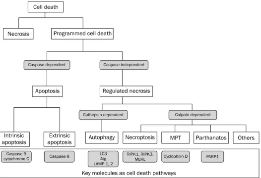
Fig. 1. Types of cell death. Programmed cell death can be divided into apoptosis and regulated necrosis. There are two subtypes of apoptosis:
intrinsic apoptosis mediated by caspase 9 vs. extrinsic apoptosis mediated by caspase 8. As for regulated necrosis, there are several subtypes including necroptosis regulated by RIPK1, RIPK3 and MLKL, and MPT mediated by cyclophilin D, In addition to necroptosis and MPT, we suspect that there would be another regulated necrosis pathway.
Note: The classification shown is not a confirmative one but has been presented for descriptive purposes.
MPT, mitochondrial permeability transition; LC3, light chain 3; Atg, autophagy-related protein; LAMP, lysosomal associated membrane protein;
RIPK, receptor-interacting protein kinase; MLKL, mixed lineage kinase domain-like; PARP1, poly [ADP-ribose] polymerase 1.
형태의 변형을 유발하게 된다.2 이와 다르게 세포괴사의 경우 세포사멸의 일련의 과정이 프로그램화되어 있지 않다.3 그러 나, 최근 이러한 괴사성 세포사멸 중 지금까지 알려진 세포괴 사가 아닌 정해진 분자생물학 기전을 통해서 ‘프로그램화된 세포괴사(programmed necrosis)’가 이루어진다는 것이 알려 졌다.4-6 최근 이러한 괴사성 세포 사멸의 한 종류로 프로그램 화된 세포괴사를 “necroptosis”라 정의하였다(Fig. 1). Ne- croptosis는 괴사와 유사한 예비 세포사멸 기전으로 세포자멸 사가 차단되었을 때 개시된다. 세포자멸사의 경로는 카스파제 에 의존하는 반면 necroptosis의 경로는 키나아제(kinase)에 의존하므로, kinase activity containing protein의 일종인 receptor interacting protein (RIP)이 세포 스트레스를 감지 하는 데 필수적이다. Necroptosis에 대한 정의는 2012년에 Galluzzi 등6,7에 의해 발표된 권고에 따라 RIPK3 (receptor- interacting protein kinase 3)에 의한 괴사성 세포사멸로 기 술하고자 한다(Fig. 1). Necroptosis에 대한 분자생물학 기전 이 밝혀짐에 따라서 이전에는 조절할 수 없었던 세포괴사와 연관된 질환에 대한 치료접근이 가능해질 것으로 생각된다.
최근 심근경색,8 뇌경색, 동맥경화, 허혈-재관류로 인한 조직 손상,9 궤사성 췌장염,10,11 염증성 장질환,12 지방간염13 등 다 양한 질환에서 necroptosis에 대한 이해를 바탕으로 한 병리
기전 및 치료방법에 대한 연구들이 활발히 진행되고 있다. 이 종설에서는 necrotpsis에 대한 소개 및 간, 췌장질환과 관련 된 연구에 대해 설명하고자 한다.
본 론
1. Necroptosis에 대한 소개
세포자멸사는 카스파제의 활성화를 일으키는 일련의 분자 생물학 기전에 의한 세포죽음을 의미하고 궁극적으로는 세포 형태의 변화를 유발한다.2,14 반면, necrotopsis는 발견 초기 카스파제의 작용이 아닌 tumor necrotic factor (TNF)에 의 해 유발되는 일종의 “caspase-independent form”의 세포 사 멸로 알려졌다.4,6,8,14,15
Necroptotsis의 개념이 알려지기 전에 TNF는 카스파제 8을 활성화시키는 단백질 상호작용을 통해 세포자멸사를 유발하는 것으로 알고 있었다.16 이러한 nec- roptosis와 세포 자멸사를 유발시키는 분자생물학적인 신호 전달 소자(upstream signaling elements)는 일정부분을 공 유하고 있다.17,18 공유하는 신호전달 경로에 반응하는 결과에 따라 necroptosis가 유발될 수도 있고, 세포자멸사가 유도될 수도 있다. 아직 어떠한 요인에 의한 세포의 운명이 세포자멸 사가 유도될 것인지 아니면 necroptosis가 발생될 것인지에
Fig. 2. Signaling pathway of apoptosis and necroptosis induced by death receptor such as TNF receptor. Stimulation of cells with TNF leads to recruitment of TRADD, FADD, and RIPK1 to TNF receptor. The different outcomes are determined by distinct TNF receptor-associated signaling complexes. FADD and caspase-8 are the essential adapter proteins involved in apoptosis. Under conditions of impaired apoptosis, TNF receptor-1 can induce necroptosis, which involves RIPK1 and RIPK3 kinases. RIPK1 and RIPK3 engage in physical and functional interactions with MLKL to form a multiprotein complex called necrosome. The necrosome stimulates regulated necrosis at the mitochondrial level by inhibiting adenine nucleotide transferase, by exacerbating glutaminolysis (not shown) and hence, inducing the overgeneration of reactive oxygen species (ROS), and by promoting mitochondrial fragmentation. Consequently, cell death is induced by necroptosis.
TNFR, tumor necrosis factor (TNF) receptor; TRAILR, TNF-related apoptosis-inducing ligand receptor; NF, nuclear factor; NEMO, NF-B essential modulator; FADD, Fas associated death domain; cIAP, cellular inhibitor of apoptosis; RIPK, receptor-interacting protein kinase; TRADD, TNF-recptor-associated death domain; zVAD fluoromethyl ketone, benzyloxycarbonyl-Val-Ala-Asp[OMe] fluoromethyl ketone; MLKL, mixed lineage kinase domain-like; MPT, mitochondrial permeability transition; FLIP, Fas-associated death domain-like interleukin-1-converting enzyme [FLICE]-like inhibitory proteins.
대한 연구는 부족하다. 다만 몇 개의 선행 연구에서 이러한 반응 정도는 FLIP (Fas-associated death domain-like in- terleukin-1-converting enzyme [FLICE]-like inhibitory proteins),18,19 deubiquitinase A20, cIAP1 (cellular inhibi- tor of apoptosis I)과 cIAP2의 세포내 억제제 등과 같은 조절 인자 등에 의해 조절된다는 것이 밝혀졌다.20 Necroptosis는 tumor necrosis factor receptor (TNFR) 이외에 다른 죽음 수용체(death receptors; Fas, TNF-related apoptosis-in- ducing ligand [TRAIL] receptor)나21 toll-like receptors를 통해서 유발되기도 한다(Fig. 2).22 최근에는 DAI (DNA-de- pendent activator of interferon regulatory factors)나 pro- tein kinase R같은 세포내 necroptosis 유발인자가 밝혀져
있다.23 TNF수용체에 TNF가 결합하면 RIPK1 및 NEMO (nuclear factor [NF-B] essential modulator)의 poly- ubiquitination을 포함하는 NF-B를 통한 신호전달을 유도 한다(Fig. 2).24 RIP1은 세포의 생사를 결정짓는 데 중요한 작 용을 하는 키나아제로 생각되고 있다. RIP1에는 necroptosis 에 필요한 serine/threonine kinase domain, homotypic in- teraction motif를 포함하는 intermediate domain, 그리고 apoptosis 활성을 위한 death domain까지 총 3개의 domain 이 있다. RIP1의 ubiquitination은 세포생존을 촉진하고, deubiquitination은 반대로 세포내에서 세포사멸을 일으키게 된다.15,17,18,25
RIP1에는 여러 개의 domain이 존재하기에 RIP1의 활성화는 NF-B, mitogen-activated protein kin-
ases (MAPK), apoptosis나 necrosis 등 다양한 결과를 초래 할 수 있다. Necrostatin-1 (nec-1)은 RIP1 kinase 활성을 차 단하여 death receptor에 의해 유도되는 necroptosis를 막는 다. RIP3은 programmed necrosis를 조절하고, necrosome 에서의 RIP1 recruitment를 늘인다. RIP1과 RIP3 키나아제 외에도 여러 다른 키나아제들이 RIP1과 RIP3의 인산화에 관 여한다.
TNF가 수용체에 결합하면, adapter protein인 FADD (Fas associated death domain)와 TRADD (TNF-recptor-asso- ciated death domain)가 모이게 되고 이후 procaspase 8 (caspase의 inactive form)과 결합한다.18 프로카스파제(Pro- caspase) 8이 homodimer 형태에서 활성화되어 세포자멸사 를 유발하게 된다.26 이와 달리, FLIP이라 알려진 카스파제 8과 비슷한 구조를 가지나 프로테아제(protease)로 기능은 없 는 단백질이 많을 경우 프로카스파제 8과 heterodimer를 형 성하여 카스파제 8을 통한 세포자멸사나 RIPK3를 통한 nec- roptosis가 일어나지 않게 된다.26,27 NF-B의 활성에 의해 FLIP의 발현은 증가된다. FLIP이나 카스파제 8이 없어지거나 기능을 하지 못하게 되면 RIPK1이 RIPK3와 세포내에서 결합 하여 아밀로이드(amyloid)와 유사한 구조체인 necrosome을 형성하여 necroptosis를 유발하게 된다(Fig. 2).18,19
활성화된 RIP3는 necroptosis에 기여하는 몇 가지의 down- stream 신호들을 활성화시키는 것으로 알려져 있다. 이 중에 는 necrosome의 형성, mixed-lineage kinase domain-like (MLKL)의 활성, phosphoglycerate mutase 5 (PGAM5), 그 리고 활성산소(reactive oxygen species, ROS)의 생성과 막 투과를 유도하는 dynamin-related protein (Drp1) 등이 있
다.28,29 미토콘드리아의 ROS의 생성은 미토콘드리아의 에너
지 대사가 괴사가 발생하는 데 중요하게 작용하기 때문에 necroptosis의 강력한 결정인자로 생각된다(Fig. 2). 모든 종 류의 necroptosis에서 ROS의 생성이 필수적인 것은 아니지 만 RIP3 인산화 활성도는 미토콘드리아의 에너지 대사와 ROS의 과도한 생성과 관계가 있다. 따라서 미토콘드리아 막 을 손상시켜 결국 세포내 ROS가 증가하므로 necroptosis가 유발된다. 또, JNK (c-Jun N-terminal kinases) 활성을 통해 RIP1/RIP3 카스파제 cascade는 미토콘드리아의 산화스트레 스를 조절하는 것으로 알려졌다. RIP3 발현의 증가는 pro-in- flammatory cytokine 발현의 증가 및 inflammasome 활성 과도 관련된 것으로 생각된다. 또한 RIP3는 에너지 대사에도 관계하며, 세포자멸사와 괴사 사이의 ‘스위치를 누르는’ 역할 을 할 수 있고, 최근 RIP3의 억제가 허혈-재관류 신장손상 (renal ischemic-reperfusion injury) 모델에서의 이형이식 (allograft) 후 생존율을 증가시키는 것이 보고되었다.
Oberst 등27과 Dillon 등30은 mouse를 이용한 동물실험에
서 FADD, FLIP, 카스파제 8을 결손시킨 모델을 통하여 RIPK3의 necroptosis 기전을 규명하였다. 조직에 따른 FADD 혹은 카스파제 8 유전자의 선택적 결손이 장기별 질환을 유발 할 수도 있다.12,31 따라서 FADD-caspase 8-FLIP 복합체가 RIPK3와 연관된 necroptosis를 예방하거나 유발할 수 있을 것이다. 그러나 necroptosis의 분자생물학적 신호전달 과정 은 아직 명확히 알려져 있지는 않다. 세포 내 면역 단백질 물 질들이 사멸한 세포 내에 격리되는 세포자멸사와 달리, nec- roptosis는 강력한 선천적, 후천적 면역 반응 유발효과를 보인 다.32 RIPK3는 RHIM (receptor-interacting protein homo- typic interacting motif) domain을 가지고 있는 다른 단백질 과 상호작용을 할 수 있다.18 인간 유전체중 RHIM domain을 가지고 있는 단백질은 현재까지 RIPK1, RIPK3, DAI, TRIF (toll-interleukin-1 recptor [TIR]-domain-containing adap- ter-inducing interferon-)가 알려져 있다.15 TRIF는 toll-like receptor 3과 4에 의해 necroptosis를 유발할 수 있다.33 DAI 는 세포질 내에서 바이러스를 인식 후 necroptosis를 일으키 는 데 작용하는 것으로 알려져 있다.34 또한 interferon type I과 II에 의해 세포자멸사와 necroptosis가 모두 유발될 수 있고, 이를 통해 바이러스에 감염된 세포를 제거시킬 수 있 다.35 Enteropathogenic Escherichia coli 등 몇몇 바이러스 및 세균은 카스파제 8의 활성은 억제하고 necroptosis를 유발 시키기도 한다.36 이와 같은 현재까지의 이해를 토대로 nec- roptosis는 세포내로 침입하는 바이러스 및 세균 등에 대한 방어기전으로 추정할 수도 있다. 앞서 언급한 FLIP의 세포내 발현이 감소하면 세포자멸사와 necroptosis가 유발될 수 있
다.18,19 FLIP은 NF-B의 활성에 의해 발현되고 빠른 turn-
over를 보이기 때문에 FLIP의 합성, 발현이 방해받거나 NF-B의 작용이 억제되면 세포는 세포자멸사 혹은 nec- roptosis를 통해 사멸할 수 있다.15 이러한 necroptosis의 과 정을 현재까지 정확하게 이해할 수는 없지만, 일부 바이러스 감염 등의 질환에서는 방어기전으로 추측할 수 있으나 다른 질환에서는 병리기전의 일부로 작용할 수도 있을 것이다.
2. 췌장 질환에서 necroptosis
Cerulein 유발 췌장염 동물실험이 소화기 관련 necrop- tosis 중 처음으로 발표되었다.4 궤사성 췌장염은 현재까지는 수액투여, 진통제, 항생제 등을 이용한 집중치료, 일부 내시경 을 이용하거나 혹은 수술로 궤사조직의 제거를 하는 치료 등 으로 제한적인 치료만 가능하고 근본적으로 궤사를 막는 치료 는 개발된 것이 없기 때문에, 궤사성 췌장염에서 necroptosis 를 억제할 수 있는 방법이 있다면 획기적인 치료법으로 사용 될 수 있을 것이다. Ripk3−/− mice를 이용한 cerulein 유발 췌장염 동물모델 실험에서 necroptosis를 억제하여 괴사성
췌장염에서 췌장 보호효과를 명확히 관찰할 수 있었다.4,37 그 러나, nec-1 (RIPK1 inhibitor)의 투여는 췌장염 발생을 억제 하지 못했고, 반면에 췌장조직 손상 및 혈청 리파아제, 아밀라 아제 상승을 유발했다.10 최근 Mlkl−/− mice에서도 cerulein 에 의한 췌장염에 대한 보호효과가 보고되었지만, 아직 기전 및 치료제로서의 가능성에 대해 이견들이 있다.11 이는 현재 실험에 사용되는 RIP1 inhibitor인 nec-1의 반감기가 너무 짧 아서 necroptosis의 억제 효과가 미약한 것으로 추정된다.15 향후 새로운 nec-1이 개발된다면 이러한 의혹을 풀 수 있을 것으로 생각한다.
최근에는 인돌구조를 기반으로 한 necroX가 활성산소, 활 성질소 생성을 억제하여 과다한 ROS 발생으로 인한 nec- roptosis의 downstream을 조절하고 세포괴사를 효과적으로 억제할 수 있어 허혈 재관류 간손상 실험, 아세타아미노펜 과 량 투여 간손상, celulein 유발췌장염 모델 등에서 실험이 진 행 중이다.38-40 또한 췌장염에서 자가포식의 역할에 대해 최근 많이 연구되고 있다.41 자가포식은 lysosome에 의해 손상된 세포의 미토콘드리아를 포함하는 세포내 기관 및 단백질들을 분해하는 일련의 과정을 의미한다.14 췌장염은 long-lived 단 백질의 분해율 감소 등의 자가포식 기능이 저하되고,42 유비퀴 틴이 부착된 응집된 단백질의 자가포식에 관여하는 단백질인 sequestosome 1으로 알려질 p62의 발현이 증가한다.43 자가 포식과 necroptosis는 복잡하게 연결되어 있을 것으로 생각 되나 분자생물학적인 기전은 거의 알려진 바가 없다. 몇몇 연 구에서는 L929 cells, 림프구 같은 세포주에서 TNF- 작용에 의해 자가포식이 활성화되고, necroptosis는 억제된다고 하 였다.44 췌장염과 췌장암 등의 병리기전 및 치료물질 개발을 위해 향후 자가포식과 necroptosis의 상호 연관성에 대한 연 구가 도움이 될 것으로 생각된다.
3. 간질환에서 necroptosis
간질환에서 NF-B에 의한 세포자멸사의 유발 및 억제 기 전은 간내 TNF의 의한 세포사멸 과정에 대한 여러 연구들에 의해 많이 밝혀져 있다.45 최근에는 세포사멸과 지방간염, 독 성간염 등 간질환과의 관계에 있어 세포자멸사와 세포괴사 외 에 necroptosis의 역할에 대해 새롭게 연구되고 있다. 다른 연구에서 밝혀진 허혈-재관류 조직손상에서 발생하는 nec- roptosis의 경로처럼, 간질환에서도 TNF에 의한 necroptosis 를 유발하는 경로일 것으로 추정된다.15 최근 들어 necrop- tosis의 표지자(surrogate marker)에 대한 연구가 활발하게 진행되고 있으며, 동물실험 모델과 약인성 간손상(drug in- duced liver injury) 환자에서 인산화된 MLKL을 necroptosis 의 가능성 있는 지표로 소개하기도 하였다.46
현재까지 간질환과 necroptosis의 연관성에 대한 실험자료
들은 주로 쥐를 이용한 실험을 통해 얻은 결과들이고, 인체 간세포를 이용한 실험은 없다. Mice를 이용한 실험에서 보면 폐나 비장 같은 조직에서는 RIPK3가 발현되지 않고, 간조직 에서는 풍부하게 발현된다.47 또한 정상적인 조건에서는 RIP3 의 발현이 없거나 적으나, 카스파제를 억제하여 세포자멸사를 억제시킨 세포에서 RIPK3의 발현이 증가하는 것을 관찰할 수 있다.48 아세트아미노펜이나 에탄올로 유발시킨 독성 간손상 mice 실험모델에서 RIPK3의 발현이 억제되면 간세포 사멸이 감소하는 연구결과도 있다. 이를 통해 간접적으로 독성 간손 상에서 necroptosis가 병리기전의 일부로 관여함을 추정할 수 있다.49,50 그러나 necroptosis에 함께 관여하는 RIP1과 MLKL의 경우, RIP1 및 MLKL1 억제가 세포사멸에 미치는 연구결과는 다소 다양하게 나타나고 있다. 일부 연구에서는 nec-1을 이용한 RIPK1의 억제나 MLKL의 necroptosis의 억 제는 손상초기에만 효과가 있는 것으로 추정된다.49,51 RIPK3 결핍 mice 모델 실험에서 24시간에서는 아세트아미노펜 유 발 독성 간손상을 억제할 수 없었다.49 이는 nec-1의 반감기가 짧아서 생기는 현상일 수도 있고 RIPK3의 결핍을 보상하는 다른 기전이 존재할 수도 있다. 향후 간손상의 기전에 있어 세포자멸사와 necroptosis의 상호 관계 및 RIPK3를 통한 necroptosis의 분자생물학적 기전에 대한 연구가 필요할 것 이다.
결 론
최근 괴사성 세포사멸 중 지금까지 알려진 세포괴사가 아 닌 정해진 분자생물학 기전을 통해서 ‘프로그램화된 세포괴사 (programmed necrosis)’가 존재함이 밝혀졌다. 이러한 프로 그램화된 세포괴사는 “necroptosis”라 정의되었고, 최근 소화 기 질환 중에 궤사성 췌장염, 염증성 장질환, 에탄올 유발 간 손상 등에서 necroptosis의 역할에 대한 연구들이 보고되었 다. 아직 자세한 분자생물학적인 기전 및 다른 세포사멸의 종 류인 세포자멸사, 자가포식 등과의 관계는 밝혀져 있지 않아 향후 연구를 통해 밝혀야 할 것이다. Necroptosis의 기전 연 구를 통해 앞서 언급한 소화기 질환에서 병리기전의 이해를 높일 수 있고, 치료제 개발에 큰 영향을 줄 것으로 생각한다.
REFERENCES
1. Kerr JF. Shrinkage necrosis: a distinct mode of cellular death. J Pathol 1971;105:13-20.
2. Bortner CD, Cidlowski JA. Cellular mechanisms for the re- pression of apoptosis. Annu Rev Pharmacol Toxicol 2002;42:
259-281.
3. Kitanaka C, Kuchino Y. Caspase-independent programmed cell
death with necrotic morphology. Cell Death Differ 1999;6:508- 515.
4. Zhang DW, Shao J, Lin J, et al. RIP3, an energy metabolism regu- lator that switches TNF-induced cell death from apoptosis to necrosis. Science 2009;325:332-336.
5. Degterev A, Huang Z, Boyce M, et al. Chemical inhibitor of non- apoptotic cell death with therapeutic potential for ischemic brain injury. Nat Chem Biol 2005;1:112-119.
6. Galluzzi L, Kroemer G. Necroptosis: a specialized pathway of pro- grammed necrosis. Cell 2008;135:1161-1163.
7. Galluzzi L, Vitale I, Abrams JM, et al. Molecular definitions of cell death subroutines: recommendations of the Nomenclature Committee on Cell Death 2012. Cell Death Differ 2012;19:
107-120.
8. Smith CC, Davidson SM, Lim SY, Simpkin JC, Hothersall JS, Yellon DM. Necrostatin: a potentially novel cardioprotective agent?
Cardiovasc Drugs Ther 2007;21:227-233.
9. Linkermann A, Bräsen JH, Himmerkus N, et al. Rip1 (receptor-in- teracting protein kinase 1) mediates necroptosis and contrib- utes to renal ischemia/reperfusion injury. Kidney Int 2012;
81:751-761.
10. Linkermann A, Bräsen JH, De Zen F, et al. Dichotomy between RIP1- and RIP3-mediated necroptosis in tumor necrosis factor-- induced shock. Mol Med 2012;18:577-586.
11. Wu J, Huang Z, Ren J, et al. Mlkl knockout mice demonstrate the indispensable role of Mlkl in necroptosis. Cell Res 2013;23:
994-1006.
12. Günther C, Martini E, Wittkopf N, et al. Caspase-8 regulates TNF- α-induced epithelial necroptosis and terminal ileitis. Nature 2011;477:335-339.
13. von Montfort C, Matias N, Fernandez A, et al. Mitochondrial GSH determines the toxic or therapeutic potential of superoxide scav- enging in steatohepatitis. J Hepatol 2012;57:852-859.
14. Smith CC, Yellon DM. Necroptosis, necrostatins and tissue injury. J Cell Mol Med 2011;15:1797-1806.
15. Linkermann A, Green DR. Necroptosis. N Engl J Med 2014;370:
455-465.
16. Vercammen D, Beyaert R, Denecker G, et al. Inhibition of cas- pases increases the sensitivity of L929 cells to necrosis medi- ated by tumor necrosis factor. J Exp Med 1998;187:1477-1485.
17. Nikoletopoulou V, Markaki M, Palikaras K, Tavernarakis N.
Crosstalk between apoptosis, necrosis and autophagy. Biochim Biophys Acta 2013;1833:3448-3459.
18. Han J, Zhong CQ, Zhang DW. Programmed necrosis: backup to and competitor with apoptosis in the immune system. Nat Immunol 2011;12:1143-1149.
19. Silke J, Strasser A. The FLIP side of life. Sci Signal 2013;6:pe2.
20. Vanlangenakker N, Vanden Berghe T, Bogaert P, et al. cIAP1 and TAK1 protect cells from TNF-induced necrosis by preventing RIP1/RIP3-dependent reactive oxygen species production. Cell Death Differ 2011;18:656-665.
21. Holler N, Zaru R, Micheau O, et al. Fas triggers an alternative, cas- pase-8-independent cell death pathway using the kinase RIP as effector molecule. Nat Immunol 2000;1:489-495.
22. Schworer SA, Smirnova II, Kurbatova I, et al. Toll-like re- ceptor-mediated down-regulation of the deubiquitinase cylin-
dromatosis (CYLD) protects macrophages from necroptosis in wild-derived mice. J Biol Chem 2014;289:14422-14433.
23. Khan N, Lawlor KE, Murphy JM, Vince JE. More to life than death:
molecular determinants of necroptotic and non-necroptotic RIP3 kinase signaling. Curr Opin Immunol 2014;26:76-89.
24. Gerlach B, Cordier SM, Schmukle AC, et al. Linear ubiquitination prevents inflammation and regulates immune signalling. Nature 2011;471:591-596.
25. Mevissen TE, Hospenthal MK, Geurink PP, et al. OTU deubiquiti- nases reveal mechanisms of linkage specificity and enable ubiquitin chain restriction analysis. Cell 2013;154:169-184.
26. Oberst A, Green DR. It cuts both ways: reconciling the dual roles of caspase 8 in cell death and survival. Nat Rev Mol Cell Biol 2011;12:757-763.
27. Oberst A, Dillon CP, Weinlich R, et al. Catalytic activity of the cas- pase-8-FLIP(L) complex inhibits RIPK3-dependent necrosis.
Nature 2011;471:363-367.
28. Zhao J, Jitkaew S, Cai Z, et al. Mixed lineage kinase domain-like is a key receptor interacting protein 3 downstream component of TNF-induced necrosis. Proc Natl Acad Sci U S A 2012;
109:5322-5327.
29. Sun L, Wang H, Wang Z, et al. Mixed lineage kinase domain-like protein mediates necrosis signaling downstream of RIP3 kinase. Cell 2012;148:213-227.
30. Dillon CP, Oberst A, Weinlich R, et al. Survival function of the FADD-CASPASE-8-cFLIP(L) complex. Cell Rep 2012;1:401-407.
31. Welz PS, Wullaert A, Vlantis K, et al. FADD prevents RIP3-medi- ated epithelial cell necrosis and chronic intestinal inflammation.
Nature 2011;477:330-334.
32. Kaczmarek A, Vandenabeele P, Krysko DV. Necroptosis: the re- lease of damage-associated molecular patterns and its physio- logical relevance. Immunity 2013;38:209-223.
33. Kaiser WJ, Upton JW, Long AB, et al. RIP3 mediates the embry- onic lethality of caspase-8-deficient mice. Nature 2011;471:
368-372.
34. Kaiser WJ, Upton JW, Mocarski ES. Viral modulation of pro- grammed necrosis. Curr Opin Virol 2013;3:296-306.
35. Thapa RJ, Nogusa S, Chen P, et al. Interferon-induced RIP1/RIP3-mediated necrosis requires PKR and is licensed by FADD and caspases. Proc Natl Acad Sci U S A 2013;110:E3109- E3118.
36. Li S, Zhang L, Yao Q, et al. Pathogen blocks host death receptor signalling by arginine GlcNAcylation of death domains. Nature 2013;501:242-246.
37. He S, Wang L, Miao L, et al. Receptor interacting protein kinase-3 determines cellular necrotic response to TNF-alpha. Cell 2009;
137:1100-1111.
38. Shirinzadeh H, Eren B, Gurer-Orhan H, Suzen S, Ozden S. Novel indole-based analogs of melatonin: synthesis and in vitro anti- oxidant activity studies. Molecules 2010;15:2187-2202.
39. Kim HJ, Koo SY, Ahn BH, et al. NecroX as a novel class of mi- tochondrial reactive oxygen species and ONOO⁻ scavenger. Arch Pharm Res 2010;33:1813-1823.
40. Choi JM, Park KM, Kim SH, et al. Effect of necrosis modulator necrox-7 on hepatic ischemia-reperfusion injury in beagle dogs.
Transplant Proc 2010;42:3414-3421.
41. Gukovsky I, Li N, Todoric J, Gukovskaya A, Karin M. Inflammation, autophagy, and obesity: common features in the pathogenesis of pancreatitis and pancreatic cancer. Gastroenterology 2013;
144:1199-1209.e4.
42. Mareninova OA, Hermann K, French SW, et al. Impaired auto- phagic flux mediates acinar cell vacuole formation and trypsi- nogen activation in rodent models of acute pancreatitis. J Clin Invest 2009;119:3340-3355.
43. Moscat J, Diaz-Meco MT. p62: a versatile multitasker takes on cancer. Trends Biochem Sci 2012;37:230-236.
44. Farkas T, Daugaard M, Jäättelä M. Identification of small mole- cule inhibitors of phosphatidylinositol 3-kinase and autophagy.
J Biol Chem 2011;286:38904-38912.
45. Luedde T, Kaplowitz N, Schwabe RF. Cell death and cell death responses in liver disease: mechanisms and clinical relevance.
Gastroenterology 2014;147:765-783.e4.
46. Wang H, Sun L, Su L, et al. Mixed lineage kinase domain-like pro- tein MLKL causes necrotic membrane disruption upon phos-
phorylation by RIP3. Mol Cell 2014;54:133-146.
47. Luedde M, Lutz M, Carter N, et al. RIP3, a kinase promoting nec- roptotic cell death, mediates adverse remodelling after my- ocardial infarction. Cardiovasc Res 2014;103:206-216.
48. Vucur M, Reisinger F, Gautheron J, et al. RIP3 inhibits inflam- matory hepatocarcinogenesis but promotes cholestasis by con- trolling caspase-8- and JNK-dependent compensatory cell proliferation. Cell Rep 2013;4:776-790.
49. Ramachandran A, McGill MR, Xie Y, Ni HM, Ding WX, Jaeschke H. Receptor interacting protein kinase 3 is a critical early media- tor of acetaminophen-induced hepatocyte necrosis in mice.
Hepatology 2013;58:2099-2108.
50. Roychowdhury S, McMullen MR, Pisano SG, Liu X, Nagy LE.
Absence of receptor interacting protein kinase 3 prevents etha- nol-induced liver injury. Hepatology 2013;57:1773-1783.
51. Sharma M, Gadang V, Jaeschke A. Critical role for mixed-lineage kinase 3 in acetaminophen-induced hepatotoxicity. Mol Phar- macol 2012;82:1001-1007.