I. Introduction
Nitric oxide (NO) is a short lived, highly reactive free radical gas that is synthesized from L-arginine by the nitric oxide synthase (NOS) of which three isoforms. Periodontal tissues such as gingival fibrob- lasts and periodontal fibroblasts express at least one of these isoforms; inducible NOS (iNOS) activated by bacterial lipopolysaccharide or cytokines1,2). NO acts as an intracellular messenger at physiological level, whereas it can be cytotoxic at high concentra- tion, resulting in cell death such as necrosis and apoptosis3,4).
Necrosis is often characterized by cell swelling fol- lowed by rupture of the plasma membrane.
Apoptosis is characterized by early condensation of nuclear chromatin, cell shrinkage, and DNA frag- mentation. Apoptosis is driven from the activation of a family of cysteine protease called caspases, which then cleave a critical set of cellular proteins to initiate apoptotic cell death5). These family are expressed as proenzymes and are activated by upstream stimuli.
Among mammalian caspases of at least 14 known members, those involved with apoptosis can be fur- ther subdivided into the initiator caspases (-2, -8, -9, and -10) and the effector caspases(-3, -6, and -7)6,7).
There are two main pathways activating caspases such as death receptor-mediated and mitochondria- mediated mechanism. Both pathways share the acti- vation of caspase-3 as an executioner caspase, which activates caspase-activated DNase, causing apoptotic DNA fragmentation. Death receptor path- way is stimulated by cell surface death receptors such as tumor necrosis factor(TNF) receptor and Fas8. The receptors activated by ligands lead to cas- pase-8 activation, with subsequent activation of cas- pase-3. The mitochondrial pathway is initiated from release of cytochrome cfrom mitochondria into cytosol, subsequently resulting in caspase-9 activa- tion which causes caspase-3 activation.
Besides the caspases, members of the Bcl-2 protein family are also critical for the regulation of apoptosis.
Bcl-2 family control the release of mitochondrial cytochrome cby regulating the permeability of the
A study on the mechanism of NO-induced apoptosis in human gingival fibroblast
Kang-Moon Kim1, Hyun-Ju Chung1,3, Won-Jae Kim2,3
1Dept. of Periodontology, 2Dept. of Oral Phsiology, College of Dentistry and
3Dental Science Research Institute, Chonnam National University
대한치주과학회지 : Vol. 34, No. 4, 2004
Corresponding Author: Won-Jae Kim, Dept. of Oral Phsiology, College of Dentistry, 300 Yongbongdong, Bukku, Gwangju, 500-757, Email: [email protected]
outer mitochondrial membrane. Bcl-2 family mem- bers are functionally divided into anti-apoptotic mole- cules (Bcl-2, Bcl-XL, Bcl-W, Mcl-1, A1) and pro-apop- totic molecules (Bax, Bcl-1s, Bid, Bad, Bim, Bik)6,7). Among the Bcl-2 protein family, Bcl-2 and Bcl-XLare prominent anti-apoptotic family whereas Bax, Bid and Bak are prominent pro-apoptotic family9).
There are increasing body of evidences that NO may give rise to cytotoxicity on human gingival fibroblasts because it is recently known that the larg- er amount of NO is produced by bacterial lipopolysaccharide and cytokines in periodontal tis- sues1,10). However, NO-induced cytotoxicity and its underlying mechanisms have been still not studied in periodontal tissues.
The purpose of this study was to investigate the roles of molecules associated with mitochondria- and death receptor-mediated pathway in NO- induced apoptosis of the human gingival fibroblasts.
II. Materials and methods
1. Cell culture and cell viability assayHuman gingival fibroblast (HGF) cells were obtained from healthy gingival tissue of patient in the Department of Periodontics, Chonmam National University Hospital. HGF cells were maintained in DMEM medium supplemented with 10% fetal bovine serum(Gibco, USA) under 5% CO2at 37℃.
Sodium nitroprusside (SNP, Sigma, USA) was dis- solved in distilled DMEM and sterilized through 0.2
㎛ filter. Cell viability was determined using MTT(3- (4,5-dimethylthiazol-2-yl) -2,5-diphenyltetra zolium bromide, Sigma, USA) assay.
2. Nuclear staining with Diff-Quick
Morphological changes of apoptotic cells were
investigated by Diff-Quick stain(Imeb Inc. CA, USA). Cells were plated in 8-well chamber slide at a density of 1x10⁵ , incubated for 18 h, and subse- quently followed by treatment of 5 mM SNP for 12 h. The cells were then washed with 1×PBS and fixed with acetone and methanol(1:1). After being incubated for 20 min at -20℃, cells were stained with 10 ㎍/ml PBS containing Diff-Quick stain solu- tion and observed under fluorescence micro- scope(Olympus, USA).
3. Detection of reactive oxygen species (ROS) production and caspase activity
ROS production was monitored by fluoresence spectrometer(Hitachi F-4500, Japan) using 2', 7'- dichlorofluorescein diacetate (DCF-DA). Cells were plated on 96-well plate and treated with N-acetyl-cys- teine (NAC, Sigma, USA) and SNP. DCF-DA (25 μM) was added into the media and incubated for further 10 min at 37℃. Emission was measured at 530 nm.
Caspase activities were assayed by spectrometer using the caspase-3,-9 activity assay kit(Calbiochem, CA) and caspase-8 activity Kit(Santa Cruz, USA) according to the manufacturer's instructions.
4. Isolation of total RNA and reverse tran- scription polymerase chain reaction (RT-PCR)
For extraction of total RNA, cells were homoge- nized in Trizol reagent(Gibco-BRL, USA). RNA sam- ples were quantified by spectrophotometry at 260 nm wavelength. For synthesis of cDNA, 2 ㎍ of total RNA and 2 ㎕ of Oligo-dT(10 pmoles) were mixed with 50 ㎕ RNase-free water, and then incubated at 42℃ for 1 h and 94℃ for 5 min. PCR products were generated in PCR buffer containing 10 pmoles of each primer using PCR-premix kit(Bioneer, Korea).
After the first denaturation step(5 min at 95℃), sam- ples were subjected to 30 cycles consisting of 40 sec at 95℃, 40 sec at 55℃, and 1 min 30 sec at 72℃, with a final extention step of 10 min, on a GeneAmp PCR system(Perkin-Elmer 2400, USA). The following primer pairs were used: for Bax, 5'-GTT TCA TCC AGG ATC GAG CAG-3'(senseprimer) and 5'-CAT CTT CTT CCA GAT GGT GA-3'(antisense primer);
for Bcl-2, 5'-CCT GTG GAT GAC TGG TAC C- 3'(sense primer), 5'-GAG ACA GCC AGG AGA AAT CA-3'(antisense primer); for Fas, 5'-CAA GGG ACT GAT AGC ATC TTT GAG G-3' (sense primer), 5'- TCC AGA TTC AGG GTC ACA GGT TG-3'(antisense primer). The amplified products were analyzed on 1.5% agarose gels containing ethidium bromide and visualized by UVP Transilluminator /Polaroid camera System(UVP Laboratories, CA). RT-PCR was per- formed with primers for the housekeeping gene, GAPDH, as a control. The following primer pairs for GAPDH were used: 5'-TGC ATC CTG CAC CAC CAA CT-3'(sense primer) and 5'-CGC CTG CTT CAC CAC CTT C-3'(antisense primer). The intensity of the obtained bands was determined using the NIH Scion Image Software.
5. Western blotting
From cells washed with PBS, proteins were solu- bilized in the lysis buffer(500 mM Tris-HCl, pH 7.4, 150 mM NaCl, 5 mM EDTA, 1 mM Benzamiden, 1
㎍/ml Trypsin inhibitor) containing a cocktail of pro- tease inhibitor (Complete, Germany). To determi- nate cytosolic cytochrome c11), pellet was resus- pended in extraction buffer(pH 7.4) containing 220 mM mannitol, 68 mM sucrose, 50 mM PIPES-NaOH, 50 mM KCl, 5 mM EGTA, 2 mM MgCl2, and 1 mM DTT. Lysates were incubated for 30 min at 4℃, and centrifuged at 11000xg for 20 min, and protein con- centrations were determined by BCA protein assay(Pierce, IL). Protein extracts(100∼500 ㎍) were subjected to electrophoresis on 12% polyacrylamide gel, electroblotted onto nitrocellulose membrane (Amersham Pharmacia Biotech, UK) and blocked with 5% skim milk(Becton Dickinson, USA) in Tris- buffered saline-0.1%Tween 20(TBS-T). As primary antibodies, rat monoclonal anti-cytochrome c(Pharmingen, CA), and Bid(Santa Cruz, USA) were used. Blots were subsequently washed in TBS-T for 5 min and incubated with specific peroxidase-cou-
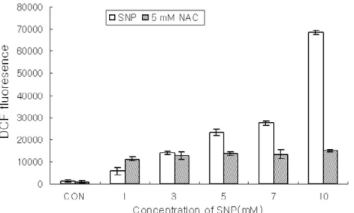
Figure 1. ROS production was enhanced in SNP-treated HGF cells. HGF cells loading DCF were incubated for 12 h with SNP alone or co-incubation with 5 mM N-acetyl-L-cysteine (NAC) for 1 h. The intracellular levels of ROS were detected by measuring the DCF-DA fluorescence (Data are mean±SD from 5 independent experiments.). SNP augment- ed the production of ROS in a dose-dependent manner and NAC, a free radical scavenger, ameliorated the increments of ROS produced by SNP.
pled secondary antibodies(Sigma, USA). Bound antibodies were visualized using an enhanced chemiluminescent detection system(Amersham Pharmacia Biotech, UK).
III. Results
1. NO-induced ROS production and apoptosis in HGF cells
To detetermine the involvement of ROS in NO-
induced cell death of the HGF cells, ROS production was measured using DCF-DA. Figure 1 showed that SNP, a NO donor, enhanced the ROS production in a dose-dependent manner in the HGF cells.
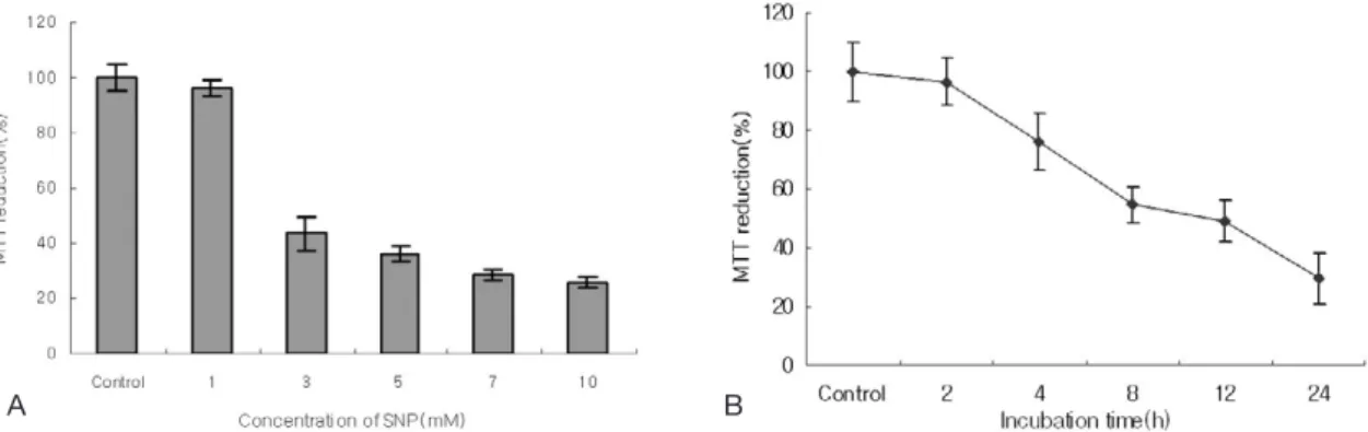
Pretreatment of cells with 5 mM NAC, a ROS scav- enger, inhibited the increment of ROS produced by SNP. The cell viability determined by MTT assay was gradually reduced in a dose- and time-depen- dent manner when HGF cells were exposed to SNP(Figure 2A, 2B). The cell survival rate was less than 80% when the cells were treated with 5 mM
Figure 2. NO induced cell death in HGF cells. Cell viability was determined by MTT assay. HGF cells were incubated with SNP for indicated concentrations(A) and 5 mM SNP for indicated times (B). Viability of the cells without SNP treatment(Control) was defined as 100%. The viability was reduced in dose- and time-dependent manners in SNP-treated HGF cells (Data are mean±SD from 5 independent experiments).
Figure 3. The morphologic changes of NO-induced apoptosis in HGF cells. HGF cells were treated with 5 mM SNP for 12 h and fixed with ethanol and cells were stained using Diff-Quick. Cell shrinkage, chromatin condensation, and DNA fragmentation were shown in SNP-treated HGF cells.
Control 5 mM SNP
A B
SNP for 24 h.
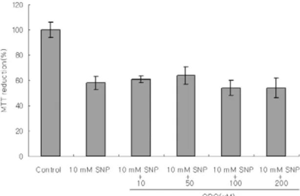
In the presence of 5 mM SNP for 12 h, Diff-Quick staining revealed apoptotic morphological changes, including chromatin condensation and nuclear frag- mentation(Figure 3B). Since, NO usually targets sol- uble guanylate cyclase, effects of soluble guanylate cyclase inhibitor(1H-[1,2,4] oxadiatolo[4,3-a] cluinox- aline-1-one, ODQ, 10-4M) on NO-induced cell death were examined. ODQ, a gunanylate cyclase inhibitor, did not recover the cell viability decreased by SNP (Figure 4), indicating that NO-induced apop- tosis is not mediated through cyclic GMP.
2. Mitochondria-mediated pathway in NO- induced apoptosis of HGF cells
To evaluate whether mitochondria are involved in NO-induced apoptosis of HGF cells, the amount of cytochrome c released from mitochondria into cytosol was determined using cytosolic fractions by western blot after HGF cells were incubated with varying concentrations of SNP for different periods.
Cytosolic cytochrome cwas enhanced in dose- dependent manner in response to exposure of SNP (Figure 5A). Cytochrome cshowed a peak value at Figure 4. Cyclic GMP is not involved in NO-induced cell death in HGF cells. ODQ, a soluble guanylate cyclase
inhibitor, did not recover the cell viability decreased by SNP. Results are mean±SD from 5 independent experiments.
Figure 5. Enhancement of cytochrome c released from mitochondria into cytosol in SNP-treated HGF cells.
Cytosolic cytochrome c was analyzed by immunoblotting with antibody against cytochrome c. SNP enhanced the cytochrome c released from mitochondria into cytosol in a dose-dependent manner (A) and at 4 h as peak time (B).
4 h of SNP incubation and remained higher than the control values even after 24h of incubation (Figure 5B). This result demonstrates that cytochrome cis released from mitochondria into cytoplasm during NO-induced apoptosis in HGF cells.
3. Bax and Bcl-2 expression in NO- induced apoptosis
Generally, expression ratio of Bax to Bcl-2 has been known to be significant for apoptosis deter- mination, since a high ratio denotes a low apoptot- ic threshold, whereas a low ratio indicates a higher
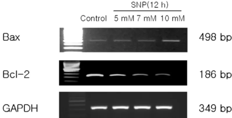
apoptotic threshold. After the treatment of HGF cells with 10 mM SNP for 12 h, the changes in the mRNA level of Bax and Bcl-2 in HGF cells were determined using RT-PCR. Figure 6 showed that SNP upregulated Bcl-2 expression and downregu- lated the Bax expression in a dose-dependent manner.
4. Involvement of caspases in the NO- induced apoptosis in HGF cells
Since it is important to identify the intracellular apoptotic pathways induced by NO in HGF cells, Figure 6. Altered expression of Bax and Bcl-2 in SNP-treated HGF cells. After incubation of HGF cells with SNP for 12 h, RT-PCR was performed for Bax and Bcl-2 expression. SNP upregulated Bax expression and downregulated Bcl-2 expression in a dose-dependent manner.
Figure 7. Caspase-9 was activated in SNP-treated HGF cells. Absorbance for caspase-9 activity was measured at 405 nm after incubation with LEHD-pNA substrate(200 μM) for 2 h at 37℃. SNP at 5 mM enhanced the caspase-9 activity in a time-dependent manner ( Results are mean±SD from 5 experiments).
Incubation time (h)
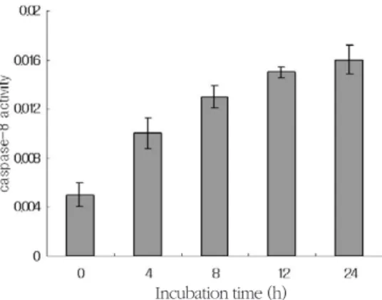
caspases activities were measured on the basis that active caspases consequently cleave their substrate at a specific site. LEHD-pNA (200 μM), IETD--pNA (200 μM), and DEVD-pNA (200 μM) were used as substrates for caspase-9, -8, and -3, respectively. SNP at 5 mM enhanced caspase-8, -9 and -3 activities in a time-dependent manner. It is speculated that both mitochondria and death receptor-dependent apoptotic pathways are involved in NO-induced apoptosis of the HGF cells (Figure 7, 8, 9).
5. Fas and Bid expression in NO-induced apoptosis in HGF cells
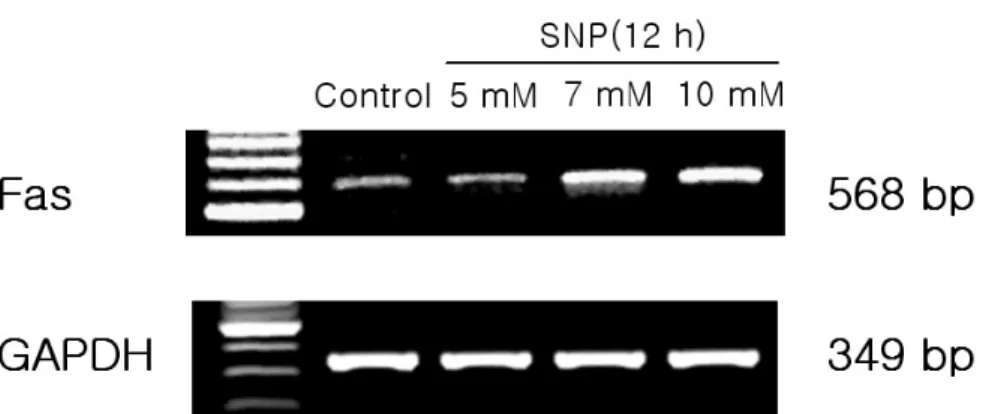
To know whether death recepter-mediated apop- totic pathway is activated in HGF cells, the mRNA levels of Fas, a death receptor assembly, were deter- mined using RT-PCR. SNP at 5 mM upregulated the expression of Fas in a dose-dependent manner (Figure 10). In addition, Bid level was determined since activated caspase-8 is known to cleave pro- form Bid (pro-Bid) into truncated Bid (tBid). Pro- Bid protein was on the decrease by addition of SNP Figure 8. Caspase-8 was activated in SNP-treated HGF cells. Absorbance for caspase-8 activity was measured at 405 nm after incubation with IETD-pNA substrate(200 μM) for 2 h at 37℃. SNP at 5 mM enhanced the caspase-8 activity in a time-dependent manner ( Results are mean±SD from 5 experiments.)
Figure 9. Caspase-3 was activated in SNP-treated HGF cells. Absorbance for caspase-3 activity was measured in the wells at 405 nm after incubation with DEVD-pNA substrate(200 μM) for 24 h at 37℃. SNP at 5 mM enhanced the cas- pase-3 activity in a time-dependent manner ( Results are mean±SD from 5 experiments).
Incubation time (h)
Incubation time (h)
in a dose- and time-dependent manner (Figure 11).
These results suggest that death receptor-mediated pathway plays a crucial role in NO-induced apopto- sis of the HGF cells.
IV. Discussion
Periodontal tissues such as gingival fibroblast and periodontal fibroblast have a nitric oxide syntheses and mass produce NO of high concentration by bacterial lipopolysaccharide and cytokines1,2. However there are none of experiments for NO- induced cell death in periodontal tissues.
NO-induced cell death has been classified as
apoptosis and necrosis, on the basis of changes in morphology, enzymatic activity, ATP concentration and adjacent cellular effects3,4. The characteristic morphology in apoptotic cell is distinct, including cellular shrinkage, internucleosomal DNA fragmen- tation and chromatin condensation12,13.
In the present study, SNP, a NO donor, decreased the cell viability and gave rise to morphogical changes of apoptosis including chromatin conden- sation, DNA fragmentation, and cell shrinkage in HGF cells. These present results demonstrate the first evidence that nitric oxide induces apoptosis in HGF cells. Previous studies have shown that NO elicit apoptosis through the production of ROS such Figure 10. Upregulated expression of Fas in SNP-treated HGF cells. mRNA levels of Fas, one component of death
receptor assemblies, was determined by RT-PCR. SNP upregulated Fas expression in a dose-dependent manner.
Figure 11. Bid level in SNP-treated HGF cells. SNP reduced the level of the proform Bid (pro-Bid) in a dose- and time- dependent manner.
as H2O2in mitochondria and reaction with superox- ide, resulting in formation of peroxynitrite14,15). In the present study, SNP enhanced the production of ROS ameliorated by NAC, a free radical scavenger in HGF cells. From these results, it is speculated that NO-induced apoptosis may be in part mediated by ROS in HGF cells, in consistent with those of previ- ous reports in other tissues.
A variety of free radicals such as ROS and perox- ynitrite are known to impair mitochondrial function, subsequently resulting in loss of mitochondrial trans- membrane potential and release of mitochondrial pro-apoptotic molecules including cytochrome c, Smac, apoptosis-inducing factor (AIF)16,17). The pre- sent study shows that SNP resulted in an increment of cytochrome c release from mitochondria into cytoplasm in a dose-dependent manner. Besides SNP enhanced the activity of caspase-9 activated by mitochondrial cytochrome c, subsequently resulting in activation of caspase-3 in concert with Apaf-1 and dATP. Taken together, mitochondria-dependent apoptotic pathway is definitely proven to be involved in NO-induced apoptosis of the HGF cells since cytochrome cand caspase-9 are major mole- cules associated with mitochondria-dependent path- way.
In general, caspase-3 is a key and common pro- tease in both mitochondria- and death receptor- dependent pathways and particularly important in free radical-induced apoptosis18,19). Previous stud- ies19,20). have shown that caspase-3 is activated in response to various ROS. The present study showes that caspase-3 activity was enhanced in SNP-treated HGF cells, which is consistent with that of the previ- ous report in other tissues and cells. From the pre- sent study and previous reports, it is assumed that caspase-3 plays a pivotal role in NO-induced apop- tosis in HGF cells, even if caspase-independent cell death is proposed to involve in NO-induced cell
death in PC12 cells14).
Another possible mechanism for activating cas- pase-3 is caspase-8 mediated process activated by Fas and TNF receptor-1. Recent studies have report- ed that ROS such as H2O2directly induces upregula- tion of death receptor assembly such as Fas and Fas- ligand, subsequently activating caspase-816,21. From these previous reports, it is proposed a possibility that death receptor-dependent apoptosis pathway may be involved in caspase-3 activation in NO- induced apoptosis of HGF cells. In the present study, Fas, a death receptor assembly, was upregulated and caspase-8 activity was enhanced in SNP-treated HGF cells. The present study shows the first evidence that death receptor-dependent pathway may be involved in NO-induced apoptosis of HGF cells. From the present results, NO-induced apoptosis is likely to be mediated by both mitochondria and death receptor- mediated pathways in HGF cells.
On the otherhand, the Bcl-2 family of proteins are known to be well-characterized regulators of cytochrome c release from mitochondria into cytosol. The Bcl-2 subfamily contains anti-apoptotic proteins such as Bcl-2 and Bcl-XL, which reduce cytochrome c release and a loss of mitochondrial trans-membrane potential (Δψm)22,23) . The Bax subfamily contains pro-apoptotic proteins such as Bax and Bak, which induce cytochrome crelease and a loss of Δψm24). Bcl-2 proteins such as Bid, Bik and Bim are another subfamily of pro-apoptotic pro- teins, which are activated by caspase-8. Thus, ratio of pro-apoptotic and anti-apoptotic caspases may be pivotal cue to release of cytochrome cfrom mito- chondria into cytosol. Therefore, expression of Bcl-2 family was examined in the present study to eluci- date the involvement of Bcl-2 family in NO-induced apoptosis. In the present study, Bcl-2 mRNA was downregulated, whereas Bax mRNA was upregulat- ed in SNP-treated HGF cells. Even in previous
reports25,26, NO has been reported to directly or indi- rectly regulate Bcl-2 family expression in other tissue and cells. These present results suggest that Bcl-2 proteins are involved in NO-induced apoptosis of HGF cells. An interesting result is that Bid was acti- vated by SNP in the present study since Bid is known to be activated by caspase-8, unlike other Bcl-2 family. From the present data, it is speculated that there is an interrelation lineage between the death receptor-mediated apoptotic signals and the mitochondria-mediated apoptotic signals. However, roles of Bcl-2 family may be a debate in NO- induced apoptosis of HGF cells since Bcl-2 family regulates the production of ROS and cytochrome c release from mitochondria into cytosol22,24and ROS could conversely regulates the expression of Bcl-XL
mRNA17. Further researches are required to deter- mine the roles of the Bcl-2 family in NO-induced apoptosis of HGF cells.
In general, NO acts a variety of physiological function through activating soluble guanylate cyclase, subsequently resulting in synthesis of cyclic GMP. Thus, it was examined whether cyclic GMP pathway might be involved in NO-induced apopto- sis of HGF cells. In the present study, ODQ, a solu- ble guanylate cyclase inhibitor, did not ameliorated the cell viability reduced by SNP in HGF cells. This result suggests that NO-induced apoptosis is not mediated by cyclic GMP pathway. In summary, the results from this study suggest that NO induces apoptosis through activation of both the mitochon- dria- and death receptor-dependent pathways medi- ated by ROS and Bcl-2 family in HGF cells.
V. References
1. Dahigh F., Borghaei R.C., Thornton R.D. and Bee J.H. Human gingival fibroblasts produced nitric oxide in response to proinflammatory
cytokines. J Periodontal 2002;73:392-400.
2. Kendall H.K., Haase H.R., Li H., Xiao Y. and Bartold P.M. Nitric oxide synthase type-II is syn- thesized by human gingival and cultured human gingival fibroblasts. J. Periodontal Res. 2000;35:
194-200.
3. Gross S.S. and Wolin M.S. Nitric oxide: patho- physiological mechanisms. Annu. Rev. Physiol 1995;57:737-69.
4. Dawson V.L. and Dawson T.M. Nitric oxide neurotoxicity. J Chem Neuroanat. 1996;10:179- 90.
5. Roth J.A., Feng L., Walowitz J. and Browne R.W. Manganese -induced rat pheochromocy- toma (HGF) cell death is independent of cas- pase activation. J Neurosci Res 2000;61:162-71.
6. Adams J.M. and Cory S. The Bcl-2 protein fami- ly: arbiters of cell survival. Science 1998;281:
1322-6.
7. Tsujimoto Y. and Shimizu S. Bcl-2: Life-or-death switch. FEBS Lett. 2000;466:6-10.
8. Beer R., Frenz G., Schopf M., Reindl M., Zelger B., Schmutzhard E., Poewe W. and Kampfl A.
Expression of Fas and Fas ligand after experi- mental traumatic brain injury in the rat. J Cereb Blood Flow Metab 2000;20:669-77.
9. Cheng E.H., Nicholas J., Bellows D.S., Hayward G.S., Guo H.G., Reitz M.S. and Hardwick J.M. A Bcl-2 homolog encoded by kaposi sarcoma-asso- ciated virus, human herpesvirus 8, inhibits apop- tosis but does not heterodimerize with Bax or Bak. Proc. Natl. Acad. Sci. USA 1997;94:690-4.
10. Susilowati H., Santoso A.L., Barid I. and Sosroseno W. Rat periodontal fibroblast respons- es to bacterial lipopolysacchraide in vitro. J Microbiol Immunol Infect 2002;35:203-6
11. Boulares A.H., Zoltoski A.J., Sherif Z.A., Yakovler A. and Smulson M.E. Roles of DNA fragmentation factor and poly(ADP-ribose) poly-
merase-1 in sensitization of fibroblasts to tumor necrosis factor-induced apoptosis. Biochem Biophys Res Commun 2002;290:796-801.
12. Fujimura M., Morita-Fujimura Noshita N., Sugawara T., Kawase M. and Chan P.H. The cytosolic antioxidant copper/zinc-superoxide dismutase prevents the early release of mito- chondrial cytochrome c in ischemic brain after transient focal cerebral in mice. J Neurosci 2000;20:2817-24.
13. Oppenheim R.W. Cell death during develop- ment of the nervous system. Annu. Rev.
Neurosci. 1991;14:453-501.
14. Brown G.C. Nitric oxide and mitochondrial res- piration. Biochim. Biophys. Acta. 1999;1411:
351-69.
15. Yuyama K., Yamamoto H., Nishozaki I., Kato T., Sora I. and Yamamoto T. Caspase-indepen- dent cell death by low concentrations of nitric oxide in PC12 cells: Involvement of cytochrome c oxidase inhibition and the production of reac- tive oxygen species in mitochondria. J neurosci Res 2003;73:351-63.
16. Fleury C., Mignotte B. and Vayssiere J.L.
Mitochondrial reactive oxygen species in cell death signaling. Biochimie 2002:84:131-41.
17. Herrera B., Alvarez A.M., Sanchez A., Fernandez M., Roncero C., Benito M. and Fabregat I. Reactive oxygen species (ROS) medi- ates the mitochondrial-dependent apoptosis induced by transforming growth factor (beta) in fetal hepatocytes. FASEB J. 2001;15:741-51.
18. Earnshaw W.C., Marins L.M. and Kaufmann S.H. Mammalian caspases: structure, activation, substrates, and functions during apoptosis.
Annu. Rev. Biochem. 1999;68:383-424.
19. Bal-Price A. and Brown G.C. Nitric-oxide- induced necrosis and apoptosis in PC12 cells mediated by mitochondria. J Neurochem
2000;75:1455-64.
20. Leist M., Single B., Naumann H., Fava E., Simon B., Kuhnle S. and Nicotera P. Nitric oxide inhibits execution of apoptosis at two distinct ATP-dependent steps upstream and downstream of mitochondrial cytochrome c release.
Biochem. Biophys. Res. Commun. 1999;258:
215-21.
21. Facchinetti F., Furegato S., Terrazzino S. and Leon A. H2O2 induces upregulation of Fas and Fas ligand expression in NGF-differentiated HGF cells: Modulation by cAMP. J Neurosci Res 2002;69:178-88.
22. Gottlieb E., Vander Heiden M.G. and Thompson C.B. Bcl-xl prevents the initial decrease in mitochondrial membrane potential and subsequent reactive oxygen species produc- tion during tumor necrosis factor alpha-induced apoptosis. Mol Cell Biol 2000;20:5680-9.
23. Howard S., Bottino C., Brooke S., Cheng E., Giffard R.G. and Sapolsky R. Neuroprotective effects of Bcl-2 overexpression in hippocampal cultures: interactions with pathways of oxidative damage. J Neurochem 2002;83:914-23.
24. Starkov A., Polster B. and Fiskum G. Regulation of hydrogen peroxide production by brain mito- chondria by calcium and Bax. J Neurochem 2002;83:220-8.
25. Hemish J., Nakaya N., Mittal V. and Enikolopov G. Nitric oxide activates diverse signaling path- ways to regulate gene expression. J Biol Chem 2003;278:42321-9.
26. Huerta-Yepez S., Vega M., Jazirehi A., Garban H., Hongo F., Cheng G. and Bonavida B. Nitric oxide sensitizes prostate carcinoma cell lines to TRAIL-mediated apoptosis via inactivation of NF- κB and inhibition of Bcl-xL expression.
Oncogene 2004;23:4993-5003
-국문초록-
사람 치은 섬유아세포에서 산화질소 유도 세포고사에 대한 연구
김강문1, 정현주1,3, 김원재2,3
전남대학교 치과대학1치주과학교실, 2구강생리학교실 및3치의학연구소
산화질소는 생리적 농도에서 세포내 신호전달자로 작용하지만 높은 농도에서는 세포독성을 일으킨다. 최근 치은 섬유아세포와 치주인대 섬유아세포는 산화질소 합성효소를 가지고 있고 세균의 lipopolysaccharide나 cytokine에 의해 대량의 높은 농도의 산화질소가 합성된다는 보고가 있음에도 지금까지 치은 조직에서 산화질 소의 세포독성에 대한 연구는 아직 이루어 지지않고 있다. 본 연구는 사람의 치은 섬유아세포에서, 산화질소유 도세포 고사기전을 밝히는데 목적이 있다.
세포 생장력은 MTT 방법으로 측정하였고, 세포의 형태적 변화는 Diff-Quick 염색법으로 조사하였다. Bcl-2 family와 Fas 발현 정도는 RT-PCR 방법에 의해 확인하였으며, caspase-3, -8 와 -9의 활성은 spectrophotometer 로 reactive oxygen species (ROS)는 형광분광계에 의해 측정되었다. 미토콘드리아에서 세포질로 분비된 cytochrome c는 western blot으로 조사하였다.
산화질소 유리제인 sodium nitroprusside (SNP) 처리는 사람 섬유아세포의 생존률을 시간과 농도 의존적으로 감소시켰고, 세포용적축소, 염색사 응축, DNA 절편화를 일으켰다. 또한, SNP 처리로 미토콘드리아에서 세포질 로 유리되는 cytochrome c양이 증가되었고, caspase-9 과 caspase-3 의 활성이 증가되었다. 한편, SNP 처리에 의해 death receptor 구성요소인 Fas 발현이 증가되었고, caspase-8의 활성이 증가되었다. Bcl-2 family 에 대한 RT-PCR 분석결과, 세포고사를 억제하는 Bcl-2 발현은 감소되었으나 세포고사를 자극하는 Bax와 Bid의 발현은 증가되었다. Soluble guanylate cyclase 억제제인 ODQ는 SNP에 의한 세포 생존율 감소를 차단하지 못했다. 따 라서, 본 실험의 결과들은 사람 섬유아세포에서 산화질소유도 세포고사에 Bcl-2 family나 ROS가 매개하는 미토 콘드리아 의존 및 death receptor 의존 세포고사기전이 관여함을 시사하였다.
주요어 : 치은 섬유아세포, 산화질소, 세포고사, 신호경로