Effect of Activated Protein C (APC) on Apoptosis of Cancer Cells
Kyoung-jin Min
1, Jong-Sup Bae
2and Taeg Kyu Kwon
1*
1
Department of Immunology, School of Medicine, Keimyung University, Daegu 704-701, Korea.
2
College of Pharmacy, Research Institute of Pharmaceutical Sciences, Kyungpook National University, Daegu 702-701, Korea
Received March 12, 2012 /Revised March 26, 2012 /Accepted March 27, 2012Activated protein C (APC) has an anticoagulant effect and a non-hemostatic effect such as regulation of cell metastasis and modulation of inflammation. In this study, we investigated whether APC could modulate apoptosis in cancer cells. Tumor necrosis factor (TNF)-α , cyclohexamide, and FAS markedly induced apoptosis in human renal carcinoma Caki cells. When Caki cells were pretreated with APC, the percentage of death receptor-induced apoptosis did not change. Furthermore, we checked the effect of APC on tumor necrosis factor-related apoptosis-inducing ligand (TRAIL)-induced apoptosis in hu- man glioma T98G and human breast carcinoma MDA231 cells. APC also had no effect on TRAIL-in- duced apoptosis in both cell lines. However, pretreatment with APC inhibited combination treatment (kahweol plus TRAIL and kahweol plus melatonin)-induced apoptosis and PARP cleavage in Caki cells.
Taken together, our results suggest that APC can modulate anti-cancer therapeutic efficiency.
Key words : Activated protein C (APC), apoptosis, TRAIL, Caki cells, kahweol
*Corresponding author
*Tel:+82-53-580-3882, Fax:+82-53-580-3795
*E-mail : [email protected]
서 론
과거 종양을 가진 환자에서 정맥혈전(venous thrombosis) 이 나타나는 것을 확인하였고, 그 이후 지속된 연구를 통하여 종양과 정맥혈전에 대한 상관관계를 밝히게 되었는데, 이는 종양을 가진 환자에서 종양이 없는 사람들과 비교 하였을 때 약 6~7배 이상으로 빈번하게 정맥혈전이 나타나는 것을 확인 하였다[5,6,17]. 따라서, 최근 종양과 정맥혈전을 가진 환자들 을 치료하기 위하여 항응고제(anticoagulant)를 처리하는 방법 을 사용하고 있는데, 이는 정맥혈전뿐 아니라 종양치료에 효 과적인 결과를 나타내고 있다. 예를 들어 저분자량 헤파린 (low molecular heparin)을 종양을 가진 환자에게 처치를 하 면, 환자의 생명연장을 유도하고 사망률을 낮추는 결과를 가 져왔다[1,10]. 이는 혈액의 응고와 항응고에 관여하는 여러 가 지 인자들이 종양치료에 영향을 줄 수 있다는 것을 의미한다.
혈액의 항응고 작용에 관여하는 요소 중 activated protein C (APC)는 혈액응고에 관여하는 thrombin이 thrombomo- dulin과 세포막에서 결합체를 만들게 되면 이로 인해 pro- tein C로부터 APC가 만들어지게 된다[8]. 이렇게 형성된 APC는 factor Va와 VIIIa의 활성을 억제시킴으로써 혈액응 고를 억제하게 된다[7]. 최근에는 혈액응고과정에서의 APC 기능뿐 아니라, 혈액응고와는 상관없는 다른 역할들이 밝혀 지고 있다. 예를 들면, APC는 다양한 세포의 이동을 조절하 는 것으로 알려져 있는데 APC를 인간 각질세포 (keratinocyte)에 처리하게 되면 세포의 분화와 이동을 증가
시키고, 이러한 효과는 metalloprotease-2의 활성을 증가시킴 으로써 나타난다[18]. 또한, 암세포에서의 APC의 역할이 잘 알려져 있는데, 자궁암세포와 융모암세포, 그리고 유방암세 포에서 세포의 이동을 증가시킨다는 보고가 있다[2,11]. 하지 만, 최근 Bezuhly 등에 의해 수행된 연구를 보면 APC가 en- dothelial protein C receptor (EPCR)이 과발현 된 경우 in vivo에서 오히려 흑생종암세포의 전이를 억제한다는 보고가 있어[3], APC의 역할에 대한 연구는 그 기능을 이해하기에 미흡하여 더 많은 연구가 필요한 실정이다.
본 연구에서는 항응고제로 알려져 있는 APC가 다양한 종 양치료 약물에 의한 종양세포의 사멸에 있어서의 그 기능을 파악하기 위하여 인간 신장암세포인 Caki 세포를 사용하여 그 효과를 확인하였다.
재료 및 방법
세포 배양 및 시약
본 연구에 사용한 신장암세포주인 Caki, 뇌종양세포인 T98G, 유방암세포인 MDA231세포는 American Type Culture Collection (ATCC, Rockville, MD, USA)에서 구입하 였다. 세포주 배양을 위한 배지는 10% 태아우혈청(fetal bo- vine serum, Hyclone laboratories, Lagan, Utah, USA)과 1%
Antibiotics, 0.2% Gentamycin을 첨가한 DMEM (Dulbecco’s
Modified Eagle’s Medium, Gibco BRL, Grand Island, NY)을
사용하였으며 37℃로 유지되는 5% CO
2배양기를 이용하여
배양하였다. 실험에 사용된 약제인 TRAIL과 cyclohexamide
는 Sigma (St. Louis, MO, USA), anti-FAS는 Millipore
(Bedford, MA), TNF-α는 R&D Systems (Minneapolis, MN)
- Note -
를 이용하여 측정하였다.
Western Blotting
단백질의 발현을 알아보기 위해서 Western blotting을 실시 하였다. 세포를 0.4×10
6cells/well로 12시간 배양 후에, FBS를 첨가하지 않은 무혈청 배지로 교체하여 시약을 처리하여 배양 후, 세포를 모아 32 μl lysis buffer (137 mM NaCl, 15 mM EGTA, 0.1 mM sodium orthovanadate, 15 mM MgCl
2, 0.1%
Triton X-100, 25 mM MOPS, 100 mM phenylmethylsulfonyl fluoride, and 20 mM leupeptin, pH 7.2)를 첨가하고 5분 간격 으로 15초 동안 3번 vortex하여 세포를 파쇄한 후 13,000 rpm, 4℃, 15분간 원심 분리하여 시료를 준비하였다. 시료는 562 nm에서 흡광도를 측정하여 단백질을 정량하였으며, 10% so- dium dodecyl sulfate polyacrylamide gel electrophoresis (SDS-PAGE)에서 단백질을 분리한 후, immobilon membrane (Milipore, Bedford, MA)으로 transfer하였다. Membrane은 5% milk/TBST (20 mM Tris-HCL, 137 mM NaCl, 0.1%Tween 20, pH 7.4)로 실온에서 1시간 유지한 후, PARP (Santa Cruz, CA, USA)와 actin (Sigma, Louis, MO)을 희석한 5%
milk/TBST로 실온에서 12시간 유지하였다. Anti-mouse 또는 rabbit Ig horseradish peroxidase/TBST (Amersham Buckinghamshire, England)로 1시간 반응 후 Enhanced Chemiluminoscence (ECL, Pierce, IL, USA)용액을 가하여 발 색시켜서 단백질을 확인하였다.
결과 및 고찰
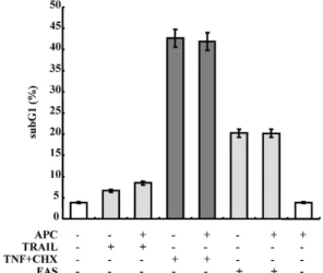
신장암세포에서 외인성 세포사멸 신호기전에 의한 세포사멸 에서의 APC의 효과 확인
TRAIL, TNF-α와 cyclohexamide 그리고 FAS는 외인성 신호전달을 통해 종양세포의 사멸을 일으키는 것으로 알려 져 있다[8,12,15]. 본 연구에서 외인성 신호전달을 통한 세포 사멸에 있어서의 APC의 역할을 확인하였다. 인간의 신장암 세포에 APC를 30분간 전처리 한 후, 각각의 세포사멸을 일 으키는 100 ng/ml TRAIL, 20 ng/ml TNF-α와 10 μg/ml
인 MDA231세포에서의 APC 세포사멸 조절 여부
APC에 의한 human umbilical vein endothelial (HUVE) 세포의 세포보호(cytoprotective) 능력은 APC에 의한 Egr1/ERK 활성화를 통한 TRAIL 발현 억제 때문에 야기된다 [13]. TRAIL에 의한 세포사멸에 APC의 역할을 규명하기 위하 여 TRAIL 에 민감성을 가지는 인간 뇌암세포인 T98G세포와 인간 유방암세포인 MDA231세포를 사용하여 확인하였다.
APC를 30분간 전처리 한 후, 30 ng/ml TRAIL을 24시간 동안 처리하였다. TRAIL은 두 가지 세포에서 모두 세포사멸을 유 의적으로 증가시켰으나 APC는 두 가지 세포에서 모두 세포사 멸을 억제하지는 못하였다(Fig. 2). 본 결과는 TRAIL 매개의 세포사멸에 APC가 아무런 영향을 미치지 못함을 제시한다.
암세포사멸을 위한 약물병합처리에 의한 세포사멸에 있어서 의 APC의 조절
외인성 신호전달체계를 통한 종양세포의 사멸 이외의 항암
Fig. 1. Effect of APC on extrinsic pathway activated cell death.
Caki cells were pretreated with 100 nM activated protein C (APC) for 30 min, and then treated with 100 ng/ml TRAIL, 20 ng/ml TNF-α plus 10 μg/ml cyclohexamide (CHX), and 500 ng/ml agonistic FAS antibody for 24 hr.
Apoptosis was analyzed as a subG1 fraction by FACS.
The data represent three independent experiments.
Fig. 2. APC has no effect on TRAIL-induced apoptosis in human glioma T98G and human breast carcinoma MDA231 cells. T98G and MDA231 cells were pretreated with 100 nM APC for 30 min, and then added with 30 ng/ml TRAIL for 24 hr. Apoptosis was analyzed as a subG1 fraction by FACS. The data represent three independent experiments.
Fig. 3. APC inhibits Kahweol plus TRAIL and Kahweol plus melatonin-induced apoptosis in human renal carcinoma Caki cells.
Caki cells were pretreated with 100 nM APC for 30 min, and then stimulated with 20 μM kahweol plus 100 ng/ml TRAIL and 10 μM kahweol plus 1 mM melatonin for 24 hr. Apoptosis was analyzed as a subG1 fraction by FACS (A). PARP and actin protein expression were determined using Western blotting. Relative levels of each protein in cleaved PARP were shown as a relative densitometric value of each protein using the Bio-Rad Gel Doc System (B). The data represent three independent experiments.
약물의 병합처리를 통한 종양세포의 사멸에는 APC가 어떠한 영향을 미치는 가를 확인해 보았다. 이전 연구에서, 커피의 추출물 중 한 성분인 kahweol과 TRAIL의 병합처리는 신장암 세포에서 유의적인 세포사멸을 일으키는 것으로 본 연구실에 서 발표한 바 있다[15]. 또한, kahweol과 생체시계 호르몬으로 알려진 melatonin을 병합 처리하면 역시 종양세포의 사멸을 유도한다고 본 연구실에서 발표하였다[16]. 따라서 이러한 두
가지 약물을 병합처리를 통한 종양세포의 사멸에서의 APC의
역할을 규명하고자 실험하였다. 이전 보고와 같이, 신장암세
포에서 두 가지 약물의 병합처리는 유의적인 세포사멸을 일으
켰고, 흥미롭게도 APC의 전처리는 이러한 세포사멸을 유의적
으로 감소시키는 것을 확인하였다(Fig. 3A). Apoptosis의 bio-
marker인 caspase-3의 기질로 알려진 PARP의 분절을 확인함
으로써 APC의 anti-apoptotic 효과를 확인하였다. Kahweol과
만 cell death receptor 매개의 세포 사멸에서는 APC의 세포 보호작용을 확인 할 수 없었다. 하지만 항암제와의 병합처리 시 APC의 세포보호 효과 즉 anti-apoptotic 효과를 처음으로 확인 하였다. APC에 의한 항암작용 증진 기전 및 효과에 대한 연구는 더 많은 실험을 통하여 이루어 져야 한다고 생각된다.
References
1. Altinbas, M., Coskun, H. S., Er, O., Ozkan, M., Eser, B., Unal, A., Cetin, M. and Soyuer, S. 2004. A randomized clinical trial of combination chemotherapy with and without low-molecular-weight heparin in small cell lung cancer.
J.
Thromb. Haemost
. 2, 1266-1271.2. Beaulieu, L. M. and Church, F. C. 2007. Activated protein C promotes breast cancer cell migration through interactions with EPCR and PAR-1.
Exp. Cell Res
. 313, 677-687.3. Bezuhly, M., Cullen, R., Esmon, C. T., Morris, S. F., West, K. A., Johnston, B. and Liwski, R. S. 2009. Role of activated protein C and its receptor in inhibition of tumor metastasis.
Blood
113, 3371-3374.4. Cheng, T., Liu, D., Griffin, J. H., Fernández, J. A., Castellino, F., Rosen, E. D., Fukudome, K. and Zlokovic, B. V. 2003.
Activated protein C blocks p53-mediated apoptosis in ische- mic human brain endothelium and is neuroprotective.
Nat.
Med
. 9, 338-342.5. Chew, H. K., Wun, T., Harvey, D., Zhou, H. and White, R. H. 2006. Incidence of venous thromboembolism and its effect on survival among patients with common cancers.
Arch. Intern. Med
. 166, 458-464.6. Donati, M. B. 2009. Thrombosis and cancer: Trousseau syn- drome revisited.
Best Pract. Res. Clin. Haematol
. 22, 3-8.7. Esmon, C. T., Vigano-D'Angelo, S., D'Angelo, A. and Comp, P. C. 1987. Anticoagulation proteins C and S.
Adv. Exp.
11. Kobayashi, H., Moniwa, N., Gotoh, J., Sugimura, M. and Terao, T. 1994. Role of activated protein C in facilitating basement membrane invasion by tumor cells.
Cancer Res
. 54, 261-267.12. Nagata, S. 1994. Apoptosis regulated by a death factor and its receptor: Fas ligand and Fas.
Philos. Trans. R. Soc. Lond.
B. Biol. Sci
. 345, 281-287.13. O'Brien, L. A., Richardson, M. A., Mehrbod, S. F., Berg, D.
T., Gerlitz, B., Gupta, A. and Grinnell, B. W. 2007. Activated protein C decreases tumor necrosis factor related apopto- sis-inducing ligand by an EPCR- independent mechanism involving Egr-1/Erk-1/2 activation.
Arterioscler. Thromb.
Vasc. Biol.
27, 2634-264114. Pajak, B., Gajkowska, B. and Orzechowski, A. 2005.
Cycloheximide-mediated sensitization to TNF-alpha-in- duced apoptosis in human colorectal cancer cell line COLO 205; role of FLIP and metabolic inhibitors.
J. Physiol.
Pharmacol
. 56, 101-118.15. Um, H. J., Oh, J. H., Kim, Y. N., Choi, Y. H., Kim, S. H., Park, J. W. and Kwon, T. K. 2010. The coffee diterpene kah- weol sensitizes TRAIL-induced apoptosis in renal carcinoma Caki cells through down-regulation of Bcl-2 and c-FLIP.
Chem. Biol. Interact
. 186, 36-42.16. Um, H. J., Park, J. W. and Kwon, T. K. 2011. Melatonin sensi- tizes Caki renal cancer cells to kahweol-induced apoptosis through CHOP-mediated up-regulation of PUMA.
J. Pineal Res
. 50, 359-366.17. Varki, A. 2007. Trousseau's syndrome: multiple definitions and multiple mechanisms.
Blood
110, 1723-1729.18. Xue, M, Thompson, P., Kelso, I. and Jackson, C. 2004.
Activated protein C stimulates proliferation, migration and wound closure, inhibits apoptosis and upregulates MMP-2 activity in cultured human keratinocytes.