385
Original Article
Korean Circulation J 2007;37:385-387
ISSN1738-5520
ⓒ 2007, The Korean Society of Circulation CASE REPORT
Very Late Stent Thrombosis Related to Fracture of a Sirolimus-Eluting Stent
Chang-Wook Nam, MD1, Kwon-Bae Kim, MD1 and In-Sung Chung, MD2
1Department of Internal and 2Industrial Medicine, College of Medicine, Keimyung University, Daegu, Korea
ABSTRACT
Although late stent thrombosis is a very rare complication after a drug-eluting stent implantation, this could in- duce significant sequelae. Several mechanisms for very late stent thrombosis have been suggested. We experi- enced a new possible mechanism of very late stent thrombosis after the implantation of a sirolimus-eluting stent.
A sirolimus-eluting stent fracture might induce mechanical injury of the endothelium and then thrombotic occ- lusion at 24 months after the stent procedure. (Korean Circulation J 2007;37:385-387)
KEY WORDS:Thrombosis;Stents;Sirolimus.
Introduction
Drug-eluting stents(DESs) are commonly used in daily practice during percutaneous coronary intervention(PCI).
Several significant complications after DES implant- ations have been recently reported. Among them, stent thrombosis(ST) is a rare and serious complication. A stent fracture is also a very rare event after a DES im- plantation. We report here on a case of very late stent thrombosis that occurred in a patient 24 months after a sirolimus-eluting stent(SES) implantation, and this might have been related to a stent fracture.
Case
A 58-year old male was admitted with an inferior ST segment elevation myocardial infarction(STEMI). He had been transferred to our hospital due to 4 hours of sustained chest pain. The initial ECG exhibited ST se- gment elevation in the inferior leads during sinus rh- ythm. Two years prior to his admission, he had unde- rgone two stent implantations to the right coronary artery(RCA) because of an inferior STEMI.
Two years previously, he was transferred to our ho- spital for the management of an inferior wall STEMI after thrombolytic therapy had been performed for tr- eating the inferior STEMI. As the cardiovascular risk
factors, he had a history of hypertension and hyperli- pidemia and he was a current smoker. The coronary angiogram revealed two focal stenotic lesions in the proximal and mid RCA(Fig. 1A). After predilation with a 3.0 mm balloon, a 3.5×23 mm SES (Cypher, Cordis, Miami Lakes, Florida, USA) was implanted at 16 atm in the mid RCA, and a 4.0×18 mm BX- Velocity(Cypher, Cordis, Miami Lakes, Florida, USA) was implanted at 16 atm, without overlapping, in the proximal RCA after predilation with a 3.0 mm balloon.
There were no events during the 8 month clinical follow- up. He underwent a follow-up coronary angiogram 8 months later as part of a workup for noncardiac surgery.
There was minimal neointimal hyperplasia around both stents(Fig. 1B). Two months later, his antiplatelet the- rapy was discontinued for performing a left nephrectomy that was due to multiple renal stones and obstructive uropathy. After the operation, he received aspirin 100 mg daily as antiplatelet therapy. The 14 month clinical follow-up after the operation was uneventful.
Primary PCI was then performed. The angiogram revealed total occlusion of the mid RCA at the previous SES implantation site(Fig. 1C). A floppy wire was easily passed. A large amount of thrombi was noted after pr- edilation with a 2.5 mm balloon. After repeated 3.5 mm ballooning, intravascular ultrasound was performed to obtain more information on the occluded site. During the automatic pull-back, a sudden loss of a stent strut was noted. There was a stent fracture in the mid por- tion of the SES and also minimal neointimal hyper- plasia(Fig. 2). Because we had already obtained opti- mal results and there was minimal proliferation of the neointima, we did not perform any additional stent implantations(Fig. 1D). The patient was discharged with
Received:May 3, 2007 Revision Received:May 22, 2007 Accepted:June 27, 2007
Correspondence:Kwon-Bae Kim, MD, Department of Industrial Medicine, College of Medicine, Keimyung University, 194 Dongsan-dong, Jung-gu, Daegu 700-821, Korea
Tel: 82-53-250-8040, Fax: 82-53-250-7034 E-mail: [email protected]
386·Korean Circulation J 2007;37:385-387
taking a triple antiplatelet medication regimen(aspirin 200 mg, clopidogrel 75 mg, cilostazol 200 mg). There were no adverse cardiac events noted during the 9 months clinical follow-up. A second follow-up coronary angio- graphy was performed 7 months after the stent thro- mbosis had occurred; the angiography did not show any evidence of restenosis(Fig. 1E).
Discussion
The DES has revolutionized the treatment of coronary artery disease, and the randomized studies have show that DES has brought about a significant reduction in
the need for repeated revascularizations and a reduced rate of major adverse cardiac event.1) However, concerns have been raised about the safety of those stents in re- lation to the occurrence of stent thrombosis.2-4) ST with DESs may be associated with significantly higher mortality rates that are related to myocardial infarctions.
This rare, but serious complication has drawn the atten- tion of interventional cardiologists. A large portion of STs occur acutely(within 24 hours after the index pr- ocedure) or subacutely(between 1 and 30 days after).5) However, the focus of concern for the STs was that no end point for late stent thrombosis(LAST: 30 days after) could be observed, even though the incidence of this complication was small. A recent report showed signi- ficantly higher rates of death and myocardial infarc- tions between 6 to 18 months after the procedure in the DES group compared to the BMS group(4.9% in the DES group vs. 1.3% in the BMS group; p=0.01).4) This astonishing result was interpreted as the clinical consequence of late thrombotic events in the DES im- planted patients. ST is mostly related to the disconti- nuation of the antiplatelet therapy.6) However, several different additional factors such as a delayed endothe- lialization,7) chronic inflammation or hypersensitivity to the polymer,8)9) late stent malappositioning,7)10)11) and stent underexpansion12) also affect this serious complication.
Most of the mechanisms are hypothetical, and especially those that are concerned with very late stent thrombosis, and so more experience and data are needed to understand this phenomenon.
Stent fractures have recently been suggested as a new mechanism of the adverse cardiac events after DES implantation. SES fractures have also been observed by interventional cardiologists. According to the reports, stent fractures might occur with a very tortuous or gre- atly moved vessels that induce excessive mechanical str- ess during the contractions and flexion of the heart,
Fig. 1. Angiographic images of the right coronary artery (RCA). A: The initial angiogram showed two focal stenotic lesions at the proximal and mid RCA. B: The follow-up angiogram at 8 months showed a patent stents with minimal luminal narrowing. C: Thrombotic total occlusion in the mid RCA was noted, which was the previous sirolimus-eluting stent implanted site. D: After ballooning, there was minimal residual stenosis. E: The 2nd follow-up angiogram at 6 months showed that the previous ballooning site was patent.
A B C D E
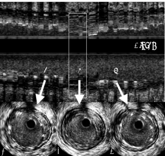
A B C
a b
Distal
c
Fig. 2. Top: Longitudinal IVUS reconstruction of the RCA; the white vertical square corresponds to the point of stent fracture. Bottom: A, B, and C are cross-sectional images corresponding the positions a, b and c on the longitudinal reconstruction. A and C; There are stent struts visible with minimal intimal hyperplasia. B; There is mild concentric plaque with loss of the stent strut, which indicates stent fracture.
Chang-Wook Nam, et al:Very Late Stent Thrombosis·387
heavy calcified lesions, long stents, overlapping sites of the stents, larger-sized ballooning sites and especially balloonmg with high pressures.13) Several cases have been reported in which the stent fractures might have been related to the restenosis after the SES implantation.13)14) However, the data regarding stent fractures and res- tenosis has been very limited up to the present. We ex- perienced eight cases with SES stent fractures, and three of these cases experienced adverse cardiac events.
Because of the uncertainty of the related mechanism between stent fractures and adverse cardiac events, delicate attention should be paid to the fracture site.
The case we report here suggests the existence of a new potential mechanism for very late stent thrombosis (occurring more than 1 year after the procedure) after the implantation of an SES. Stent fractures might in- duce mechanical injury to the endothelium, which could produce a stent thrombosis. Because our patient’s fra- cture developed in the late period of the clinical follow- up, the antiplatelet therapy may have been insufficient to prevent thrombosis formation. Therefore, longer dual antiplatelet therapy may be needed for the groups of patients with a high risk for stent fractures.
Because no IVUS examination was performed during the first follow-up coronary angiogram, the stent fracture may have existed prior to that event. If so, the two events may have developed simultaneously apart from each other. Another possible mechanism of this event is that chronic inflammation, which developed at the fracture site, might have induced atherosclerotic plaque growth and rupture. However, there was minimal intimal hy- perplasia observed on the follow-up angiogram. The possibility of plaque formation at the fracture site after the follow-up angiogram was slight. Even though the fracture had previously existed, thrombotic occlusion developed at the fracture site. The stent fracture may have affected or caused some part of the very late stent th- rombosis in this patient. However, further experience is required to understand the definite mechanism.
In conclusion, we would like to suggest a new possible mechanism for very late stent thrombosis associated with a stent fracture. In the era of DESs, several special conditions might exist such as the possible need for a longer course of dual antiplatelet therapy than the usual recommended course for the patient groups with a high risk for stent thromboses or stent fractures. Careful se- lection of the stent length, type and positioning may also
be required for special coronary situations. Further st- udies and collection of more data should be done to cl- arify whether this is an important issue or only a spor- adic observation.
REFERENCES
1) Morice MC, Serruys PW, Sousa JE, et al. A randomized compa- rison of a sirolimus-eluting stent with a standard stent for coron- ary revascularization. N Engl J Med 2002;346:1773-80.
2) Colombo A, Corbett SJ. Drug-eluting stent thrombosis: increas- ingly recognized but too frequently over-emphasized. J Am Coll Cardiol 2006;48:203-5.
3) Pfisterer ME. Presentation of the results of the Basel Stent Cost- effectiveness Trial-Late thrombotic events (BASKET-LATE). 55th Annual Scientific Sessions of the American College of Cardio- logy. Atlanta, Gerogia, March 14; 2006.
4) Ong AT, McFadden EP, Regar E, Jaegere PT, van Domburg RT, Serruys PW. Late angiographic stent thrombosis (LAST) events with drug-eluting stents. J Am Coll Cardiol 2005;45:2088-92.
5) Kuchulakanti PK, Chu WW, Torguson R, et al. Correlates and long- term outcomes of angiographically proven stent thrombosis with sirolimus-and paclitaxel-eluting stents. Circulation 2006;113:
1108-13.
6) Park DW, Park SW. Stent thrombosis in the era of the drug- eluting stent. Korean Circ J 2005;35:791-4.
7) Joner M, Finn AV, Farb A, et al. Pathology of drug-eluting stents in humans: delayed healing and late thrombotic risk. J Am Coll Cardiol 2006;48:193-202.
8) Azarbal B, Currier JW. Allergic reactions after the implantation of drug-eluting stents: is it the pill or the polymer? J Am Coll Cardiol 2006;47:182-3.
9) Nebeker JR, Virmani R, Bennett CL, et al. Hypersensitivity cases associated with drug-eluting coronary stents: a review of available drug events and reports (RADAR) project. J Am Coll Cardiol 2006;47:175-81.
10) Virmani R, Farb A, Guagliumi G, Kolodgie FD. Drug-eluting stents: caution and concerns for long-term outcome. Coron Artery Dis 2004;15:313-8.
11) Choi BR, Lee CW, Park SW. Late stent thrombosis associated with late stent malapposition after drug-eluting stenting: a case report.
Korean Circ J 2006;36:472-5.
12) Fujii K, Carlier SG, Mintz GS, et al. Stent underexpansion and residual reference segment stenosis are related to stent thrombosis after sirolimus-eluting stent implantation: an intravascular ultr- asound study. J Am Coll Cardiol 2005;45:995-8.
13) Sianos G, Hofma S, Ligthart JM, et al. Stent fracture and reste- nosis in the drug-eluting stent era. Catheter Cardiovasc Interv 2004;61:111-6.
14) Wilczynska J, Rdzanek A, Kochman J, Horszczaruk GJ, Pietr- asik A, Opolski G. Sirolimus eluting stent fracture following angioplasty of diffuse in-stent restenosis in the right coronary artery. Int J Cardiol 2007;118:126-7.