1
I
ntermediate coronary stenosis is frequently encountered during diagnostic angiography. It is well known that the decision to perform revascularization should be guided by evidence of myocardial ischemia.1 Fractional flow reserve (FFR) is a lesion-specific physiological index to evaluate thefunctional significance of coronary stenosis and can be easily measured in a cardiac catheterization laboratory. Although its benefit has been proven by many clinical studies,1–6 the use of FFR-guided revascularization is still low and there is still some room to apply FFR in more patients.7
Background—We aimed to compare the long-term clinical outcomes between fractional flow reserve (FFR)–guided and routine drug-eluting stent (DES) implantation in patients with an intermediate coronary stenosis.
Methods and Results—A total of 229 patients with an angiographically intermediate coronary stenosis were randomly assigned to FFR-guided or Routine-DES implantation group. For FFR-guided group (n=114), treatment strategy was determined according to the target vessel FFR (FFR<0.75: DES implantation [FFR-DES group]; FFR≥0.75: deferral of stenting [FFR-Defer group]). Routine-DES group underwent DES implantation without FFR measurement (n=115).
The primary end point was the incidence of major adverse cardiac events, a composite of cardiac death, myocardial infarction, and target lesion revascularization. Of lesions assigned to FFR-guided strategy, only one quarter had functional significance (FFR<0.75). At 2-year follow-up, the cumulative incidence of major adverse cardiac events was 7.9±2.5%
in the FFR-guided group and 8.8±2.7% in Routine-DES group (P=0.80). At 5-year follow-up, the cumulative incidence of major adverse cardiac events was 11.6±3.0% and 14.2±3.3% for the FFR-guided group and the Routine-DES group (P=0.55). There was no difference in major adverse cardiac events rates between the 2 groups ≤5-year follow-up (hazard ratio, 1.25; 95% confidence interval, 0.60–2.60).
Conclusions—In lesions with angiographically intermediate stenosis, FFR guidance provides a tailored approach, which is at least as good as an angiography-guided routine-DES implantation strategy and avoids unnecessary DES-stenting in a considerable part of the patients.
Clinical Trial Registration—URL: http://www.clinicaltrials.gov. Unique identifier: NCT00592228.
(Circ Cardiovasc Interv. 2015;8:e002442. DOI: 10.1161/CIRCINTERVENTIONS.115.002442.)
Key Words: coronary artery disease ◼ coronary stenosis ◼ drug-eluting stents ◼ myocardial fractional flow reserve
◼ percutaneous coronary intervention
© 2015 American Heart Association, Inc.
Circ Cardiovasc Interv is available at http://circinterventions.ahajournals.org DOI: 10.1161/CIRCINTERVENTIONS.115.002442 Received January 27, 2015; accepted October 26, 2015.
From the Department of Internal Medicine, Eulji University Hospital, Daejeon, Korea (S.H.P., K.T.J.); Department of Internal Medicine, Sejong General Hospital, Bucheon, Korea (K.-H.J.); Department of Internal Medicine (J.M.L., H.-Y.L., B.-K.K.) and Institute of Aging (B.-K.K.), Seoul National University Hospital, Seoul, Korea; Department of Internal Medicine, Keimyung University Dongsan Medical Center, Daegu, Korea (C.-W.N.); Department of Internal Medicine, Inje University Ilsan Paik Hospital, Goyang, Korea (J.-H.D.); Department of Internal Medicine, Kangwon National University Hospital, Chuncheon, Korea (B.-K.L.); Department of Internal Medicine, Korea University Guro Hospital, Seoul, Korea (S.-W.R.); Department of Internal Medicine, St Vincent’s Hospital, The Catholic University of Korea College of Medicine, Suwon, Korea (K.-d.Y.); Department of Internal Medicine, Seoul National University Bundang Hospital, Seongnam, Korea (Y.-S.C., T.-J.Y.); and Department of Internal Medicine, Seoul National University Boramae Medical Center, Korea, Seoul, Korea (W.-Y.C.).
*Drs Park and Jeon contributed equally to this work.
The Data Supplement is available at http://circinterventions.ahajournals.org/lookup/suppl/doi:10.1161/CIRCINTERVENTIONS.115.002442/-/DC1.
Correspondence to Bon-Kwon Koo, MD, PhD, Department of Internal Medicine, Cardiovascular Center, Seoul National University Hospital, 101 Daehang-ro, Chongno-gu, Seoul, 110–744, Korea. E-mail [email protected]
Long-Term Clinical Outcomes of Fractional Flow Reserve–
Guided Versus Routine Drug-Eluting Stent Implantation in Patients With Intermediate Coronary Stenosis
Five-Year Clinical Outcomes of DEFER-DES Trial
Sang Hyun Park, MD*; Ki-Hyun Jeon, MD*; Joo Myung Lee, MD, MPH;
Chang-Wook Nam, MD, PhD; Joon-Hyung Doh, MD, PhD; Bong-Ki Lee, MD, PhD;
Seung-Woon Rha, MD, PhD; Ki-dong Yoo, MD, PhD; Kyung Tae Jung, MD, PhD;
Young-Seok Cho, MD, PhD; Hae-Young Lee, MD, PhD; Tae-Jin Youn, MD, PhD;
Woo-Young Chung, MD, PhD; Bon-Kwon Koo, MD, PhD
by guest on April 24, 2017http://circinterventions.ahajournals.org/Downloaded from by guest on April 24, 2017http://circinterventions.ahajournals.org/Downloaded from by guest on April 24, 2017http://circinterventions.ahajournals.org/Downloaded from
In the Fractional Flow Reserve to Determine the Appropriateness of Angioplasty in Moderate Coronary Stenosis, A Randomized Trial (DEFER trial), patients with intermediate stenosis without definite evidence of myocardial ischemia were randomized to deferral of percutaneous coro- nary intervention (PCI) and performance of PCI arms.1 Five- year follow-up data showed that the event-free survival was not different between the defer group and the perform group.2 However, this study was performed in the bare-metal stent era, and the outcomes of drug-eluting stents (DES) for intermedi- ate stenosis were reported to be excellent.8 We sought to com- pare the long-term clinical outcomes between FFR-guided DES implantation and routine DES implantation strategy in patients with intermediate stenosis.
Methods Study Population
Patients with angiographically intermediate stenosis (40%–70% di- ameter stenosis by visual estimation) in a native coronary artery with a reference diameter >2.5 mm were included in this study. Included lesions did not had objective proof of ischemia, and noninvasive stress test results were negative, inconclusive, not evaluable for a target le- sion, or simply not performed. Patients who had angiographically sig- nificant left main disease, cardiogenic shock, chronic kidney disease (serum creatinine >2 mg/dL), a life expectancy of <2 years, conduc- tion disturbance more than first-degree AV block or contraindication to adenosine was excluded. The study protocol was approved by the institutional review board of each participating center, and written informed consent was obtained from all patients.
Study Design
The Proper Fractional Flow Reserve Criteria for Intermediate Lesions in the Era of Drug-Eluting Stent (DEFER-DES) Trial was a
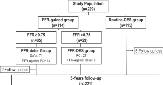
prospective, randomized study conducted in 6 university hospitals in Korea. Eligible patients with an intermediate coronary stenosis were randomly assigned to either FFR-guided or Routine-DES group (Figure 1). For FFR-guided group, treatment strategy was deter- mined according to the target vessel FFR (FFR<0.75: DES implan- tation [FFR-DES group], FFR≥0.75: deferral of PCI [FFR-Defer group]). Routine-DES group underwent DES implantation for the target lesion without FFR measurement. This study was designed to include 325 patients with 2-year clinical follow-up as DEFER study,1 but was prematurely terminated in December 2007 because of the concern of late stent thrombosis after DES implantation. The steer- ing committee decided to stop the enrollment and extend the clinical follow-up out to 5 years.
Study Procedures
The target vessel was engaged using a guiding catheter of 5F to 7F either by radial or femoral approach. Angiographic images were ac- quired after intracoronary administration of 100 to 200 μg of nitro- glycerin. FFR was measured using a 0.014-inch pressure guidewire (PressureWire; Radi Medical Systems, Uppsala, Sweden) as previ- ously described.9 Hyperemia was induced with an intracoronary bo- lus administration (80 μg in left coronary artery and 40 μg in right coronary artery) or intravenous infusion (140 μg/kg per minute) of adenosine. FFR was calculated as the ratio of the mean distal coro- nary pressure measured by the pressure wire to the mean aortic pres- sure measured by the guiding catheter under maximal hyperemia.10
Quantitative Coronary Angiography
Quantitative coronary angiography was performed by an indepen- dent core laboratory at Seoul National University Cardiovascular Center. Using the guiding catheter for calibration and an edge detec- tion system (CAAS 5.7; Quantitative Coronary Angiography System;
Pie Medical, Maastricht, the Netherlands), the reference diameters, minimum lumen diameter, and lesion length were measured, and the
% diameter stenosis was calculated. Lesion location was determined according to the American Heart Association classification.11
End Point and Clinical Follow-Up
The primary end point was the rate of major adverse cardiac events (MACE), and the results at 2 and 5 years were of special interest.
MACE was defined as a composite of cardiac death, myocardial in- farction (MI), and target lesion revascularization. All deaths were considered cardiac unless there was a clear noncardiac cause. MI was defined as an elevated cardiac enzyme with ischemic symptoms or new pathological Q waves on ECG. Periprocedural MI was not includ- ed. Clinical follow-up data were obtained from a Web-based reporting system. Additional information was obtained from hospital records and telephone contact if needed. An independent study monitor veri- fied all information on electronic case report forms. All clinical events were adjudicated by a clinical events committee in a blinded fashion using original source documents and angiographic images.
WHAT IS KNOWN
•
Fractional flow reserve (FFR) represents the ratio of the myocardial blood flow to the normal maximal myocardial flow and reflects the functional signifi- cance of coronary stenosis.•
FFR-guided revascularization strategy is known to be better than angiography-guided revascularization.•
The DEFER study demonstrated that the revascular- ization of the lesion with FFR≥0.75 did not improve the clinical outcomes of the patients with intermedi- ate coronary artery stenosis. However, this study was performed in the bare-metal stent era.WHAT THE STUDY ADDS
•
In patients with intermediate stenosis, FFR-guided drug-eluting stent (DES) implantation had compa- rable long-term clinical outcomes when compared with routine DES implantation strategy, but with much less use of DES.•
Functionally significant lesions (FFR<0.75) had the worst clinical outcomes, even after DES implantation.FFR can be helpful to define high-risk groups after DES implantation among patients with intermediate stenosis.
Figure 1. Study flow. DES indicates drug-eluting stent; FFR, frac- tional flow reserve; and PCI, percutaneous coronary intervention.
by guest on April 24, 2017http://circinterventions.ahajournals.org/Downloaded from
Statistical Analysis
All comparisons were made on an intention-to-treat principle.
Categorical variables were expressed as proportions. The χ2 test or Fisher exact test was used to compare differences between the groups. Continuous variables are expressed as mean and SD and were compared by Student t test or 1-way ANOVA, and Bonferroni correction was used as post hoc analysis. The log-rank test was used to compare hazard rates for the 2 groups. For the compari- sons at 2 and 5 years, we directly compared the Kaplan–Meier es- timates at these time points using the estimated time-specific SEs
with Bonferroni correction for multiplicity adjustment. A 2-sided P<0.05 was considered to indicate statistical significance. All sta- tistical analyses were performed using SPSS version 18.0 (SPSS Inc, Chicago, IL).
Results
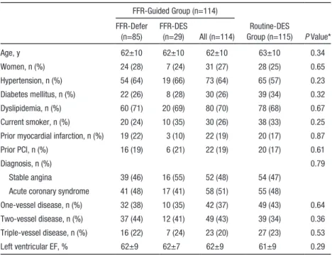
From November 2006 to December 2007, a total of 229 patients were enrolled and 114 patients were assigned to the FFR-guided group and 115 patients to the Routine-DES group Table 1. Baseline Clinical Characteristics of Study Population
FFR-Guided Group (n=114)
Routine-DES
Group (n=115) P Value*
FFR-Defer (n=85)
FFR-DES
(n=29) All (n=114)
Age, y 62±10 62±10 62±10 63±10 0.34
Women, n (%) 24 (28) 7 (24) 31 (27) 28 (25) 0.65
Hypertension, n (%) 54 (64) 19 (66) 73 (64) 65 (57) 0.23
Diabetes mellitus, n (%) 22 (26) 8 (28) 30 (26) 39 (34) 0.32
Dyslipidemia, n (%) 60 (71) 20 (69) 80 (70) 78 (68) 0.67
Current smoker, n (%) 20 (24) 10 (35) 30 (26) 38 (33) 0.25 Prior myocardial infarction, n (%) 19 (22) 3 (10) 22 (19) 20 (17) 0.87
Prior PCI, n (%) 16 (19) 6 (21) 22 (19) 20 (17) 0.61
Diagnosis, n (%) 0.79
Stable angina 39 (46) 16 (55) 52 (48) 54 (47) Acute coronary syndrome 41 (48) 17 (41) 58 (51) 55 (48)
One-vessel disease, n (%) 32 (38) 10 (35) 42 (37) 49 (43) 0.64 Two-vessel disease, n (%) 37 (44) 12 (41) 49 (43) 39 (34) 0.36 Triple-vessel disease, n (%) 16 (22) 7 (24) 23 (20) 27 (23) 0.53
Left ventricular EF, % 62±9 62±7 62±9 61±9 0.29
DES indicates drug-eluting stent; EF, ejection fraction; FFR, fractional flow reserve; and PCI, percutaneous coronary intervention.
*Comparison between FFR-guided group vs Routine-DES group.
Table 2. Baseline Lesions Characteristics
FFR-Guided Group (n=114) Routine-DES Group
(n=115) P Value*
FFR-Defer (n=85) FFR-DES (n=29) All (n=114)
Location of target lesion, % 0.72
Left anterior descending artery 52 66 55 56
Left circumflex 21 10 21 15
Right coronary artery 27 24 27 30
Lesion characteristics
Proximal reference diameter, mm 3.29±0.50 3.25±0.40 3.28±0.48 3.38±0.48 0.07 Distal reference diameter, mm 3.00±0.45 2.77±0.43 2.94±0.46 3.07±0.51 0.04
% diameter stenosis 53±9 58±10 54±10 56±9 0.13
Minimal lumen diameter, mm 1.46±0.34 1.23±0.33 1.41±0.35 1.41±0.35 0.97
Lesion length, mm 15.6±10.0 29.1±15.9 18.9±13.0 18.4±10.6 0.77
Fractional flow reserve 0.88±0.06 0.68±0.06 0.83±0.10 … …
Total stent number, n … 53 … 187 …
First-generation stents, n (%) … 31 (58) … 126 (67) 0.25†
DES indicates drug-eluting stent; and FFR, fractional flow reserve.
*Comparison between FFR-guided group vs Routine-DES group.
†Comparison between FFR-DES group vs Routine-DES group.
by guest on April 24, 2017http://circinterventions.ahajournals.org/Downloaded from
(Figure 1). FFR was successfully measured in all patients of the FFR-guided group without complication and FFR was
<0.75 in only 29 patients. Stents were implanted in nonstudy vessels in 24 patients and 44 patients in the FFR-guided group and the Routine-DES group, respectively.
Baseline Clinical and Angiographic Characteristics Baseline clinical characteristics were similar between the FFR-guided group and the Routine-DES group (Table 1).
There was also no difference in patients characteristics between the FFR-Defer and the FFR-DES groups.
Baseline lesion characteristics are shown in Table 2.
Angiographic lesion parameters such as angiographic % diameter, minimal lumen diameter, and lesion length were similar between the 2 groups. Distal reference vessel diameter was larger in the Routine-DES group than in the FFR-guided group (3.07±0.51 versus 2.94±0.46 mm; P=0.04). The post hoc analysis showed that % diameter stenosis of FFR-DES
group was significantly higher than that of the FFR-Defer group (P=0.03). However, there was a wide overlap of angio- graphic % diameter stenosis among the 3 groups (Figure 2).
Furthermore, there was no statistically significant difference in angiographic % diameter stenosis between FFR-Defer and Routine-DES groups (P=0.07). The mean FFR in FFR- Defer and FFR-DES groups were 0.88±0.06 and 0.68±0.06, respectively.
Clinical Outcomes Between FFR-Guided Group and Routine-DES Group
Kaplan–Meier survival curves for MACE by groups at 5-year follow-up are displayed in Figure 3. The incidence of clinical events at 5-year follow-up is shown in Table 3. At 2-year follow- up, the cumulative incidence of MACE (1-Kaplan–Meier esti- mate) was 7.9±2.5% in the FFR-guided group and 8.8±2.7% in Routine-DES group (P=0.80). At 5-year follow-up, the cumu- lative incidence of MACE was 11.6±3.0% and 14.2±3.3% for
A B
Figure 2. Angiographic % diameter stenosis. There was no difference in % diameter stenosis between fractional flow reserve (FFR)–
guided group and. Routine-drug-eluting stent (DES) group (A). However, % diameter stenosis of FFR-Defer group was lower than that of other groups (B).
A B
Figure 3. Kaplan–Meier survival curve for major adverse cardiac events (MACE). There was no difference in MACE between fractional flow reserve (FFR)–guided group and Routine-drug-eluting stent (DES) group at 2-y follow-up (A) and 5-y follow-up (B).
by guest on April 24, 2017http://circinterventions.ahajournals.org/Downloaded from
the FFR-guided group and the Routine-DES group (P=0.55).
There was no difference in MACE rates between the 2 groups up to 5-year follow-up (hazard ratio, 1.25; 95% confidence interval, 0.60–2.60). There was also no difference in the inci- dence of cardiac death, MI, target lesion revascularization, or any repeated revascularization between the 2 groups.
Clinical Outcomes Among FFR-Defer, FFR-DES, and Routine-DES Groups
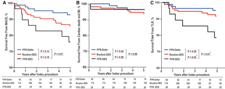
When the clinical outcomes among FFR-Defer, FFR-DES, and Routine-DES groups were compared, there was a difference in MACE rates at 5-year follow-up, which was mainly driven by high target lesion revascularization of FFR-DES group (Figure 4; Table I in the Data Supplement). At 5-year follow- up, the MACE rate was not statistically different between Routine-DES and FFR-Defer groups (13.9±3.3% versus 7.1±2.8%; P=0.13) and between Routine-DES and FFR-DES groups (13.9±3.3% versus 24.1±7.9%; P=0.19). The FFR- DES group had significantly higher rates of MACE (7.2±2.8%
versus 24.1±7.9%; P=0.01) than the FFR-Defer group. These results were reproduced by per-protocol analyses.
When the incidence of each event was compared, FFR- DES group had the highest incidence of MACE (P=0.05), target lesion revascularization (P=0.03), and any repeated
revascularization (P=0.03). The incidence of cardiac death or MI was not significantly different among the 3 groups (Table 3).
When the DES implanted patients were divided into 2 groups according to angiographic lesion severity (50% diam- eter stenosis), there was no difference in the rate of MACE at 5-year follow-up between the 2 groups (15.6% in diame- ter stenosis <50% group, 16.1% in diameter stenosis ≥50%
group; P=1.00).
Discussion
This study investigated the long-term clinical outcomes of FFR-guided revascularization versus routine PCI with DES implantation in patients with intermediate stenosis without objective evidence of ischemia and found that FFR-guided DES implantation had comparable long-term clinical out- comes than routine DES implantation strategy, but with much less use of DES. The present study showed that there was no benefit of routine stent implantation for a functionally insignificant lesion, even if DES was used. The prognosis of lesions with high FFR was excellent with medical treatment, whereas those with low FFR had the worst outcomes even after DES implantation because they had the most severe disease.
Table 3. Five-Year Clinical Outcomes According to Therapeutic Strategy
FFR-Guided Group (n=114) Routine-DES Group
(n=115) P Value*
P Value Among 3 Groups FFR-Defer (n=85) FFR-DES (n=29) All (n=114)
Cardiac death or MI 3 (3.5) 1 (3.4) 4 (3.5) 7 (6.1) 0.54 0.66
Target lesion revascularization 4 (4.7) 6 (20.7) 10 (8.8) 9 (7.8) 0.82 0.03
All revascularization 7 (8.2) 8 (27.6) 15 (13.2) 15 (13.0) 1.00 0.03
MACE 6 (7.1) 7 (24.1) 13 (11.4) 16 (13.9) 0.69 0.05
DES indicates drug-eluting stent; FFR, fractional flow reserve; MACE, major adverse cardiac events; and MI, myocardial infarction.
*Comparison between FFR-guided group vs Routine-DES group.
A B C
Figure 4. Kaplan–Meier survival curve for clinical events among 3 groups at 5-y follow-up. The rate of major adverse cardiac events (MACE; A) and target lesion revascularization (TLR; C) was higher in fractional flow reserve (FFR)-drug-eluting stent (DES) group than in other groups, and no significant difference of cardiac death and myocardial infarction (MI; B) rate was observed. FFR-Defer means FFR>0.75 and FFR-DES means FFR<0.75. Routine-DES means no FFR. *P<0.017 is significant according to Bonferroni correction.
by guest on April 24, 2017http://circinterventions.ahajournals.org/Downloaded from
The primary population in our study were patients with intermediate stenosis, which is the preferred indication by cli- nicians for FFR use. Our study population is different from that in the Fractional Flow Reserve Versus Angiography for Multivessel Evaluation (FAME) study.4 FAME included patients with multivessel coronary artery disease and more than half of lesions had angiographic stenosis of >70%. Mean FFR in FAME study was 0.71. Mean % diameter stenosis of all studied lesions was 53% in our study, which was similar to that of DEFER study (52%). Mean % diameter stenosis and FFR of defer group were 53% and 0.88 in our study and 48%
and 0.87 in the DEFER study,1 respectively.
In the DEFER study, 325 patients were randomized to deferral of PCI and performance of PCI groups, and then FFR was measured. When FFR was <0.75, PCI was performed in all patients and the randomization was executed only in patients with an FFR ≥0.75. This study was performed in the pre-DES era and the PCI performed was balloon angioplasty or bare-metal stent implantation. Our study design was a lit- tle bit different from the DEFER study. After introduction of DES into clinical practice, PCI was performed in a wide range of coronary lesions with high success, low complication rate, and excellent long-term patency.12–14 Therefore, we random- ized the patients into FFR-guided DES implantation group and routine DES implantation group to compare the outcomes of 2 different strategies in patients with intermediate stenosis.
FFR was measured only in the FFR-guided group in our study, in contrast to the original DEFER study where FFR was avail- able for all patients.
Moses et al8 reported that DES implantation in patients with intermediate stenosis (% diameter stenosis <50% [mean 44%] by quantitative coronary angiography) resulted in a marked reduction in clinical and angiographic restenosis.
Lavi et al15 investigated the 2-year outcomes of patients with intermediate lesions (0.75≤FFR<0.9) and found that DES implantation resulted in better outcomes than bare-metal stent implantation. Therefore, it is natural to postulate that the results would have been different if DES was used in the DEFER study. In our study, there was no difference in both 2- and 5-year clinical outcomes between the FFR-guided and the Routine-DES groups. There was also no difference in the rate of death or MI. Our study results again confirmed that the use of FFR reduced unnecessary intervention, and PCI even with DES, cannot improve patient outcomes unless ischemia is indicated. In our study, there was no difference in angio- graphic lesion severity between the 2 groups, and only one quarter of patients in the FFR-guided group had functionally significant stenosis (FFR<0.75). This finding is important, considering the fact that visual estimation still dominates in the treatment decision for intermediate stenosis and the low penetration rate of FFR.16
In the present study, patients with functionally signifi- cant lesions had worse clinical outcomes, even after DES implantation. The 5-year event-free survival rate was 75.9%
in the FFR-DES group (FFR<0.75) and 92.9% in the FFR- defer group (FFR≥0.75). A similar trend was observed in the DEFER study. Because FFR is determined by several angio- graphic factors such as lesion length, lesion geometry, and
% diameter stenosis, low FFR in patients with intermediate
stenosis might indicate high-risk lesions after PCI. In our study, the lesion length was longer in the FFR-DES group than in the FFR-Defer group (29.1±15.9 versus 15.6±10.0 mm; P<0.01). Furthermore, when DES implanted patients were divided into 2 groups according to 50% diameter ste- nosis, there was no difference in clinical outcomes. Although clinical outcome of angiographic intermediate stenosis is gen- erally favorable regardless of treatment strategy, the results of our study and the DEFER study suggest that the lesions with low FFR, despite intermediate degree of stenosis, might need meticulous surveillance for future clinical events.
Limitations
Our study has several limitations. First, the sample size was small, and the enrollment was interrupted early because of the concern about late stent thrombosis after DES implantation.
However, considering the favorable outcomes of intermediate stenosis, regardless of treatment strategy, it may not be easy to perform a study large enough to meet the statistical require- ment. Second, the study was not a blinded one. It is possible that the awareness of having an unstented coronary lesion had influenced the decision of the physicians during follow-up.
Third, our study was performed in first-generation DES era, and recent reports have shown that second-generation DESs have more favorable clinical outcomes than first-generation DES.17–19 Fourth, the information on the angina status and antianginal medication was not available in this study. Finally, because this study was designed before FAME studies, PCI indication in the FFR-guided group was FFR<0.75. Recent meta-analysis by Johnson et al20 found that the lesions with lower FFR were associated with higher risk of clinical events.
However, because there was no difference in clinical outcomes between the FFR-guided and Routine-DES groups, the main findings of our study would have not changed significantly, even if the criterion of FFR≤0.80 had been used.
Conclusions
In lesions with angiographically intermediate stenosis, FFR guidance provides a tailored approach, which is at least as good as an angiography-guided routine DES implantation strategy and avoids unnecessary DES in a considerable part of the patients.
Disclosures
Dr Koo received the institutional research grant from St Jude Medical.
The other authors report no conflicts.
References
1. Bech GJ, De Bruyne B, Pijls NH, de Muinck ED, Hoorntje JC, Escaned J, Stella PR, Boersma E, Bartunek J, Koolen JJ, Wijns W. Fractional flow reserve to determine the appropriateness of angioplasty in moderate coro- nary stenosis: a randomized trial. Circulation. 2001;103:2928–2934.
2. Pijls NH, van Schaardenburgh P, Manoharan G, Boersma E, Bech JW, van’t Veer M, Bär F, Hoorntje J, Koolen J, Wijns W, de Bruyne B. Percutaneous coronary intervention of functionally nonsignificant stenosis: 5-year fol- low-up of the DEFER Study. J Am Coll Cardiol. 2007;49:2105–2111. doi:
10.1016/j.jacc.2007.01.087.
3. De Bruyne B, Pijls NH, Kalesan B, Barbato E, Tonino PA, Piroth Z, Jagic N, Möbius-Winkler S, Mobius-Winckler S, Rioufol G, Witt N, Kala P, MacCarthy P, Engström T, Oldroyd KG, Mavromatis K, Manoharan G, Verlee P, Frobert O, Curzen N, Johnson JB, Jüni P, Fearon WF; FAME
by guest on April 24, 2017http://circinterventions.ahajournals.org/Downloaded from
2 Trial Investigators. Fractional flow reserve-guided PCI versus medical therapy in stable coronary disease. N Engl J Med. 2012;367:991–1001.
doi: 10.1056/NEJMoa1205361.
4. Tonino PA, De Bruyne B, Pijls NH, Siebert U, Ikeno F, van’ t Veer M, Klauss V, Manoharan G, Engstrøm T, Oldroyd KG, Ver Lee PN, MacCarthy PA, Fearon WF; FAME Study Investigators. Fractional flow reserve versus angiography for guiding percutaneous coronary interven- tion. N Engl J Med. 2009;360:213–224. doi: 10.1056/NEJMoa0807611.
5. Layland J, Oldroyd KG, Curzen N, Sood A, Balachandran K, Das R, Junejo S, Ahmed N, Lee MM, Shaukat A, O’Donnell A, Nam J, Briggs A, Henderson R, McConnachie A, Berry C; FAMOUS–NSTEMI inves- tigators. Fractional flow reserve vs. angiography in guiding management to optimize outcomes in non-ST-segment elevation myocardial infarction:
the British Heart Foundation FAMOUS-NSTEMI randomized trial. Eur Heart J. 2015;36:100–111. doi: 10.1093/eurheartj/ehu338.
6. Legalery P, Schiele F, Seronde MF, Meneveau N, Wei H, Didier K, Blonde MC, Caulfield F, Bassand JP. One-year outcome of patients submitted to routine fractional flow reserve assessment to determine the need for angio- plasty. Eur Heart J. 2005;26:2623–2629. doi: 10.1093/eurheartj/ehi484.
7. Dattilo PB, Prasad A, Honeycutt E, Wang TY, Messenger JC. Contemporary patterns of fractional flow reserve and intravascular ultrasound use among patients undergoing percutaneous coronary intervention in the United States: insights from the National Cardiovascular Data Registry. J Am Coll Cardiol. 2012;60:2337–2339. doi: 10.1016/j.jacc.2012.08.990.
8. Moses JW, Stone GW, Nikolsky E, Mintz GS, Dangas G, Grube E, Ellis SG, Lansky AJ, Weisz G, Fahy M, Na Y, Russell ME, Donohoe D, Leon MB, Mehran R. Drug-eluting stents in the treatment of intermediate le- sions: pooled analysis from four randomized trials. J Am Coll Cardiol.
2006;47:2164–2171. doi: 10.1016/j.jacc.2006.01.068.
9. Pijls NH, De Bruyne B, Peels K, Van Der Voort PH, Bonnier HJ, Bartunek J, Koolen JJ. Measurement of fractional flow reserve to as- sess the functional severity of coronary-artery stenoses. N Engl J Med.
1996;334:1703–1708. doi: 10.1056/NEJM199606273342604.
10. Pijls NH, van Son JA, Kirkeeide RL, De Bruyne B, Gould KL.
Experimental basis of determining maximum coronary, myocardial, and collateral blood flow by pressure measurements for assessing functional stenosis severity before and after percutaneous transluminal coronary an- gioplasty. Circulation. 1993;87:1354–1367.
11. Austen WG, Edwards JE, Frye RL, Gensini GG, Gott VL, Griffith LS, McGoon DC, Murphy ML, Roe BB. A reporting system on patients evalu- ated for coronary artery disease. Report of the Ad Hoc Committee for Grading of Coronary Artery Disease, Council on Cardiovascular Surgery, American Heart Association. Circulation. 1975;51(4 suppl):5–40.
12. Stone GW, Ellis SG, Cox DA, Hermiller J, O’Shaughnessy C, Mann JT, Turco M, Caputo R, Bergin P, Greenberg J, Popma JJ, Russell ME; TAXUS-IV Investigators. One-year clinical results with the slow-release, polymer-based, paclitaxel-eluting TAXUS stent: the TAXUS-IV trial. Circulation. 2004;109:1942–1947. doi: 10.1161/01.
CIR.0000127110.49192.72.
13. Moses JW, Leon MB, Popma JJ, Fitzgerald PJ, Holmes DR, O’Shaughnessy C, Caputo RP, Kereiakes DJ, Williams DO, Teirstein PS, Jaeger JL, Kuntz RE; SIRIUS Investigators. Sirolimus-eluting stents ver- sus standard stents in patients with stenosis in a native coronary artery. N Engl J Med. 2003;349:1315–1323. doi: 10.1056/NEJMoa035071.
14. Bangalore S, Kumar S, Fusaro M, Amoroso N, Attubato MJ, Feit F, Bhatt DL, Slater J. Short- and long-term outcomes with drug-eluting and bare-metal coronary stents: a mixed-treatment comparison analysis of 117 762 patient-years of follow-up from randomized trials. Circulation.
2012;125:2873–2891. doi: 10.1161/CIRCULATIONAHA.112.097014.
15. Lavi S, Rihal CS, Yang EH, Fassa AA, Elesber A, Lennon RJ, Mathew V, David HR Jr, Lerman A. The effect of drug eluting stents on cardiovascu- lar events in patients with intermediate lesions and borderline fractional flow reserve. Catheter Cardiovasc Interv. 2007;70:525–531. doi: 10.1002/
ccd.21154.
16. Toth GG, Toth B, Johnson NP, De Vroey F, Di Serafino L, Pyxaras S, Rusinaru D, Di Gioia G, Pellicano M, Barbato E, Van Mieghem C, Heyndrickx GR, De Bruyne B, Wijns W. Revascularization decisions in patients with stable angina and intermediate lesions: results of the in- ternational survey on interventional strategy. Circ Cardiovasc Interv.
2014;7:751–759. doi: 10.1161/CIRCINTERVENTIONS.114.001608.
17. Windecker S, Serruys PW, Wandel S, Buszman P, Trznadel S, Linke A, Lenk K, Ischinger T, Klauss V, Eberli F, Corti R, Wijns W, Morice MC, di Mario C, Davies S, van Geuns RJ, Eerdmans P, van Es GA, Meier B, Jüni P. Biolimus-eluting stent with biodegradable polymer versus siro- limus-eluting stent with durable polymer for coronary revascularisation (LEADERS): a randomised non-inferiority trial. Lancet. 2008;372:1163–
1173. doi: 10.1016/S0140-6736(08)61244-1.
18. Stone GW, Rizvi A, Newman W, Mastali K, Wang JC, Caputo R, Doostzadeh J, Cao S, Simonton CA, Sudhir K, Lansky AJ, Cutlip DE, Kereiakes DJ; SPIRIT IV Investigators. Everolimus-eluting ver- sus paclitaxel-eluting stents in coronary artery disease. N Engl J Med.
2010;362:1663–1674. doi: 10.1056/NEJMoa0910496.
19. Navarese EP, Kowalewski M, Kandzari D, Lansky A, Górny B, Kołtowski L, Waksman R, Berti S, Musumeci G, Limbruno U, van der Schaaf RJ, Kelm M, Kubica J, Suryapranata H. First-generation versus second- generation drug-eluting stents in current clinical practice: updated evi- dence from a comprehensive meta-analysis of randomised clinical trials comprising 31 379 patients. Open Heart. 2014;1:e000064. doi: 10.1136/
openhrt-2014-000064.
20. Johnson NP, Tóth GG, Lai D, Zhu H, Açar G, Agostoni P, Appelman Y, Arslan F, Barbato E, Chen SL, Di Serafino L, Domínguez-Franco AJ, Dupouy P, Esen AM, Esen OB, Hamilos M, Iwasaki K, Jensen LO, Jiménez-Navarro MF, Katritsis DG, Kocaman SA, Koo BK, López-Palop R, Lorin JD, Miller LH, Muller O, Nam CW, Oud N, Puymirat E, Rieber J, Rioufol G, Rodés- Cabau J, Sedlis SP, Takeishi Y, Tonino PA, Van Belle E, Verna E, Werner GS, Fearon WF, Pijls NH, De Bruyne B, Gould KL. Prognostic value of fractional flow reserve: linking physiologic severity to clinical outcomes. J Am Coll Cardiol. 2014;64:1641–1654. doi: 10.1016/j.jacc.2014.07.973.
by guest on April 24, 2017http://circinterventions.ahajournals.org/Downloaded from
Lee, Tae-Jin Youn, Woo-Young Chung and Bon-Kwon Koo
Bong-Ki Lee, Seung-Woon Rha, Ki-dong Yoo, Kyung Tae Jung, Young-Seok Cho, Hae-Young Sang Hyun Park, Ki-Hyun Jeon, Joo Myung Lee, Chang-Wook Nam, Joon-Hyung Doh,
Five-Year Clinical Outcomes of DEFER-DES Trial
Print ISSN: 1941-7640. Online ISSN: 1941-7632
Copyright © 2015 American Heart Association, Inc. All rights reserved.
Avenue, Dallas, TX 75231
is published by the American Heart Association, 7272 Greenville Circulation: Cardiovascular Interventions
doi: 10.1161/CIRCINTERVENTIONS.115.002442 2015;8:
Circ Cardiovasc Interv.
http://circinterventions.ahajournals.org/content/8/12/e002442
World Wide Web at:
The online version of this article, along with updated information and services, is located on the
http://circinterventions.ahajournals.org/content/suppl/2015/12/07/CIRCINTERVENTIONS.115.002442.DC1
Data Supplement (unedited) at:
http://circinterventions.ahajournals.org//subscriptions/
is online at:
Circulation: Cardiovascular Interventions Information about subscribing to
Subscriptions:
http://www.lww.com/reprints
Information about reprints can be found online at:
Reprints:
document.
Answer
Permissions and Rights Question and under Services. Further information about this process is available in the
permission is being requested is located, click Request Permissions in the middle column of the Web page Clearance Center, not the Editorial Office. Once the online version of the published article for which
can be obtained via RightsLink, a service of the Copyright Circulation: Cardiovascular Interventions
in
Requests for permissions to reproduce figures, tables, or portions of articles originally published Permissions:
by guest on April 24, 2017http://circinterventions.ahajournals.org/Downloaded from
groups
CI, confidence interval; MACE, major adverse cardiac events; FFR, fractional flow reserve;
DES, drug eluting stent; MI, myocardial infarction; TLR, target lesion revascularization.
Hazard ratio 95% CI
5-year MACE FFR Defer
vs. FFR DES 3.67 1.24-10.93
vs. Routine DES 2.05 0.803-5.25
5-year Cardiac death and MI FFR Defer
vs. FFR DES 0.96 0.10-9.23
vs. Routine DES 1.73 0.45-6.69
5-year TLR FFR Defer
vs. FFR DES 4.73 1.34-16.76
vs. Routine DES 1.72 0.53-5.59