© 2016 The Korean Ophthalmological Society
This is an Open Access article distributed under the terms of the Creative Commons Attribution Non-Commercial License (http://creativecommons.org/licenses /by-nc/3.0/) which permits unrestricted non-commercial use, distribution, and reproduction in any medium, provided the original work is properly cited.
Original Article
Polypoidal choroidal vasculopathy (PCV) is a disorder of the eyes characterized by branching vascular networks
and terminating polypoidal lesions on indocyanine green angiography (ICGA). This condition generally responds well to intravitreal anti-vascular endothelial growth factor (VEGF) therapy [1-4], photodynamic therapy (PDT) [5,6], or a combination of the two treatments [7]. However, limit- ed knowledge is available regarding anti-VEGF therapy treatment outcomes for PCV presenting with grape-like polyp clusters on ICGA [3,8]. It has been reported that this peculiar PCV generally has unfavorable outcomes when treated with PDT [9]. Additionally, a more recent study
Intravitreal Anti-vascular Endothelial Growth Factor for Treating Polypoidal Choroidal Vasculopathy with Grape-like Polyp Clusters
Young Suk Chang1, Jae Hui Kim2, Jong Woo Kim2, Tae Gon Lee2, Chul Gu Kim2
1Department of Ophthalmology, Konyang University College of Medicine, Daejeon, Korea
2Department of Ophthalmology, Kim’s Eye Hospital, Konyang University College of Medicine, Seoul, Korea
Purpose: To evaluate 12-month outcomes of anti-vascular endothelial growth factor (VEGF) therapy for polypoi- dal choroidal vasculopathy (PCV) with grape-like polyp clusters.
Methods: This retrospective observational study included 23 eyes of 23 patients who were newly diagnosed with PCV with grape-like polyp clusters, and who were subsequently treated with anti-VEGF monotherapy.
The study compares the best-corrected visual acuity (BCVA) of the patients at diagnosis, at 3 months, and at 12 months after diagnosis. In addition, 12-month changes in BCVA values were compared between cases with subfoveal or juxtafoveal polyps and cases with extrafoveal polyps.
Results: The baseline, 3-month, and 12-month logarithm of the minimal angle of resolution BCVA was 0.62 ± 0.35, 0.50 ± 0.43, and 0.58 ± 0.48, respectively. Compared to the baseline, patient BCVA was not significantly different at 12 months after diagnosis (p = 0.764). Six eyes (26.1%) gained ≥0.2 logarithm of the minimal angle of resolution BCVA. In cases with subfoveal or juxtafoveal polyps, BCVA values at baseline and at 12 months after diagnosis were 0.66 ± 0.37 and 0.69 ± 0.53, respectively. In cases with extrafoveal polyps, the values were 0.54 ± 0.33 and 0.37 ± 0.31, respectively. Changes in BCVA values were significantly different between the two groups (p = 0.023).
Conclusions: Although anti-VEGF therapy has favorable short-term efficacy for treating PCV with grape-like polyp clusters, long-term visual improvements are generally limited in the majority of afflicted eyes. The pres- ence of subfoveal or juxtafoveal polyps may suggest unfavorable treatment outcomes.
Key Words: Age-related macular degeneration, Anti-vascular endothelial growth factor, Choroidal neovascular- ization, Polypoidal choroidal vasculopathy
Received: July 10, 2015 Accepted: August 31, 2015
Corresponding Author: Jae Hui Kim, MD. Department of Ophthalmol- ogy, Kim's Eye Hospital, Konyang University College of Medicine, #136 Yeongsin-ro, Yeongdeungpo-gu, Seoul 07301, Korea. Tel: 82-2-2639- 7664, Fax: 82-2-2639-7824, E-mail: [email protected]
This study was presented in part at the Association for Research in Vi- sion and Ophthalmology Meeting at Orlando and Florida, USA, 2014.
shows worse visual outcomes after anti-VEGF therapy in eyes with PCV with grape-like polyp clusters than in eyes with PCV without clusters [3]. That particular study, how- ever, did not include a detailed outcome evaluation in eyes with polyp clusters. To date, only one study has separately analyzed the treatment outcomes of anti-VEGF therapy for eyes with this condition [8].
The present study evaluates 12-month visual and ana- tomical outcomes of anti-VEGF therapy for eyes newly di- agnosed with PCV with grape-like polyp clusters.
Materials and Methods
This retrospective, observational case series was per- formed at a single center. All study conduct adhered to the tenets of the Declaration of Helsinki, and the study proto- col was approved by the institutional review board.
Eyes with PCV with grape-like polyp clusters that were treated with anti-VEGF monotherapy for 12 months were included. The results of fundus photographs and ICGA were reviewed for patients who were initially diagnosed with PCV between January 2010 and March 2013 at Kim's Eye Hospital. A cluster of grape-like polyps was defined based on some modification of criteria in a previous study [9]; including (1) a hyperfluorescence corresponding to an aneurysmal dilation on late-phase ICGA; (2) appearance of curvilinear, tortuous, or looped vessels, which first branch into smaller vessels before sprouting into numerous polyps of varying size, resulting in a grape-like appearance in the early ICGA phase (Fig. 1A and 1B); and (3) a maximum greatest linear dimension of the polyp cluster of ≥1,000 µm on ICGA. The criterion of “large reddish-orange aneurys- mal dilation with the longest axis of 1,500 µm or longer on fundus photograph,” which was used in a previous study [9], was not used in the current study. The decision to ex- clude this criterion was made because, in some eyes, defi- nite reddish-orange aneurysmal dilation is not easily visi- ble on fundus photographs, despite definite ICGA findings.
Instead, we used a novel criterion, as described above (cri- terion 3). Two examiners (JHK and YSC) jointly reviewed ICGA results to determine subject eligibility.
To be included in this study, all subjects were required to undergo a comprehensive ophthalmologic examination, in- cluding measurement of best-corrected visual acuity (BCVA), 90-diopter lens slit lamp biomicroscopy, fundus
photography, fluorescein angiography, and ICGA with a confocal laser-scanning system (HRA-2; Heidelberg Engi- neering, Dossenheim, Germany). Horizontal and vertical cross-hair scans centered at the center of the fovea were also performed using spectral domain optical coherence tomography (OCT; Spectral OCT/SLO, OTI Ophthalmic Technologies, Miami, FL, USA). Exclusion criteria includ- ed a follow-up period shorter than 12 months, symptom duration greater than 6 months, severe media opacity, end- stage age-related macular degeneration (central geographic atrophy and/or disciform scarring), previous intraocular surgery (except cataract surgery), macroaneurysms, prolif- erative diabetic retinopathy, central retinal vascular occlu- sion, and any other retinal disorder that may influence macular microstructure and/or function. Eyes with a sub- macular hemorrhage of ≥3 disc diameters and involving the fovea at the time of diagnosis were also excluded.
All included eyes were treated with three consecutive monthly intravitreal ranibizumab injections as an initial Fig. 1. Indocyanine green angiography images (A,B) from eyes diagnosed with polypoidal choroidal vasculopathy with grape- like polyp clusters.
A
B
treatment. Subsequently, patients were examined every 1 to 3 months, as determined by the treating physician. In general, retreatment with an anti-VEGF agent (either ran- ibizumab or bevacizumab) was performed when intrareti- nal or subretinal fluid or retinal or subretinal hemorrhages developed with an accompanying increase in macular thickness.
Because volume scans were not routinely performed for every patient, central foveal thickness (CFT) was used for analysis. The CFT was defined as the distance between the internal limiting membrane and Bruch’s membrane at the fovea. The CFT was manually measured using the built-in calipers of the OCT software program. The greatest linear dimension of the entire PCV lesion on ICGA was also measured using the built-in calipers of the ICGA software program.
In all included eyes, BCVA and CFT values were com- pared between baseline measurements and measurements taken at 3 and 12 months after initial diagnosis. Included eyes were divided into two groups according to the loca- tion of polyp clusters. Eyes exhibiting subfoveal or juxta- foveal (<200 µm from the fovea) polyps were included in the subfoveal/juxtafoveal polyp group, whereas the re- maining eyes were included in the extrafoveal polyp group. The greatest linear dimension of the PCV lesion, the number of anti-VEGF injections during the 12-month follow-up period, the proportion of eyes exhibiting subret- inal fluids after three ranibizumab injections, and changes in BCVA and CFT values during the follow-up period were compared between the two groups.
Data are presented as mean ± standard deviations where applicable. Statistical analyses were performed with a commercially available software package (SPSS ver. 12.0;
SPSS Inc., Chicago, IL, USA). Differences in values at various time points were tested for statistical significance using a repeated-measures analysis of variance with the Bonferroni correction. Comparison between the two dif- ferent groups was performed using Mann-Whitney U-tests or Fisher’s exact tests. A p-value less than 0.05 was consid- ered statistically significant for all tests.
Results
From the 23 patients, 20 eyes satisfied all eligibility cri- teria and were included in analyses (Table 1). Fourteen pa-
tients (60.9%) were male and 9 patients (39.1%) were fe- male, and the mean patient age was 65.8 ± 6.8 years. The greatest PCV lesion linear dimension averaged 2,401.6 ± 673.2 µm. During the 12-month follow-up period, patients received an average of 4.5 ± 1.4 anti-VEGF injections (3.8
± 1.5 ranibizumab injections and 0.6 ± 1.1 bevacizumab in- jections). Subretinal fluid was present after the initial three ranibizumab injections in seven of the 23 eyes (30.4%). A submacular hemorrhage developed in three eyes (13.0%) during the 12-month follow-up period.
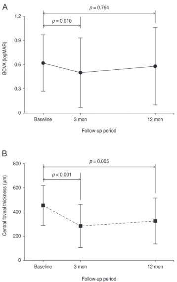
Patient BCVA values at baseline, at 3 months, and at 12 months after diagnosis were 0.62 ± 0.35 (Snellen equiva- lent = 20 / 83), 0.50 ± 0.43 (20 / 63), and 0.58 ± 0.48 (20 / 76), respectively (Fig. 2A). The BCVA values at 3 months were significantly better than the baseline values (p = 0.010), whereas BCVA values at 12 months were not sig- nificantly different from the baseline values (p = 0.764).
Six eyes (26.1%) gained two or more lines of vision (≥0.2 logarithm of the minimal angle of resolution BCVA), and five eyes (21.7%) lost two or more lines of vision. The re- maining 12 eyes (52.2%) had stable BCVA values. The CFT values at baseline, at 3 months, and at 12 months after diagnosis were 453.9 ± 164.8 µm, 282.9 ± 178.9 µm, and 325.4 ± 189.5 µm, respectively (Fig. 2B). The 3- and 12-month CFT values significantly decreased in compari- son to baseline values (p < 0.001 and p = 0.005, respective- ly).
When divided into two groups according to the location of polyp clusters, 15 eyes were included in the subfoveal/
juxtafoveal polyp group (Fig. 3A-3E), whereas the remain- ing eight eyes were included in the extrafoveal polyp group (Fig. 4A-4E). Table 2 summarizes the results of Table 1. Baseline characteristics of eyes with polypoidal cho- roidal vasculopathy with grape-like polyp clusters
Characteristics Value
Age (yr) 65.8 ± 6.8
Sex
Male 14 (60.9)
Female 9 (39.1)
Greatest lesion liner dimension (µm) 2,401.6 ± 673.2
Baseline BCVA (logMAR) 0.62 ± 0.35
Baseline central foveal thickness (µm) 453.9 ± 164.8 Values are presented as mean ± standard deviation or number (%).
BCVA = best-corrected visual acuity; logMAR = logarithm of the minimal angle of resolution.
comparisons between the two groups. There were no sig- nificant differences in the greatest PCV linear dimension (subfoveal/juxtafoveal vs. extrafoveal, 2,483.5 ± 663.9 µm vs. 2,247.9 ± 707.9 µm; p = 0.349) or number of anti-VEGF injections (4.7 ± 1.3 vs. 4.1 ± 1.6; p = 0.238) between the two groups. In the subfoveal/juxtafoveal group, subretinal fluid was present after the initial three ranibizumab injec- tions in six eyes (40.0%). The BCVA values at baseline, at 3 months, and at 12 months after diagnosis were 0.66 ± 0.37 (20 / 91), 0.54 ± 0.47 (20 / 69), and 0.69 ± 0.53 (20 / 97), respectively, while the CFT values at baseline, at 3 months, and at 12 months after diagnosis were 423.2 ±
156.7 µm, 273.9 ± 165.7 µm, and 341.9 ± 183.2 µm, respec- tively. In the extrafoveal group, subretinal fluid was pres- ent after the initial three ranibizumab injections in one eye (12.5%). The BCVA values at baseline, at 3 months, and at 12 months after diagnosis were 0.54 ± 0.33 (20 / 69), 0.42 ± 0.37 (20 / 52), and 0.37 ± 0.31 (20 / 46), respectively, while the CFT values at baseline, at 3 months, and at 12 months after diagnosis were 468.5 ± 125.7, 299.9 ± 212.7, and 294.6
± 210.1 µm, respectively. The proportion of eyes exhibiting subretinal fluids after three ranibizumab injections (p = 0.345) and changes in CFT values during the 12-month fol- low-up period (p = 0.138) were not significantly different between the two groups. On the other hand, there was a significant difference in BCVA changes across the various time points between the two groups (p = 0.023).
Discussion
In this study, CFT significantly decreased after an- ti-VEGF therapy in eyes with PCV with grape-like polyp clusters throughout the 12-month follow-up period. Al- though BCVA was not significantly improved in all eyes at 12 months after diagnosis, an improvement in BCVA of two lines or greater was observed in 26.1% of eyes, which suggests that anti-VEGF therapy has valid effects in some eyes with PCV with grape-like polyp clusters.
Grape-like polyp clusters are noted in approximately 9%
to 13% of all eyes with PCV [3,10]. The work of Uyama et al. [11] reports the natural history of PCV with grape-like polypoidal dilations. That study finds that these lesions are usually active and tend to hemorrhage or leak, resulting in severe visual loss in the eyes of patients with PCV with grape-like polypoidal dilations. A study by Lee et al. [9]
reports the results of 2-year PDT treatment for PCV with grape-like polyp clusters. Among the patients in that study, median visual acuity improved from 20 / 63 to 20 / 50 af- ter initial treatment, but visual acuity deteriorated during the 2-year follow-up period. At 1 year, 59% of eyes had a three or greater line decrease in visual acuity from base- line values. The authors hypothesized that grape-like pol- ypoidal lesions have a potent neovascular drive, and that possible negative influences of PDT may accelerate this drive. The authors also suggested the possible benefits of anti-VEGF therapy for this condition. According to their findings, however, the visual outcomes after anti-VEGF
Follow-up period Follow-up period Baseline
BCVA (logMAR)
3 mon 12 mon
1.2
0.9 0.6
0.3
0
p = 0.764 p = 0.010
Baseline
Central foveal thickness (μm)
3 mon 12 mon
800
600
400
200
0
p = 0.005 p < 0.001
Fig. 2. Changes in the mean logarithm of minimal angle of res- olution (logMAR) best-corrected visual acuity (BCVA) (A) and central foveal thickness (B) in eyes diagnosed with polypoidal choroidal vasculopathy with grape-like polyp clusters. Statistical analyses were performed using repeated measures analysis of variances with the Bonferroni correction.
A
B
therapy in eyes with PCV with grape-like polyp clusters were also unfavorable. Recently, Hikichi et al. [3] com- pared visual outcomes 12 months after anti-VEGF therapy between PCV eyes with and without grape-like polyp clus- ters, observing unfavorable outcomes in eyes with the cluster. The authors suggested that a greater incidence of
submacular hemorrhage in PCV eyes with polyp clusters was the possible cause of this difference in visual out- comes. However, detailed data about changes in visual acuity and macular thickness in PCV eyes with polyp clusters, which may provide valuable treatment response information, was not reported in that study. In the present Fig. 3. Fundus photography (A), fluorescein angiography (B), indocyanine green angiography (C), and optical coherence tomography (D,E) images at diagnosis (A-D) and at 12 months (E) from diagnosis in an eye with polypoidal choroidal vasculopathy with grape-like polyp clusters (arrow, C). In this case, the polyps were located at the fovea. Subretinal fluid was observed at 12 months after diagnosis (E).
Fig. 4. Fundus photography (A), fluorescein angiography (B), indocyanine green angiography (C), and optical coherence tomography (D,E) images at diagnosis (A-D) and at 12 months (E) from diagnosis in an eye with polypoidal choroidal vasculopathy with grape-like polyp clusters (arrow, C). In this case, the polyps were located outside the fovea. At 12 months after diagnosis (E), complete resolution of the fluid was noted.
A
D
E
C B
A
D
E
C B
study, the number of anti-VEGF injections administered over the 12-month follow-up period (a mean of 4.5 injec- tions) was comparable to the number of anti-VEGF injec- tions (mean, 4.2 injections) reported by Hikichi et al. [3].
The incidence of development of submacular hemorrhage during the 12-month follow-up period was 13.0% in the present study.
Previous studies reporting 12-month treatment outcomes of anti-VEGF therapy for PCV have revealed somewhat discrepant results. Although marked improvement in visu- al acuity is noted in many studies [1-3,6], only minimal changes in visual acuity are reported in other studies [12- 14]. In the present study, a marked decrease in CFT, ac- companied by an improvement in BCVA during the first 3 months of anti-VEGF therapy is noted. This result suggests that anti-VEGF therapy has certain short-term benefits for this condition. In contrast, BCVA values deteriorated among eyes in our study between 3 and 12 months, sug- gesting limited long-term efficacy. In addition, there were differences in response to treatment according to differing polyp cluster locations in the eyes of patients herein.
Extrafoveal polypoidal lesions are not an infrequent oc-
currence in PCV [15,16]. Although a recent study shows that anti-VEGF therapy is an effective treatment for extra- foveal exudative age-related macular degeneration [17], the efficacy of this treatment for extrafoveal PCV has not been fully elucidated. In the present study, the response to treat- ment was relatively better in the extrafoveal group than in the subfoveal/juxtafoveal group. A slight improvement in visual acuity was noted in the extrafoveal group, whereas a slight deterioration in visual acuity was noted in the sub- foveal/juxtafoveal group. As a result, there was a marked difference in the degree of change in visual acuity between the two groups. This result may suggest that anti-VEGF monotherapy is a useful treatment option in PCV with ex- trafoveal polyp clusters. When the polyps are located in the subfoveal region or close to the fovea, however, the beneficial effects of anti-VEGF therapy may be limited, suggesting the need for further studies to investigate the efficacy of other approaches. More specifically, recent studies have reported the resolution of fluid and retinal pigment epithelial detachment after intravitreal aflibercept in some PCVs that were refractory to ranibizumab therapy [18,19]. The efficacy of intravitreal aflibercept in PCV eyes with grape-like polyp clusters merits further investigation.
Recently, Lee and Lee [8] reported 24-month outcomes of anti-VEGF monotherapy in PCV eyes with grape-like polyp clusters. In that study, mean logarithm of the mini- mal angle of resolution visual acuity values at the time of diagnosis, at 3 months, at 12 months, and at 24 months af- ter diagnosis were 0.61 ± 0.28, 0.50 ± 0.30, 0.42 ± 0.27, and 0.44 ± 0.31, respectively. Values of visual acuity at diagno- sis and at 3 months after diagnosis are relatively compara- ble between this previous study [8] and the present study.
However, there is a notable difference in the visual acuity values at 12 months between the two studies. In the present study, the mean visual acuity of PCV eyes with grape-like polyp clusters slightly deteriorated from 3 months to 12 months, whereas continuous improvement in visual acuity was noted throughout the 12-month follow-up period in the previous study [8]. As a result, the visual acuity at 12 months was significantly improved relative to baseline val- ues in the previous study [8], whereas the values at the two time points were not different in the present study. Some possible reasons for this discrepancy are as follows. In the previous study [8], the mean number of anti-VEGF injec- tions was 12.5 ± 2.8 during the 2-year follow-up period.
The exact number of injections during the first year is not Table 2. Comparison of characteristics between eyes exhibit-
ing subfoveal or juxtafoveal polyps and eyes exhibiting extra- foveal polyps
Characteristics Subfoveal/
juxtafoveal group (n = 15)
Extrafoveal
group (n = 8) p-value Greatest lesion liner
dimension (µm) 2,483.5 ± 663.9 2,247.9 ± 707.9 0.349* No. of anti-VEGF
injections 4.5 ± 1.2 4.4 ± 1.5 0.463* Subretinal fluid after
initial three anti- VEGF injections
0.345†
Presence 6 (40.0) 1 (12.5)
Absence 9 (60.0) 7 (87.5)
Change in BCVA
(logMAR)‡ -0.03 ± 0.24 0.17 ± 0.18 0.023* Change in central
foveal thickness (µm)
81.3 ± 141.6 173.9 ± 197.5 0.138*
Values are presented as mean ± standard deviation or number (%).
VEGF = vascular endothelial growth factor; BCVA = best-cor- rected visual acuity; logMAR = logarithm of the minimal angle of resolution.
*Mann-Whitney U-test; †Fisher’s exact test; ‡Positive value indi- cates improvement in visual acuity, whereas negative value indi- cates deterioration in visual acuity.
presented in that study. However, considering previous PCV studies in which the number of injections is relatively greater during the first year than during the second year of therapy [3,20], we postulate that the number of injections during the first year in the study by Lee and Lee [8] is greater than the number of injections during the 12-month follow-up period in the present study. The authors of the previous study suggested that grape-like polypoidal lesions have a potent neovascular drive and characteristics of typi- cal choroidal neovascularization, thereby requiring more frequent injections than ordinary PCVs [8]. We postulate that the lower frequency of injections in our study may have caused under-treatment. The lower injection frequen- cy in the present study can be attributed primarily to our follow-up schedule. It is generally recommended that monthly follow-up examinations, including an OCT exam- ination, be conducted for prompt detection and treatment of exudation recurrence [21], because treatment delays may cause poor visual prognoses [22,23]. This strict follow-up schedule was not used in our retrospective study, however, and as a result, follow-up was performed less frequently.
Thus we suspect that our follow-up strategy may have caused the unfavorable visual results. In the study by Lee and Lee [8], rescue PDT was performed for two refractory cases. The 12-month visual acuity values of these two pa- tients were relatively good (20 / 25 and 20 / 32, respective- ly). PDT is considered to have higher efficacy than an- ti-VEGF therapy in PCV, particularly in regressing polyps [24], although the issue remains controversial [1]. Several expert groups still recommend PDT, with or without an- ti-VEGF, as the treatment of choice for PCV [25]. In the present study, all the patients were treated with anti-VEGF monotherapy regardless of their response to the therapy.
Thus it is possible that overall treatment outcomes may have been influenced by cases that were less responsive to anti-VEGF monotherapy among the patients herein.
This study has several limitations, including its retro- spective design and small sample size. Moreover, retreat- ment was performed at the discretion of the treating physi- cian rather than according to common treatment guidelines. Because ICGA was not routinely performed after anti-VEGF therapy among the patients herein, it is not possible to estimate the angiographic changes in pol- ypoidal lesions. For the same reason, development of typi- cal choroidal neovascularization during the follow-up peri- od, as shown in a previous study [9], could not be estimated.
In conclusion, the 12-month efficacy of anti-VEGF ther- apy for improving visual acuity is generally limited in PCV eyes with grape-like polyp clusters. However, defini- tive improvement in visual acuity was noted in 26.1% of the eyes included herein, which suggests that anti-VEGF therapy has valid effects in a certain proportion of eyes with this condition. In addition, the treatment is likely more beneficial when the polyps are not located in the sub- foveal or juxtafoveal regions. Further studies are required to establish more appropriate treatment strategies for PCV with grape-like polyp clusters.
Conflict of Interest
No potential conflict of interest relevant to this article was reported.
Acknowledgements
This study was supported by Kim’s Eye Hospital Re- search Center.
References
1. Oishi A, Kojima H, Mandai M, et al. Comparison of the ef- fect of ranibizumab and verteporfin for polypoidal choroi- dal vasculopathy: 12-month LAPTOP study results. Am J Ophthalmol 2013;156:644-51.
2. Cheng CK, Peng CH, Chang CK, et al. One-year outcomes of intravitreal bevacizumab (avastin) therapy for polypoi- dal choroidal vasculopathy. Retina 2011;31:846-56.
3. Hikichi T, Higuchi M, Matsushita T, et al. Results of 2 years of treatment with as-needed ranibizumab reinjection for polypoidal choroidal vasculopathy. Br J Ophthalmol 2013;97:617-21.
4. Lee SY, Kim JG, Joe SG, et al. The therapeutic effects of bevacizumab in patients with polypoidal choroidal vascu- lopathy. Korean J Ophthalmol 2008;22:92-9.
5. Chan WM, Lam DS, Lai TY, et al. Photodynamic therapy with verteporfin for symptomatic polypoidal choroidal vas- culopathy: one-year results of a prospective case series.
Ophthalmology 2004;111:1576-84.
6. Inoue M, Arakawa A, Yamane S, Kadonosono K. Long-
term outcome of intravitreal ranibizumab treatment, com- pared with photodynamic therapy, in patients with polyp- oidal choroidal vasculopathy. Eye (Lond) 2013;27:1013-20.
7. Tomita K, Tsujikawa A, Yamashiro K, et al. Treatment of polypoidal choroidal vasculopathy with photodynamic therapy combined with intravitreal injections of ranibi- zumab. Am J Ophthalmol 2012;153:68-80.e1.
8. Lee JH, Lee WK. Anti-vascular endothelial growth factor monotherapy for polypoidal choroidal vasculopathy with polyps resembling grape clusters. Graefes Arch Clin Exp Ophthalmol 2016;254:645-51.
9. Lee WK, Kim KS, Kim W, et al. Responses to photody- namic therapy in patients with polypoidal choroidal vascu- lopathy consisting of polyps resembling grape clusters. Am J Ophthalmol 2012;154:355-65.e1.
10. Sho K, Takahashi K, Yamada H, et al. Polypoidal choroidal vasculopathy: incidence, demographic features, and clini- cal characteristics. Arch Ophthalmol 2003;121:1392-6.
11. Uyama M, Wada M, Nagai Y, et al. Polypoidal choroidal vasculopathy: natural history. Am J Ophthalmol 2002;133:
639-48.
12. Lai TY, Lee GK, Luk FO, Lam DS. Intravitreal ranibizum- ab with or without photodynamic therapy for the treatment of symptomatic polypoidal choroidal vasculopathy. Retina 2011;31:1581-8.
13. Tsujikawa A, Ooto S, Yamashiro K, et al. Treatment of pol- ypoidal choroidal vasculopathy by intravitreal injection of bevacizumab. Jpn J Ophthalmol 2010;54:310-9.
14. Wakabayashi T, Gomi F, Sawa M, et al. Intravitreal bevaci- zumab for exudative branching vascular networks in pol- ypoidal choroidal vasculopathy. Br J Ophthalmol 2012;96:
394-9.
15. Gemmy Cheung CM, Yeo I, Li X, et al. Argon laser with and without anti-vascular endothelial growth factor thera- py for extrafoveal polypoidal choroidal vasculopathy. Am J Ophthalmol 2013;155:295-304.e1.
16. Hou J, Tao Y, Li XX, Zhao MW. Clinical characteristics of
polypoidal choroidal vasculopathy in Chinese patients.
Graefes Arch Clin Exp Ophthalmol 2011;249:975-9.
17. Parodi MB, Iacono P, La Spina C, et al. Intravitreal ranibi- zumab for naive extrafoveal choroidal neovascularization secondary to age-related macular degeneration. Retina 2014;34:2167-70.
18. Saito M, Kano M, Itagaki K, et al. Switching to intravitreal aflibercept injection for polypoidal choroidal vasculopathy refractory to ranibizumab. Retina 2014;34:2192-201.
19. Yamashita M, Nishi T, Hasegawa T, Ogata N. Response of serous retinal pigment epithelial detachments to intravitre- al aflibercept in polypoidal choroidal vasculopathy refrac- tory to ranibizumab. Clin Ophthalmol 2014;8:343-6.
20. Kang HM, Koh HJ. Long-term visual outcome and prog- nostic factors after intravitreal ranibizumab injections for polypoidal choroidal vasculopathy. Am J Ophthalmol 2013;
156:652-60.
21. Fung AE, Lalwani GA, Rosenfeld PJ, et al. An optical co- herence tomography-guided, variable dosing regimen with intravitreal ranibizumab (Lucentis) for neovascular age-re- lated macular degeneration. Am J Ophthalmol 2007;143:
566-83.
22. Rauch R, Weingessel B, Maca SM, Vecsei-Marlovits PV.
Time to first treatment: The significance of early treatment of exudative age-related macular degeneration. Retina 2012;32:1260-4.
23. Arias L, Armada F, Donate J, et al. Delay in treating age-related macular degeneration in Spain is associated with progressive vision loss. Eye (Lond) 2009;23:326-33.
24. Koh A, Lee WK, Chen LJ, et al. EVEREST study: efficacy and safety of verteporfin photodynamic therapy in combi- nation with ranibizumab or alone versus ranibizumab monotherapy in patients with symptomatic macular polyp- oidal choroidal vasculopathy. Retina 2012;32:1453-64.
25. Koh AH; Expert PCV Panel, Chen LJ, et al. Polypoidal choroidal vasculopathy: evidence-based guidelines for clin- ical diagnosis and treatment. Retina 2013;33:686-716.