© 2017 The Korean Ophthalmological Society
This is an Open Access article distributed under the terms of the Creative Commons Attribution Non-Commercial License (http://creativecommons.org/licenses /by-nc/3.0/) which permits unrestricted non-commercial use, distribution, and reproduction in any medium, provided the original work is properly cited.
Original Article
Polypoidal Choroidal Vasculopathy with Feeder Vessels:
Characteristics, Fellow Eye Findings, and Long-term Treatment Outcomes
Hyun Ji Hwang1, Jae Hui Kim1, Young Suk Chang2, Jong Woo Kim1, Chul Gu Kim1
1Department of Ophthalmology, Kim’s Eye Hospital, Konyang University College of Medicine, Seoul, Korea
2Department of Ophthalmology, Konyang University College of Medicine, Daejeon, Korea
Purpose: To evaluate the long-term outcomes of anti-vascular endothelial growth factor (VEGF) therapy for pol- ypoidal choroidal vasculopathy (PCV) with feeder vessels and to investigate fellow-eye findings.
Methods: This retrospective observational study included 14 eyes with treatment-naïve PCV accompanied by feeder vessels that were treated with anti-VEGF monotherapy. The best-corrected visual acuity (BCVA) at baseline was compared with that at the last follow-up. The fellow-eye indocyanine green angiography findings were also analyzed.
Results: The mean follow-up period was 28.1 ± 19.2 months (range, 12 to 60 months). During the follow-up peri- od, 5.9 ± 2.5 anti-VEGF injections were administered. The logarithm of the minimal angle of resolution (logMAR) BCVAs at the time of diagnosis, at 3 months, and at the last follow-up were 0.81 ± 0.49, 0.55 ± 0.44, and 0.71 ± 0.54, respectively. Although the BCVA at the last follow-up was not different from the baseline value (p = 0.809), an improvement of ≥0.2 logMAR BCVA was observed in seven eyes (50.0%). In 11 eyes that underwent bi- lateral indocyanine green angiography at diagnosis, PCV, branching vascular networks, and late geographic hyperfluorescence were noted in two (18.2%), five (45.4%), and three (27.3%) fellow eyes, respectively. During the follow-up period, the development of polypoidal lesions in the fellow eye was observed in three patients.
Conclusions: In this study, long-term improvement in BCVA was noted in 50% of the included patients who re- ceived anti-VEGF monotherapy. A relatively high incidence of pathological findings in the fellow eye and bilat- eral involvement suggest the need for bilateral examinations.
Key Words: Choroidal neovascularization, Macular degeneration, Polypoidal choroidal vasculopathy, Ranibizumab
Polypoidal choroidal vasculopathy (PCV) is a subtype of choroidal neovascularization (CNV) that is characterized
by polypoidal lesions and branching vascular networks (BVNs) [1,2]. Several groups have attempted to classify PCV on the basis of indocyanine green angiography (ICGA) findings. Yuzawa et al. [3] classified PCV into two sub- types according to the presence of a feeder and a draining vessel [4]. More recently, Tan et al. [5] suggested classify- ing PCV into three subtypes based on the characteristics of the BVNs and leakage on fluorescein angiography. The
Received: April 11, 2016 Accepted: June 27, 2016
Corresponding Author: Jae Hui Kim, MD. Department of Ophthalmol- ogy, Kim’s Eye Hospital, #136 Yeongsin-ro, Yeongdeungpo-gu, Seoul 07301, Korea. Tel: 82-2-2639-7664, Fax: 82-2-2639-7824, E-mail: ki- [email protected]
classification of PCV is important because treatment out- comes and genetic background may differ depending on the subtype of PCV [5-9].
Intravitreal anti-vascular endothelial growth factor (VEGF) is an effective treatment for exudative age-related macular degeneration [10]. Although the efficacy is limited in some cases [11], this therapy has been found to be gener- ally effective in the treatment of PCV [12-15]. A previous study showed that the outcomes of anti-VEGF therapy for PCV with feeder vessels were not different from those for PCV without feeder vessels [16]. However, detailed analy- ses focused on PCV with feeder vessels have not been per- formed. In addition, previous studies investigating PCV with feeder vessels primarily focused on the characteristics and treatment outcomes of the involved eye. Findings of the fellow eye in patients with this peculiar type of PCV have not been heavily studied.
The purpose of the present study was to evaluate the long- term outcomes of anti-VEGF therapy for PCV with feeder vessels (which corresponds to PCV type 1 in Yuzawa and colleagues’ classification system [3,4]) and determine the factors associated with visual outcomes. We additionally investigated the findings of the fellow and involved eyes.
Materials and Methods
This single-institutional, retrospective observational study was approved by the institutional review board and was conducted in accordance with the tenets of the Declaration of Helsinki.
The study included patients diagnosed with treat- ment-naïve PCV accompanied by feeder vessels. Eyes ex- hibiting polypoidal lesions on ICGA with or without BVNs were diagnosed with PCV. The presence of feeder vessels was determined on the basis of early-phase ICGA images.
The additional inclusion criteria for this study were as fol- lows: (1) follow-up data for at least 12 months, (2) receipt of three monthly intravitreal ranibizumab injections as ini- tial treatment, and (3) receipt of anti-VEGF monotherapy throughout the entire follow-up period. All subjects under- went a comprehensive ophthalmologic examination, includ- ing best-corrected visual acuity (BCVA) measurements, 90-diopter lens slit-lamp biomicroscopy, fundus photogra- phy, and spectral-domain optical coherence tomography (OCT; Spectral OCT/SLO, OTI Ophthalmic Technologies,
Miami, FL, USA). Fluorescein angiography and ICGA were additionally performed using a confocal laser scanning sys- tem (HRA-2; Heidelberg Engineering, Dossenheim, Germa- ny). Enhanced-depth imaging OCT [17] (Spectralis, Hei- delberg Engineering) was performed at the discretion of each physician. The exclusion criteria for this study were as follows: severe media opacity, a history of photodynam- ic therapy, a history of vitreoretinal surgery, evidence of end-stage age-related macular degeneration such as central geographic atrophy or disciform scarring, evidence of a macroaneurysm, proliferative diabetic retinopathy, and reti- nal vascular occlusion. If PCV was bilateral, the eye that was first affected was included.
The visual acuity values were converted to the logarithm of the minimal angle of resolution (logMAR) values for analysis. Central foveal thickness (CFT) was defined as the distance between the internal limiting membrane and Bruch’s membrane at the fovea. This measurement was obtained us- ing the calipers provided by an OCT software program.
The greatest linear dimension, including the entire BVN and polypoidal lesions, was measured on the ICGA imag- es. The distance between the fovea and the closest polypoi- dal lesion was also measured. In addition, the incidence of choroidal vascular hyperpermeability, punctate hyperfluo- rescent spots, and late geographic hyperfluorescence was estimated. Choroidal vascular hyperpermeability was de- fined as the presence of multifocal hyperfluorescent areas on late-phase ICGA images [18]. A punctate hyperfluores- cent spot was defined as a focal hyperfluorescent spot ob- served on mid-to-late-phase ICGA images [19]. Late geo- graphic hyperfluorescence was defined as a hyperfluorescent lesion with a clearly demarcated geographic margin, which became apparent approximately 10 minutes after the injec- tion of indocyanine green dye [20].
All patients were initially treated with three monthly in- travitreal ranibizumab injections. Following initial treat- ment, the patients were scheduled to visit the hospital once every 1 to 4 months depending on the patient’s status. Re- peat treatment was performed with intravitreal anti-VEGF (either ranibizumab or bevacizumab) if intraretinal/subret- inal fluid remained after the initial injections or if intraret- inal/subretinal fluid or retinal/subretinal hemorrhage re- curred along with an increase in macular thickness.
The baseline BCVA, BCVA at 3 and 6 months, and BCVA at the final visit were compared. The same comparison was performed with CFT values. The associations of BCVA at
the final visit with age, baseline BCVA, baseline CFT, BCVA at 3 months, greatest linear dimension, distance be- tween the fovea and the closest polypoidal lesion, number of anti-VEGF injections, and follow-up duration were also analyzed. Furthermore, the included eyes were divided into two groups according to the morphology of the lesion [4]:
an umbrella-like pattern (vessels radiating from a feeder vessel, spreading outward in a radial configuration) and a rake-like pattern (vessels extending from a feeder vessel, resembling the teeth of a rake). The treatment outcomes were evaluated for each group. The incidence of pathologi- cal ICGA findings, including PCV, BVNs, and late geo- graphic hyperfluorescence, was evaluated.
Data were recorded as mean ± standard deviation where applicable. All statistical analyses were performed using SPSS ver. 12.0 for Windows (SPSS Inc., Chicago, IL, USA).
Differences among various time points were analyzed us- ing repeated-measures analysis of variances, and individu- al comparisons were performed using Bonferroni’s meth- od. Pearson correlation analysis was performed to verify any associations between variables. Multivariable analysis was performed using multiple stepwise linear regressions.
Differences between groups were compared using the Mann-Whitney U-test with or without Bonferroni’s method.
A p-value <0.05 was considered statistically significant.
Results
Fourteen patients (nine men [60.9%] and five women [39.1%]; mean age, 66.7 ± 6.7 years) satisfied all the eligible criteria and were included in the study. Table 1 summariz- es the baseline characteristics of the included patients.
Choroidal vascular hyperpermeability and punctate hyper- fluorescent spots were observed in two (14.3%) and 10 (71.4%) eyes, respectively. Choroidal thickness measure- ments were performed for 10 eyes with available en- hanced-depth imaging OCT findings; the mean thickness was 245.4 ± 52.2 µm.
The mean follow-up period was 28.1 ± 19.2 months (range, 12 to 60 months). All included eyes were initially treated with three monthly intravitreal ranibizumab injections.
During the follow-up period, a total of 5.9 ± 2.5 injections, including 4.5 ± 2.1 ranibizumab injections and 1.4 ± 0.8 bevacizumab injections, were administered. Eleven eyes were treated with both ranibizumab and bevacizumab. The
remaining four eyes were treated with ranibizumab only.
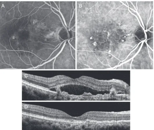
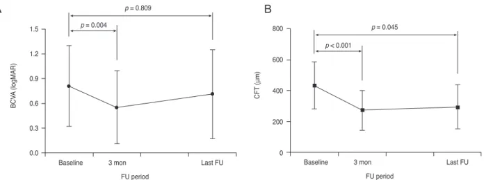
Fig. 1A-1D shows a representative case of an eye treated with intravitreal anti-VEGF therapy. The BCVAs at base- line, 3 months, and the last follow-up were 0.81 ± 0.49 (Snellen equivalent, 20 / 129), 0.55 ± 0.44 (Snellen equiva- lent, 20 / 70), and 0.71 ± 0.54 (Snellen equivalent, 20 / 102), respectively (Fig. 2A). At 3 months, complete resolution of fluid in the fovea was observed in 11 eyes (78.6%). At the last follow-up, an improvement of ≥0.2 logMAR BCVA was observed in seven eyes (50.0%), whereas deterioration by ≥0.2 logMAR BCVA was observed in four eyes (28.6%). The remaining three eyes exhibited stable visual acuity (21.4%). BCVA at 3 months was significantly im- proved compared with that at baseline (p = 0.004). However, BCVA at the last follow-up showed no significant differ- ences from the baseline value (p = 0.809). CFTs at base- line, 3 months, and the last follow-up were 435.3 ± 153.5 µm, 274.2 ± 128.7 µm, and 296.1 ± 145.0 µm, respectively (Fig. 2B). The CFT measurements obtained at the time of diagnosis were significantly different from those obtained at 3 months and the last follow-up (p < 0.001 and p = 0.045, respectively).
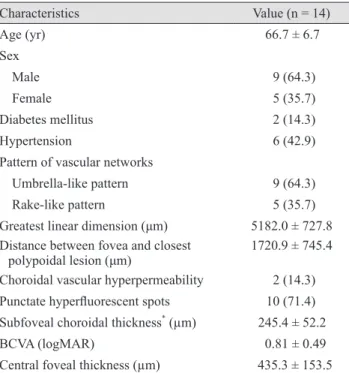
Table 1. Baseline characteristics of the patients
Characteristics Value (n = 14)
Age (yr) 66.7 ± 6.7
Sex
Male 9 (64.3)
Female 5 (35.7)
Diabetes mellitus 2 (14.3)
Hypertension 6 (42.9)
Pattern of vascular networks
Umbrella-like pattern 9 (64.3)
Rake-like pattern 5 (35.7)
Greatest linear dimension (μm) 5182.0 ± 727.8 Distance between fovea and closest
polypoidal lesion (μm) 1720.9 ± 745.4 Choroidal vascular hyperpermeability 2 (14.3) Punctate hyperfluorescent spots 10 (71.4) Subfoveal choroidal thickness* (µm) 245.4 ± 52.2
BCVA (logMAR) 0.81 ± 0.49
Central foveal thickness (µm) 435.3 ± 153.5 Values are presented as mean ± standard deviation or number (%).
BCVA = best-corrected visual acuity; logMAR = logarithm of the minimal angle of resolution.
*Values for 10 eyes with available enhanced-depth imaging opti- cal coherence tomography findings.
Table 2 summarizes the results of analyses to determine the factors associated with BCVA at the last follow-up.
Univariate analysis revealed baseline BCVA and BCVA at 3 months to be significantly associated with the final BCVA, while multivariate analysis revealed BCVA at 3 months to be the most significantly associated factor.
In total, nine eyes exhibited an umbrella-like pattern and five exhibited a rake-like pattern. The greatest linear di- mension, baseline BCVA, and BCVA at the last follow-up were not significantly different between the two groups (p
= 0.699, p = 0.380, and p = 0.596, respectively).
Pathologic ICGA findings for the fellow eyes were avail- able for 11 patients (84.6%). Table 3 shows the findings for the fellow eyes of the included patients. Two eyes (18.2%) were diagnosed with bilateral PCV, and five (45.4%) exhib- ited BVNs only, without polypoidal lesions. Late geo- graphic hyperfluorescence without definite BVNs was ob- served in three eyes (27.3%). During the follow-up period, the development of polypoidal lesions in the fellow eyes
was observed in three of the 12 patients (25.0%) with initially unilateral PCV (Figs. 3A-3F and 4A-4F). Two of these pa- tients exhibited active leakage on fluorescein angiography.
Discussion
In the present study, the short-term outcomes of an- ti-VEGF therapy for PCV with feeder vessels were favor- able. A significant improvement in visual acuity and a marked decrease in CFT were observed after three month- ly ranibizumab injections. However, the visual acuity at a mean of 28.7 months was not significantly different from baseline, even though 50% of the included eyes exhibited an improvement of ≥0.2 logMAR BCVA. The visual acui- ty measured at 3 months was found to be the most reliable predictor of the final visual outcome.
Yuzawa et al. [3] classified PCV into two types: PCV in the narrow sense (PCV type 2) and polypoidal CNV (PCV
Fig. 1. Fluorescein angiography (A), indocyanine green angiography (B), and optical coherence tomography (C,D) findings at time of di- agnosis (A,B,C) and at 57 months after diagnosis (D) in a patient with polypoidal choroidal vasculopathy. The pattern of polypoidal cho- roidal vasculopathy is classified as an umbrella-like pattern. The patient’s visual acuity was 20 / 30 at diagnosis. During the 57-month fol- low-up period, the patient was treated with 10 intravitreal anti-vascular endothelial growth factor injections, including eight ranibizumab injections and two bevacizumab injections. At 57 months, there was no fluid (D) and an improvement in the visual acuity to 20 / 20.
A
C
D
B
type 1) [4]. PCV type 1 was characterized by the presence of feeder vessels and draining venules. PCV type 2 was de- fined as vascular lesions caused by inner choroidal vessel abnormalities, not by CNV. The size of the lesion was larg- er in PCV type 1 than in type 2 [4]. In addition, plaque in the late phase ICGA, which is reported to be a characteris- tic finding of CNV [21], was more frequently noted in PCV type 1 than in PCV type 2 [4]. In OCT images, the highly
reflective line representing Bruch’s membrane was straight in PCV type 1, whereas the line exhibited irregular thick- ness and had highly reflective substances adhering to its lower portion in PCV type 2. One of the notable findings in the previous study was that the choroidal thickness was markedly thinner in PCV type 1 than PCV type 2 [4]. A thick choroid is one of the characteristics of PCV that dis- tinguishes it from exudative age-related macular degenera-
FU period
BCVA (logMAR)
Baseline 3 mon Last FU
1.5 1.2 0.9 0.6 0.3 0.0
p = 0.809 p = 0.004
CFT (μm)
Baseline 3 mon FU period
Last FU 800
600 400 200 0
p = 0.045 p < 0.001
Fig. 2. Changes in best-corrected visual acuity (BCVA, A) and central foveal thickness (CFT, B) in eyes treated with anti-vascular endo- thelial growth factor, according to the follow-up (FU) period. BCVA significantly improved, while CFT significantly decreased after the initial three monthly ranibizumab injections. However, the values at the last follow-up (mean, 29.6 ± 19.6 months) were not significantly different from those at baseline.
A B
Table 2. Results of analyses to determine factors associated with BCVA at the last follow-up
Variable p-value* p-value† 95% CI
Age 0.181 - -
Baseline BCVA 0.001 - -
Baseline central foveal thickness 0.204 - -
BCVA at 3 mon‡ <0.001 <0.001 0.571–1.444
Greatest linear dimension 0.207 - -
Distance between the fovea and the closest polypoidal lesion 0.740 - -
No. of anti-VEGF injections 0.282 - -
Duration of follow-up 0.549 - -
BCVA = best-corrected visual acuity; CI = confidence interval; VEGF = vascular endothelial growth factor.
*Statistics were analyzed by univariate linear regression analysis; †Statistically significant when tested by stepwise multiple linear regres- sion; ‡Values were measured after three monthly ranibizumab injections.
Table 3. Findings for the fellow eye in 11 patients with available indocyanine green angiography findings
Indocyanine green angiography findings No. (%)
Branching vascular networks with polypoidal lesions 2 (18.2)
Branching vascular networks without polypoidal lesions 5 (45.4)
Late geographic hyperfluorescence without definite branching vascular networks 3 (27.3)
tion [22,23]. The associated pathological process was sug- gested to be closely associated with PCV development [24].
For this reason, it is possible that the pathogenesis of the two different subtypes of PCV may differ. Recent genetic stud- ies demonstrated different associations in the genetic vari- ants of the complement factor H gene, age-related maculop- athy susceptibility 2 gene, and elastin between the different subtypes of PCV [6-8]. This result also suggests pathogenic differences between the different subtypes of PCV.
Tan et al. [5] introduced another classification system of the vascular patterns of PCV based on the characteristics of the BVNs and leakage on fluorescein angiography. In
their study, cases showing polyps supplied by interconnect- ing channels were classified as type A PCV, whereas cases showing a distinct BVN were classified as type B (cases without leakage on fluorescein angiography) or type C (cas- es showing significant late leakage on fluorescein angiog- raphy) PCV. The lesion size was smallest for type A and largest for type C. Tan et al. [5] suggested that type A and type B/C PCV may correspond to PCV type 1 and type 2 under the classification system of Yuzawa et al. [3], respec- tively. Kawamura et al. [4] suggested that efficacy of treat- ments may differ depending on the type of PCV. In a study by Honda et al. [9], a significant visual improvement after
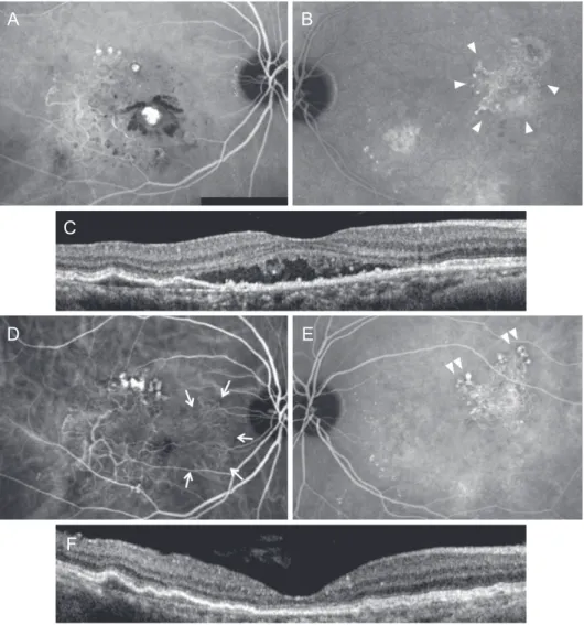
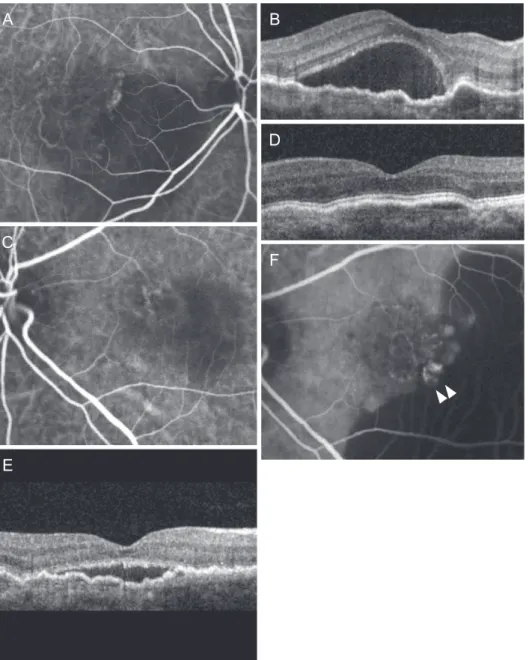
Fig. 3. A representative case of polypoidal choroidal vasculopathy (PCV) with feeder vessels. The pattern of PCV is classified as a rake- like pattern. A the time of diagnosis (A-C), polypoidal lesion (A) and subretinal fluid (C) were noted in the right eye, whereas a branching vascular network and late geographic hyperfluorescence (B, arrowheads) were noted in the left eye. The visual acuity in the right eye was 20 / 60. During the 60-month follow-up period, the right eye was treated with nine intravitreal anti-vascular endothelial growth factor injections, including seven ranibizumab injections and two bevacizumab injections. At 60 months (D-F), there is no fluid in the right eye (F) and an improvement in the visual acuity to 20 / 30. However, marked nasal extension of the PCV lesion was observed (D, arrows). In the left eye, polypoidal lesions developed at the margin of the previously noted late geographic hyperfluorescence (E, double arrowheads).
(A,B,D,E) Indocyanine green angiography, (C,F) optical coherence tomography.
A
C
D
F
E B
photodynamic therapy was noted in PCV type 2, whereas the visual improvement was limited in PCV type 1. In a study by Tan et al. [5], the treatment outcome was also as- sociated with the subtype of PCV; the best treatment out- come was noted in type A, whereas the worst treatment out- come was noted in type C. These results suggest that classifying PCV is important in establishing a treatment plan and understanding the origin of the disorder.
In a previous study reporting the outcomes of ranibizum- ab therapy in PCV, the visual prognosis was not associated with the presence of feeder vessels [25]. However, a recent study demonstrated that the treatment outcomes for PCV with a large lesion size were generally poorer than those for PCV with a small lesion size [26]. Considering that PCV with feeder vessels usually exhibits large lesions [4,5], the treatment outcomes are speculated to be limited. In the
Fig. 4. A representative case of polypoidal choroidal vasculopathy with feeder vessels. At diagnosis (A-D), polypoidal lesions (A) and subretinal fluid (B) were observed in the right eye, whereas the left eye showed type 3 neovascularization without fluid accumulation (C,D). The visual acuity in the right eye was 20 / 100. During the 42-month follow-up period, the right eye was treated with 11 intravit- real anti-vascular endothelial growth factor injections, including eight ranibizumab injections and three bevacizumab injections. At 42 months (E,F), a small amount of subretinal fluid was observed in the right eye (E). The visual acuity in the right eye improved to 20 / 50.
In the left eye, however, polypoidal lesions (F, arrowheads) developed. (A,C,F) Indocyanine green angiography, (B,D,E) optical coherence tomography.
A B
E C
D
F
present study, the short-term treatment outcomes were fa- vorable. Subretinal or intraretinal fluid in the fovea com- pletely disappeared after the initial three monthly ranibi- zumab injections in 11 of 14 patients (78.6%). Although the visual acuity at the last follow-up was not significantly dif- ferent from the baseline value, a definite improvement was observed in 50% of patients. This result suggests that an- ti-VEGF therapy has a valid effect in some proportion of PCV with feeder vessels.
A large lesion size is associated with poor treatment out- come, regardless of the treatment method [5,26]. However, the size of the lesion itself was not closely associated with the long-term visual outcomes in our patients. Instead, the visual acuity at 3 months was found to be the most signifi- cant predictor of the final visual outcome. In particular, the visual acuity at 3 months, and not the baseline visual acui- ty, was more closely associated with the final visual out- come, suggesting that the response to anti-VEGF therapy is important for predicting the long-term visual outcome.
A notable finding in the present study was the relatively high incidence of pathological findings in the fellow eye.
Two patients were initially diagnosed with bilateral PCV.
In these patients, PCV in the fellow eye showed definite feeder vessels. With regard to the remaining nine eyes with available ICGA findings for the fellow eye, 88.9%
showed BVNs, late geographic hyperfluorescence, or both.
The presence of BVNs or late geographic hyperfluores- cence is associated with the development of PCV [27]. In a study by Kim et al. [27], late geographic hyperfluorescence was observed in 23 of 47 eyes (48.9%) with unilateral PCV, six of which also exhibited BVNs. In previous cross-sec- tional studies, bilateral PCV was observed in approximate- ly 10% to 24% of patients from Asian populations [28,29].
In follow-up studies for unilateral PCV, involvement of the fellow eye was observed in 19.1% of patients during a mean follow-up period of 30.3 months [27] and in 11.1% of patients during a 5-year follow-up [30]. The incidence of involvement of the fellow eye in our patients was relatively greater than that reported previously. We postulate that the high incidence of pathological findings in the fellow eye at baseline may have contributed to this finding.
In the present study, the exact symptom duration was ei- ther not determined accurately or was longer than 3 months in eight eyes (57.1%). Although the exact reason is uncertain, previous studies have shown that PCV can de- velop from long-standing Type 1 neovascularization [31]. It
is also reported that some patients with Type 1 neovascular tissue are relatively asymptomatic [32]. We postulate that the growth and expansion of feeder vessels may not induce visual disturbances until the polypoidal lesions finally de- velop. In addition, in the present study, the mean distance between the fovea and polypoidal lesions was approximate- ly one disc diameter. This relatively peripheral location of polyps may also contribute to the delayed recognition of visual disturbances.
Choroidal vascular hyperpermeability and punctate hy- perfluorescent spots are frequently observed in PCV pa- tients. The incidence of choroidal vascular hyperpermeabili- ty and punctate hyperfluorescent spots was reported to be between 9.8% and 43.5% [33-36] and between 72.4% and 80% [35,37], respectively. In the present study, we first evaluated the incidence of these findings in PCVs with feeder vessels. As a result, the incidence of punctate hy- perfluorescent spots (71.4%) was comparable with that re- ported in previous studies, whereas the incidence of cho- roidal vascular hyperpermeability (14.3%) was relatively lower in our patients. The origin of PCV is not fully under- stood. The relatively high incidence of choroidal vascular hyperpermeability and punctate hyperfluorescent spots in both patients with PCV and those with central serous cho- rioretinopathy suggests that these two disorders share a common pathological mechanism [33,37]. A previous study showed that the mean choroidal thickness in patients with PCV accompanied by feeder vessels was only 199 µm, whereas that in patients with PCV without feeder vessels was 288 µm [4]. Although the mean choroidal thickness in our patients with available enhanced-depth imaging OCT results (245.4 µm) was relatively greater than the previous- ly reported value for patients with PCV accompanied by feeder vessels, the thickness was also relatively less than that reported for patients with PCV [22,23].
Kawamura et al. [4] classified the patterns of PCV with feeder vessels as an umbrella-like pattern and a rake-like pattern. In their study, however, a detailed comparison be- tween the two subtypes was not performed. In the present study, the greatest linear dimension and visual acuity were not different between the two subtypes. Because only a small number of eyes were included, however, further stud- ies with a larger study population are necessary to evaluate the clinical significance of this classification.
The present study had several limitations. First, it was a retrospective study with a small sample size. Second, be-
cause enhanced-depth imaging OCT was not routinely per- formed, the choroidal thickness was measured in a limited proportion of patients. Third, there was no common treat- ment protocol. Therefore, differences in treatment decisions among clinicians may have influenced the study results.
In summary, anti-VEGF therapy showed short-term ben- efits for PCV with feeder vessels. Although the long-term efficacy was limited, 50% of the included patients showed a definite improvement in the visual acuity. The visual acuity after three monthly ranibizumab injections was the most reliable predictor of long-term visual outcomes. A relatively high incidence of pathological findings in the fel- low eye and bilateral involvement suggest the need for bi- lateral angiographic and tomographic examinations at the time of diagnosis. Close monitoring of the fellow eye during follow-up is also essential.
Conflict of Interest
No potential conflict of interest relevant to this article was reported.
Acknowledgements
This study was supported by Kim’s Eye Hospital Re- search Center.
References
1. Spaide RF, Yannuzzi LA, Slakter JS, et al. Indocyanine green videoangiography of idiopathic polypoidal choroidal vasculopathy. Retina 1995;15:100-10.
2. Yannuzzi LA, Sorenson J, Spaide RF, Lipson B. Idiopathic polypoidal choroidal vasculopathy (IPCV). Retina 1990;10:1- 8.
3. Yuzawa M, Mori R, Kawamura A. The origins of polypoi- dal choroidal vasculopathy. Br J Ophthalmol 2005;89:602-7.
4. Kawamura A, Yuzawa M, Mori R, et al. Indocyanine green angiographic and optical coherence tomographic findings support classification of polypoidal choroidal vasculopathy into two types. Acta Ophthalmol 2013;91:e474-81.
5. Tan CS, Ngo WK, Lim LW, Lim TH. A novel classification of the vascular patterns of polypoidal choroidal vasculopa-
thy and its relation to clinical outcomes. Br J Ophthalmol 2014;98:1528-33.
6. Miki A, Honda S, Kondo N, Negi A. The association of age-related maculopathy susceptibility 2 (ARMS2) and com- plement factor H (CFH) variants with two angiographic subtypes of polypoidal choroidal vasculopathy. Ophthalmic Genet 2013;34:146-50.
7. Tanaka K, Nakayama T, Mori R, et al. Associations of complement factor H (CFH) and age-related maculopathy susceptibility 2 (ARMS2) genotypes with subtypes of pol- ypoidal choroidal vasculopathy. Invest Ophthalmol Vis Sci 2011;52:7441-4.
8. Yanagisawa S, Sakurada Y, Miki A, et al. The association of elastin gene variants with two angiographic subtypes of pol- ypoidal choroidal vasculopathy. PLoS One 2015;10:e0120643.
9. Honda S, Miki A, Yanagisawa S, et al. Comparison of the outcomes of photodynamic therapy between two angio- graphic subtypes of polypoidal choroidal vasculopathy.
Ophthalmologica 2014;232:92-6.
10. Rosenfeld PJ, Brown DM, Heier JS, et al. Ranibizumab for neovascular age-related macular degeneration. N Engl J Med 2006;355:1419-31.
11. Cho M, Barbazetto IA, Freund KB. Refractory neovascular age-related macular degeneration secondary to polypoidal choroidal vasculopathy. Am J Ophthalmol 2009;148:70-8.e1.
12. Oishi A, Kojima H, Mandai M, et al. Comparison of the ef- fect of ranibizumab and verteporfin for polypoidal choroi- dal vasculopathy: 12-month LAPTOP study results. Am J Ophthalmol 2013;156:644-51.
13. Lim JY, Lee SY, Kim JG, et al. Intravitreal bevacizumab alone versus in combination with photodynamic therapy for the treatment of neovascular maculopathy in patients aged 50 years or older: 1-year results of a prospective clinical study. Acta Ophthalmol 2012;90:61-7.
14. Kim JH, Lee DW, Choi SC, et al. Intravitreal anti-vascular endothelial growth factor for newly diagnosed symptomat- ic polypoidal choroidal vasculopathy with extrafoveal pol- yps. Korean J Ophthalmol 2015;29:404-10.
15. Lee SY, Kim JG, Joe SG, et al. The therapeutic effects of bevacizumab in patients with polypoidal choroidal vascu- lopathy. Korean J Ophthalmol 2008;22:92-9.
16. Hikichi T, Higuchi M, Matsushita T, et al. Factors predic- tive of outcomes 1 year after 3 monthly ranibizumab injec- tions and as-needed reinjections for polypoidal choroidal vasculopathy in Japanese patients. Retina 2013;33:1949-58.
17. Spaide RF, Koizumi H, Pozzoni MC. Enhanced depth im-
aging spectral-domain optical coherence tomography. Am J Ophthalmol 2008;146:496-500.
18. Guyer DR, Puliafito CA, Mones JM, et al. Digital indocy- anine-green angiography in chorioretinal disorders. Oph- thalmology 1992;99:287-91.
19. Tsujikawa A, Ojima Y, Yamashiro K, et al. Punctate hyper- fluorescent spots associated with central serous chorioreti- nopathy as seen on indocyanine green angiography. Retina 2010;30:801-9.
20. Kang SW, Chung SE, Shin WJ, Lee JH. Polypoidal choroidal vasculopathy and late geographic hyperfluorescence on in- docyanine green angiography. Br J Ophthalmol 2009;93:759- 64.
21. Guyer DR, Yannuzzi LA, Slakter JS, et al. Classification of choroidal neovascularization by digital indocyanine green videoangiography. Ophthalmology 1996;103:2054-60.
22. Kim SW, Oh J, Kwon SS, et al. Comparison of choroidal thickness among patients with healthy eyes, early age-re- lated maculopathy, neovascular age-related macular degen- eration, central serous chorioretinopathy, and polypoidal choroidal vasculopathy. Retina 2011;31:1904-11.
23. Koizumi H, Yamagishi T, Yamazaki T, et al. Subfoveal choroidal thickness in typical age-related macular degener- ation and polypoidal choroidal vasculopathy. Graefes Arch Clin Exp Ophthalmol 2011;249:1123-8.
24. Pang CE, Freund KB. Pachychoroid neovasculopathy. Retina 2015;35:1-9.
25. Hikichi T, Higuchi M, Matsushita T, et al. One-year results of three monthly ranibizumab injections and as-needed re- injections for polypoidal choroidal vasculopathy in Japa- nese patients. Am J Ophthalmol 2012;154:117-24.e1.
26. Tsujikawa A, Ojima Y, Yamashiro K, et al. Association of lesion size and visual prognosis to polypoidal choroidal vasculopathy. Am J Ophthalmol 2011;151:961-72.e1.
27. Kim YT, Kang SW, Chung SE, et al. Development of pol- ypoidal choroidal vasculopathy in unaffected fellow eyes.
Br J Ophthalmol 2012;96:1217-21.
28. Sho K, Takahashi K, Yamada H, et al. Polypoidal choroidal vasculopathy: incidence, demographic features, and clini- cal characteristics. Arch Ophthalmol 2003;121:1392-6.
29. Byeon SH, Lee SC, Oh HS, et al. Incidence and clinical patterns of polypoidal choroidal vasculopathy in Korean patients. Jpn J Ophthalmol 2008;52:57-62.
30. Ueta T, Iriyama A, Francis J, et al. Development of typical age-related macular degeneration and polypoidal choroidal vasculopathy in fellow eyes of Japanese patients with exu- dative age-related macular degeneration. Am J Ophthalmol 2008;146:96-101.
31. Khan S, Engelbert M, Imamura Y, Freund KB. Polypoidal choroidal vasculopathy: simultaneous indocyanine green angiography and eye-tracked spectral domain optical co- herence tomography findings. Retina 2012;32:1057-68.
32. Fung AT, Yannuzzi LA, Freund KB. Type 1 (sub-retinal pigment epithelial) neovascularization in central serous chorioretinopathy masquerading as neovascular age-related macular degeneration. Retina 2012;32:1829-37.
33. Sasahara M, Tsujikawa A, Musashi K, et al. Polypoidal choroidal vasculopathy with choroidal vascular hyperper- meability. Am J Ophthalmol 2006;142:601-7.
34. Koizumi H, Yamagishi T, Yamazaki T, Kinoshita S. Rela- tionship between clinical characteristics of polypoidal cho- roidal vasculopathy and choroidal vascular hyperperme- ability. Am J Ophthalmol 2013;155:305-13.e1.
35. Kim JH, Chang YS, Lee TG, Kim CG. Choroidal vascular hyperpermeability and punctate hyperfluorescent spot in choroidal neovascularization. Invest Ophthalmol Vis Sci 2015;56:1909-15.
36. Hikichi T, Kitamei H, Shioya S. Prognostic factors of 2-year outcomes of ranibizumab therapy for polypoidal choroidal vasculopathy. Br J Ophthalmol 2015;99:817-22.
37. Park SJ, Kim BH, Park KH, Woo SJ. Punctate hyperfluo- rescence spot as a common choroidopathy of central serous chorioretinopathy and polypoidal choroidal vasculopathy.
Am J Ophthalmol 2014;158:1155-63.e1.