서론
애플리버셉트(Aflibercept, EYLEA®; Regeneron Pharmaceuticals, Inc., Tarrytown, NY, USA)는 결절맥락막혈관병증(polypoidal choroidal vasculopathy, PCV)과 습성 나이관련황반변성 등의 질 환에서 널리 사용되고 있는 항혈관내피성장인자(anti-vascular endothelial growth factor, anti-VEGF)로 비교적 안전하게 사용
할 수 있는 치료 방법으로 알려져 있으나, 매우 드물게 유리체 내주사 후 전층황반원공이 발생한 경우가 보고된 바 있다[1-3].
특발성 황반원공의 자연 폐쇄에 대한 연구에 의하면 외상성 황반원공을 제외한 2% 정도에서 수술 치료 없이 황반원공의 폐쇄를 보인다는 보고들이 있으나 유리체내주사를 받은 눈에 서 황반원공의 발생은 그 빈도가 매우 낮으며, 발생된 황반원 공이 자연적으로 폐쇄되는 경우는 극히 드문 현상으로 현재까
결절맥락막혈관병증 환자에서 애플리버셉트 유리체내주사 후 전층황반원공의 자연발생 및 폐쇄
Spontaneous Formation and Closure of Full-thickness Macular Hole after Intravitreal Injection of Aflibercept in Patient with Polypoidal Choroidal Vasculopathy
이강민, 배건호 Kang Min Lee, Kunho Bae
동국대학교 일산병원 안과
Department of Ophthalmology, Dongguk University Ilsan Hospital, Goyang, Korea
Purpose: To report a case of spontaneous closure of full-thickness macular hole (FTMH) secondary to intravitreal aflibercept injection in a patient with polypoidal choroidal vasculopathy (PCV).
Case summary: A 64-year-old female presented with decreased visual acuity in her left eye, which had a best-corrected visual acuity (BCVA) of 0.25. PCV with a massive submacular hemorrhage was observed in the left eye. Six weeks after initial intravitreal aflibercept treatment, FTMH developed in the same eye and BCVA decreased to 0.10. Intravitreal bevacizumab treatment was administered. After 2 weeks, BCVA improved to 0.30 and OCT showed spontaneous closure of the FTMH. The patient was treated with intravitreal C3F8 gas tamponade and no recurrence of FTMH was observed.
Conclusions: FTMH may occur after intravitreal aflibercept injection during PCV treatment, and physicians should consider careful fol- low-up rather than immediate surgical treatment.
Keywords: Aflibercept; Macular hole; Polypoidal choroidal vasculopathy; Spontaneous closure
Address reprint requests to Kunho Bae, MD
Department of Ophthalmology, Dongguk University Ilsan Hospital, #27 Dongguk-ro, Goyang 10326, Korea Tel: 82-31-961-7393, Fax: 82-31-961-7393
E-mail: [email protected]
* This paper was presented at the Korean Ophthalmology Society Meeting 2018, Seoul, Korea.
Received: 2020. 1. 13 Revised: 2020. 3. 10 Accepted: 2020. 4. 6
지 국내 보고는 없는 상황이다[4]. 저자들은 결절맥락막혈관병 증에서 애플리버셉트 유리체내주사 후 전층황반원공이 발생하 고 이어서 자연 폐쇄된 증례를 경험하여 이를 문헌고찰과 함께 보고하고자 한다.
증례보고
64세 여자 환자가 좌안 시력저하를 주소로 내원하였다. 과거력 에서 특이 소견은 없었고, 최대교정시력은 우안 0.8, 좌안 0.25 였다. 세극등현미경검사에서도 특이 소견은 보이지 않았으나 안 저검사 및 빛간섭단층촬영에서 좌안 망막하출혈 및 망막색소 상피 융기 소견이 관찰되었다(Fig. 1A, B). 형광안저혈관조영술 및 인도시아닌그린혈관조영술에서 좌안에 비정상적 분지혈관망
과 확장된 형태의 원형 과형광 병변이 관찰되어 결절맥락막혈 관병증으로 진단하고 애플리버셉트 유리체내주사를 시행하였 다(Fig. 1C, D). 환자는 6주 후 내원하였는데, 좌안의 최대교정 시력은 0.1로 저하되었으며 빛간섭단층촬영검사에서 전층황반 원공이 관찰되었다(Fig. 2A). 이에 좌안 유리체절제술을 계획하 였으나 환자분이 개인적인 사유로 2주 뒤에 수술받기를 원하였 다. 그 사이 결절맥락막혈관병증으로 인한 황반부의 추가적인 손상을 막기 위해 베바시주맙 유리체내주사 치료하였다. 주사 2주 후 좌안 최대교정시력은 0.3으로 호전되었으며, 빛간섭단 층촬영에서 전층황반원공이 자연 폐쇄된 것을 확인하고 추가 적으로 유리체내 과불화프로판(C3F8) 가스 주입술을 시행하였 다(Fig. 2B). 주입술 3일 후 안저검사 및 빛간섭단층촬영검사에 서 황반하출혈 및 망막하액이 현저하게 감소한 것이 확인되었 고 황반원공의 재발도 관찰되지 않았다(Fig. 2C, D). 이후 망막
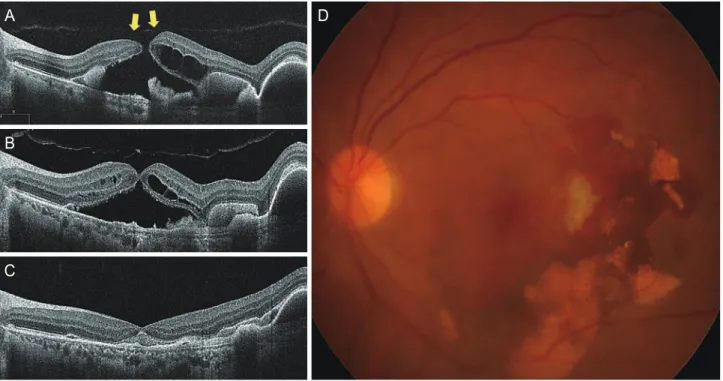
Figure 1. Fundus color photograph (A), spectral domain-optical coherence tomography (B), fluorescein (C) and indocyanine green (D) angi- ography of a 64-year-old female with unilateral polypoidal choroidal vasculopathy. (A, B) A massive submacular hemorrhage is accompanied by retinal pigment epithelial detachment. Note the incomplete posterior vitreous detachment in the parafoveal area (yellow arrows). (C, D) A hyperfluorescent polypoidal lesion and branching vascular network with leakage are visualized by fluorescein and indocyanine green angi- ography.
A
C
B
D
은 안정된 상태로 애플리버셉트 유리체내주사 치료 시행하며 경과 관찰 중이다.
고찰
황반원공의 경우 대개 수술적인 치료가 필요하나 드물게 자연 폐쇄를 보이는 증례들이 보고된 바 있다[4-6]. 2007년 빛간섭단 층촬영을 이용한 연구에서 2% 정도에서 황반원공의 자연 폐쇄 가 일어나는 것으로 나타났으며, 국내에서도 수술 치료 없이 황 반원공의 자연 폐쇄가 드물지만 보고된 바 있다[7,8]. 그러나 본 증례의 경우처럼 결절맥락막혈관병증에서 유리체내주사 치료 후 전층황반원공이 발생하고 이어서 자연 폐쇄된 증례는 아직 국내에 보고된 바 없다.
황반원공의 발생이 결절맥락막혈관병증으로 인한 직접적인 합병증은 아니지만, 동반된 망막하출혈 혹은 항혈관내피성장 인자항체와의 상호작용이 황반원공의 발생을 야기할 수 있는 것으로 알려져 있다[6,9]. 결절맥락맥혈관병증은 분지혈관망과 활동성 결절로 특징지을 수 있는데, 활동성 결절은 반복적인 출 혈과 삼출성 변화를 유발하고 때로는 이로 인해 대량의 출혈이 유발되기도 한다. Baskaran and Pan [9]은 황반하출혈의 급격한
발생이 얇아진 중심와를 파열시켜 황반원공을 일으킬 수 있다 고 보고하였으며, 황반원공 기저에 존재하는 출혈, 잔존 망막 조직의 가교 및 유리체출혈(breakthrough vitreous hemorrhage) 을 그 근거로 제시하였다. 또한, 이전의 연구들을 통해 습성 나 이관련황반변성에 대한 항혈관내피성장인자 유리체내주사 후 황반원공이 발생하는 기전에 대한 여러 가설이 제시되었는데, 후유리체막 혹은 망막에 가해진 견인력이나 맥락막신생혈관의 활성도 감소로 인한 망막색소상피의 수축과 전단력이 그 상부 의 망막에 영향을 주어 황반원공의 발생에 영향을 주었을 것 으로 생각된다[10-12]. 추가적으로, 유리체절제술 후 전층황반 원공의 발생 기전으로 낭포황반부종이 제시되고 있는데, 낭포 병변이 주변 망막조직의 장력을 증가시켜 내층망막 및 외층망 막의 파열을 유발함으로써 황반원공의 발생을 야기할 수 있다 고 알려져 있다[13].
본 증례의 경우 결절맥락막혈관병증에 동반된 대량의 황반하 출혈이 발생하면서 얇아진 중심와에 일차적으로 손상이 유발 되었을 가능성이 있고, 이어진 유리체내주사가 결절성 맥락막혈 관의 수축을 일으켜 후유리체박리가 유발되며 황반부에 견인력 이 가해졌을 것으로 생각된다. 또한, 초진시에는 황반부에 낭포 병변이 관찰되지 않으나 애플리버셉트 유리체내주사 6주 뒤에 는 중심와주변에 낭포병변들이 관찰되며, 베바시주맙 유리체내
Figure 2. (A) A full-thickness macular hole is observed six weeks after initial intravitreal aflibercept treatment. Note the incomplete posterior vitreous detachment (PVD) around the hole (yellow arrows). (B) Two weeks after additional intravitreal bevacizumab treatment, spontaneous closure of the macular hole is observed on optical coherence tomography. (C, D) Submacular hemorrhage and subretinal fluid were significantly decreased three days after C3F8 gas tamponade. Complete PVD was observed and the closed macular hole remained stable.
A
B
C
D
주사 시행 후에는 낭포병변이 감소하고 황반원공의 내벽이 서로 접촉하는 것으로 보아 낭포황반부종이 황반원공의 발생 및 폐 쇄에 관여하였을 가능성이 있다(Fig. 2). 그러나 후유리체박리가 완전히 유발되고, 추가적인 유리체내주사 후에 중심와가 정상적 인 구조를 회복하며, 남아있는 망막하출혈 내의 플라즈미노겐, 섬유아세포 또는 조직인자와 같은 조직 재생 및 응고에 관여하 는 인자들이 황반원공의 자연 폐쇄에 관여했을 가능성이 있다.
결절맥락막혈관병증에서 애플리버셉트 유리체내주사 후 전층 황반원공이 발생하고 이어서 자연 폐쇄된 증례를 경험하였다.
유리체내주사 후 전층황반원공이 발생할 가능성이 있음을 염 두에 두어야 하며, 특히 후유리체박리가 아직 일어나지 않은 경 우에는 위험성이 더 크므로 환자에게 충분한 설명과 교육이 뒷 받침되어야 하겠다. 또한, 결절맥락막혈관병증 환자에서 애플리 버셉트 유리체내주사 후 전층황반원공이 발생하는 경우 자연적 인 폐쇄를 보일 수 있어 즉각적인 수술보다는 단기간의 주의 깊 은 경과 관찰을 고려해볼 수 있겠다.
Conflicts of Interest
The authors declare no conflicts of interest relevant to this article.
References
1. Heier JS, Brown DM, Chong V, et al. Intravitreal aflibercept (VEGF trap-eye) in wet age-related macular degeneration. Ophthal- mology 2012;119:2537-48.
2. Lee G, Lee S. Full-thickness macular hole after intravitreal afliber- cept injection in a patient with wet age-related macular degen- eration. J Korean Ophthalmol Soc 2017;58:875-8.
3. Oshima Y, Apte RS, Nakao S, et al. Full thickness macular hole case after intravitreal aflibercept treatment. BMC Ophthalmol 2015;15:30.
4. Privat E, Tadayoni R, Gaucher D, et al. Residual defect in the fo- veal photoreceptor layer detected by optical coherence tomog- raphy in eyes with spontaneously closed macular holes. Am J Ophthalmol 2007;143:814-9.
5. Lo WR, Hubbard GB. Macular hole formation, spontaneous closure, and recurrence in a previously vitrectomized eye. Am J Ophthalmol 2006;141:962-4.
6. Sethia A, Sheth J, Gopalakrishnan M, Anantharaman G. Sponta- neous formation and closure of full thickness macular hole after treatment with anti-vascular endothelial growth factor therapy in polypoidal choroidal vasculopathy. Indian J Ophthalmol 2019;67:1756-8.
7. Lee S, Lee G, Yoon S, Kim YY. A case of spontaneous closure of macular hole in a previously vitrectomized eye. J Korean Oph- thalmol Soc 2013;54:1626-9.
8. Kim M, Kim D, Kim YJ, et al. A case of spontaneous closure of macular hole in infectious posterior uveitis involving the fovea. J Korean Ophthalmol Soc 2016;57:155-60.
9. Baskaran P, Pan U. Macular hole secondary to polypoidal choroi- dal vasculopathy. Middle East Afr J Ophthalmol 2017;24:159-61.
10. Carvounis PE, Kopel AC, Benz MS. Retinal pigment epithelium tears following ranibizumab for exudative age-related macular degeneration. Am J Ophthalmol 2007;143:504-5.
11. Raiji VR, Eliott D, Sadda SR. Macular hole overlying pigment epithelial detachment after intravitreal injection with ranibizu- mab. Retin Cases Brief Rep 2013;7:91-4.
12. Querques G, Souied EH, Soubrane G. Macular hole following intravitreal ranibizumab injection for choroidal neovascular membrane caused by age-related macular degeneration. Acta Ophthalmol 2009;87:235-7.
13. Zhang W, Grewal DS, Jaffe GJ, et al. Spontaneous closure of full-thickness macular hole with epiretinal membrane in vitrec- tomized eyes: case series and review of literature. Ophthalmic Surg Lasers Imaging Retina 2017;48:183-90.
결절맥락막혈관병증 환자에서 애플리버셉트 유리체내주사 후 전층황반원공 의 자연발생 및 폐쇄
목적: 결절맥락막혈관병증 환자에서 애플리버셉트 유리체내주사 후 발생한 전층황반원공이 자연 폐쇄된 증례를 경험하여 이를 보고하 고자 한다.
증례 요약: 64세 여자 환자가 갑작스러운 좌안 시력저하를 주소로 내원하였다. 좌안의 최대교정시력은 0.25였다. 인도시아닌그린혈관 조영술에서 비정상적 분지혈관망과 폴립이 관찰되어 결절맥락막혈관병증으로 진단하고 애플리버셉트 유리체내주사를 시행하였다. 주사 6주 후 최대교정시력이 0.1로 저하되었으며, 빛간섭단층촬영검사에서 전층황반원공이 발생한 것을 확인하였다. 유리체절제술 계획하 에 이에 앞서 베바시주맙 유리체내주사 치료하였다. 주사 2주 후 좌안 최대교정시력은 0.3으로 호전되었으며, 전층황반원공이 자연 폐 쇄된 것을 확인하였고 추가적으로 유리체내 과불화프로판(C3F8) 가스 주입술을 시행하였다. 주입술 후 1개월째 좌안 최대교정시력은 0.3이었고 황반원공의 재발도 관찰되지 않았다.
결론: 결절맥락막혈관병증 환자에서 애플리버셉트를 포함한 항혈관내피성장인자의 유리체내주사는 황반원공을 유발할 수 있어 주의를 요하며, 전층황반원공이 발생하는 경우 비수술적인 치료로 폐쇄를 보일 수 있어 즉각적인 수술보다는 경과 관찰을 고려해볼 수 있겠다.
국문초록