Received: September 6, 2007 Accepted: April 10, 2008
Reprint requests to Young Hee Yoon, MD, PhD, Department of Ophthalmology, University of Ulsan, Asan Medical Center, 388‐1, Poongnap‐dong, Songpa‐gu, Seoul 138‐040, Korea. Fax: 82‐02‐470‐
6440, Tel: 82‐02‐3010‐3675, E‐mail: [email protected]
* This study was partially supported by Asan Institute for Life &
Science Grant # 07‐084 Competing interests: We declare that we have no conflicting financial interests.
The Therapeutic Effects of Bevacizumab in Patients with Polypoidal Choroidal Vasculopathy
Sun Young Lee, MD, June‐Gone Kim, MD, Soo Geun Joe, MD, Hyewon Chung, MD, Young Hee Yoon, MD
Department of Ophthalmology, Asan Medical Center, University of Ulsan, College of Medicine, Seoul, Korea
Purpose: To evaluate the efficacy and safety of intravitreal bevacizumab for polypoidal choroidal vasculopathy (PCV).
Methods: In this retrospective interventional pilot study, 12 eyes of 11 patients with active PCV were treated with intravitreal bevacizumab (1.25 mg) alone or in combination with photodynamic therapy (PDT) depending on the informed patient’s choice. Intravitreal bevacizumab was repeated at 6‐week intervals until the regression of active lesion was detected on fluorescein angiography (FA) which was done on a regular basis, Indocyanine green angiography (ICGA) and optical coherence tomography (OCT) analyses.
Results: Intravitreal bevacizumab was given alone in 8 eyes (Group 1) and in combination with PDT in 4 eyes (Group 2). Mean follow‐up duration was 17 weeks in group 1 and 15 weeks in group 2 after bevacizumab treatment. The mean number of bevacizumab injections was 2.2 in group 1 and 2.5 in group 2. Mean BCVA improved from 20/63 to 20/40 in group 1 and 20/63 to 20/32 in group 2. Of all eyes, the BCVA improved by ≥2 lines in seven (58%) eyes and resolution of fluid and hemorrhages in clinical examination, an absence of leakage on repeat FAs, or resolved pigment epithelial detachment (PED) and/or subretinal fluid (SRF) on OCT exam was confirmed in 10 (83%) eyes. Partial or complete regression of the polypoidal vessels and interconnecting vessels was reported for most cases at the last follow‐up. No significant ocular or systemic side effects were observed in both groups.
Conclusions: Short‐term results indicate that intravitreal bevacizumab (1.25 mg) alone or in combination with PDT is well tolerated and associated with improvement in BCVA and reduced angiographic leakage in most patients. Further evaluation of intravitreal bevacizumab therapy for the treatment of PCV is warranted.
Korean Journal of Ophthalmology 22(2):92-99, 2008
Key Words: Intravitreal bevacizumab, Polypoidal choroidal vasculopathy, Photodynamic therapy
Polypoidal choroidal vasculopathy (PCV) is a choroidal vascular disease characterized by an inner choroidal vascular network ending in an aneurysmal bulge or outward projection visible clinically as a reddish orange, spheroid, polyp‐like structure.
1‐3PCV can remain clinically asymptomatic in its quiescent form, with nonleaking asymptomatic polyps. Occasionally, PCV causes insidious visual loss owing to serosanguinous detachment of the retinal pigment epithelium and neurosensory retina affecting the macula, or causes acute and
severe visual loss secondary to massive submacular or vitreous hemorrhage due to spontaneously ruptured vessels.
4,5Treatment for PCV is not yet well established. Asymptomatic PCV is recommended for observation and the polyps may resolve spontaneously over time.
5,6Although various treatment modalities for PCV with exudative and hemorrhagic complications such as direct thermal laser photocoagulation, tissue plasminogen activator (t‐PA) injection with gas displacement, submacular surgery, and macular translocation surgery have been proposed, the beneficial effects are still in doubt owing to recurrence or poor long‐term results.
7‐11Recently, photodynamic therapy (PDT) has been proposed as a standard treatment modality with its favorable outcome, nevertheless its application has been found to be limited owing to difficulty in treating all wide spread multiple polyps and the possibility of subsequent massive submacular hemorrhage.
12‐14Favorable results have been reported with intravitreal
injection of bevacizumab (Avastin
®, Genentech, Inc. South
San Francisco, CA) to treat choroidal neovascularization.
15‐18Table 1. Clinical Data and Treatment Results in Patients with Polypoidal Choroidal Vasculopathy Treated by Intravitreal Bevacizumab (Group 1)
PatientNo./
Gender/
Age (years) Location Prior
Tx Treatment* BCVA
Pre** BCVA Final
Change in lines
Duration of F/U
†(week)
Clinical and angiographic features at last F/U
OCT features at last F/U
Additional event 1/M/66 Subfoveal P(‐36) A(0),A(6),A(12) 20/100 20/160 ‐2 17 Dry and
quiescent Resolved PED 2/M/80 Juxtafoveal A(0),A(6),A(12) 20/50 20/32 +2 13 Dry and
quiescent Reduced PED and SRF 3/M/63 Subfoveal A(0),A(6),A(12) 20/63 20/32 +3 17 Dry and
quiescent Resolved SRF 4/M/63 Subfoveal P(‐6),
T/G A(0),A(6) 20/200 20/320 ‐ 2 12 Dry and
scarring Resolved PED and RPE scarring
Massive subretinal hemorrhage after PDT
5/M/57 Extrafoveal A(0) 20/25 20/25 0 22 Dry and
quiescent Resolved PED
6/M/58 Juxtafoveal A(0),A(6) 20/100 20/32 +5 27 Dry and
quiescent Resolved PED and SRF
7/M/58 Extrafoveal A(0) 20/40 20/40 0 20 Dry and
quiescent Resolved SRF 8/M/62 Subfoveal P(‐16) A(0),A(6),A(12) 20/50 20/32 +2 13 Dry and
quiescent Resolved PED
M=male; F=female; Tx=treatment; F/U=follow‐up; P=photodynamic therapy, PDT; T/G=t‐PA with gas injection; L=focal laser photocoagulation, A=intravitreal bevacizumab; BCVA=best corrected visual acuity; PED=pigment epithelial detachment;
SRF=subretinal fluid;RPE=retinal pigment epithelium.
* Treatment (time, weeks); ** BCVA before intravitreal bevacizumab;
†follow‐up duration after intravitreal bevacizumab injection;
‡Localized bleeding at 6 weeks after PDT plus intravitreal bevacizumab.
Although the pathogenesis of PCV is still not fully understood, it has been suggested that vascular endothelial growth factor (VEGF) may have a similar role in PCV as it does in choroidal neovascularization (CNV) owing to marked increases in VEGF concentration in aqueous humor and histologic examination in active PCV eyes.
19,20The aim of the present study was to determine the efficacy and safety of intravitreal bevacizumab, alone or in combination with PDT, for the treatment of PCV.
Materials and Methods
The retrospective interventional case series study included medical records of 12 eyes of 11 patients with symptomatic PCV who were either newly diagnosed or failed in previous treatment and treated PCV at Asan Medical Center, Seoul, Korea, from January 2006 to October 2006. The study was approved by the Institutional Review Board at the Asan Medical Center and informed consent was obtained from all patients. Patients with new or recurrent subretinal pigment epithelial orange‐red vascular lesions associated with exudative changes were included. To confirm the diagnosis of symptomatic PCV, all patients underwent fluorescein angiography (FA), indocyanine green angiography (ICGA), and optical coherence tomography (OCT) analyses. All patients also underwent a comprehensive ocular examination, including best‐corrected visual acuity (BCVA), slit‐lamp biomicroscopy with intraocular pressure measurement and
indirect ophthalmoscopy.
Patients received either an intravitreal injection of bevacizumab alone (Group 1) or an intravitreal injection of bevacizumab combined with PDT on the same day (Group 2) according to informed patient’s choice. For intravitreal injection, 1.25 mg of bevacizumab was given using a 30‐
gauage needle after topical anesthesia. For combination therapy, PDT was performed to cover the entire lesion of symptomatic PCV lesions in a standard manner and the 1.25 mg intravitreal bevacizumab injection was then administered.
Follow‐up evaluations were made at 1 week, 6 weeks and then every 2–3 months after treatment. BCVA, funduscopic exam, and OCT tests were performed at every visit, and FA/ICGA was assessed at the ~3 month visit or earlier if necessary. FA/ICGA findings were reviewed to confirm the regression of active PCV, and visual acuity and tomographic findings were recorded to assess the efficacy of the treatment.
Side effects related to the procedure were also evaluated.
Results
The patient characteristics and clinical data of group 1 and
group 2 are listed in Table 1 and Table 2. The 11 patients
(ten men and one woman) were aged 45–80 years (mean
63±7 years in group 1 and 53±7 years in group 2). Cases
1 and 8 in group 1 and case 9 in group 2 received prior PDT
(9, 4, and 22 months before treatment of bevacizumab,
Table 2. Clinical Data and Treatment Results in Patients with Polypoidal Choroidal Vasculopathy Treated by Intravitreal Bevacizumab combined with photodynamic therapy (Group 2)
PatientNo./
Gender/
Age (years)
Location Prior
Tx Treatment* BCVA
Pre** BCVA Final Change
in lines Duration of F/U
†(week)
Clinical and angiographic features at last F/U
OCT features at last F/U
Additional event 9/F/51 Subfoveal P(‐88) P+A(0),A(6),A(12) 20/250 20/250 0 13 Persistent
serous PED Persistent
serous PED Localized bleeding(6)
‡10/M/57 Subfoveal P+A(0),A(6),A(12) 20/63 20/32 +3 13 Persistent
serous PED Persistent serous PED 11/M/45 Extrafoveal T/G P+A(0),L(4) 20/100 20/25 +6 22 Dry and
quiescent Resolved PED and SRF 12/M/62 Subfoveal P+A(0),A(6),A(12) 20/32 20/20 +2 13 Quiescent Resolved PED
M=male; F=female; Tx=treatment; F/U=follow‐up; P=photodynamic therapy, PDT; T/G=t‐PA with gas injection;
A=intravitreal bevacizumab; BCVA=best corrected visual acuity; PED=pigment epithelial detachment; SRF=subretinal fluid;RPE=retinal pigment epithelium.
* Treatment (time, weeks); ** BCVA before intravitreal bevacizumab;
†follow‐up duration after intravitreal bevacizumab injection;
‡Localized bleeding at 6 weeks after PDT plus intravitreal bevacizumab.
respectively). Case 11 in group 2 had received prior gas tamponade with t‐PA due to massive submacular hemorrhage at presentation. Case 4 had previously received gas tamponade with t‐PA due to massive submacular hemorrhage followed by prior PDT. All patients who had received prior treatment switched treatment to intravitreal injection of bevacizumab with/without combined PDT owing to persistent or recurrent PCV. All patients except cases 5, 7 and 11 received repeated intravitreal bevacizumab at 6 week intervals owing to persistent subretinal fluid, as shown by OCT analysis, or leakage, as shown by FA/ICGA analysis.
Intravitreal bevacizumab was well tolerated in all patients.
There were no procedure‐related complications in any of the patients.
Mean follow‐up duration was 17 weeks (range, 12-27) in group 1 and 15 weeks (range, 13-22) in group 2. The mean number of intravitreal bevacizumab injections was 2.2 in group 1 and 2.5 in group 2. Mean BCVA improved in both groups from 20/63 to 20/40 in group 1 and 20/63 to 20/32 in group 2. Of all eyes, BCVA improved by ≥2 lines in seven (58%) eyes, and there was a moderate gain in vision in four (33%) of these eyes (BCVA improved by ≥3 lines).
Three (25%) eyes had stable vision, with BCVA at the last follow‐up being within 1 line of the initial BCVA level.
Resolution of fluid and hemorrhages in clinical examination, and absence of leakage on repeat FAs or resolved pigment epithelial detachment (PED) and/or subretinal fluid (SRF) on OCT examinations could be seen in 10 (83%) eyes.
Regression of the polypoidal vessels and interconnecting vessels were seen in most cases at the last follow‐up.
Case Report (Cases 3 and 4)
A 63‐year‐old man was referred to our clinic with a 5 month history of reduced visual acuity in the right eye. At the first visit, his visual acuity was 20/63 in the right eye and 20/20 in the left eye. Based on the findings in FA and
ICGA, he was diagnosed as having PCV in his right eye and was treated with PDT covering whole active lesions. Four weeks after PDT, funduscopic examination of the right eye revealed extensive subretinal hemorrhage with a reduced visual acuity of 20/320. The right eye with massive subretinal hemorrhage was treated with intravitreal t‐PA (50 µg) and 0.3 ml of 100% C
3F
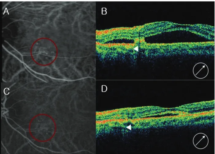
8. Postoperative prone positioning was maintained for 72 hours. Two weeks after administration of t‐PA injection and gas, funduscopic examination of the right eye showed dispersed and reduced subretinal hemorrhage (Fig. 1). At one month follow up, he complained a decreased vision to 20/50 in his untreated, left eye. The OCT examination demonstrated subfoveal serous elevation and FA/ICGA confirmed new active PCV in his left eye (Fig. 2).
Both eyes were treated with an intravitreal injection of 1.25 mg bevacizumab, two injections in the right eye and three injections in the left eye, at 6 week intervals. The final visual acuity was 20/320 in the right eye with fibrovascular scarring and 20/32 in the left eye with minimal subfoveal serous elevation.
D iscussion
Polypoidal choroidal vasculopathy (PCV) was first described by Yannuzzi et al.
1and is characterized by an abnormal vascular network of choroidal vessels with polyp‐
like dilations at the terminals of the branches. It is unclear
whether PCV represents abnormal vessels from the choroidal
circulation or is a variant of CNV. However, it is generally
considered distinct from CNV of age‐related macular
degeneration (AMD) for several reasons. First, PCV mainly
affects Asians and pigmented individuals and is associated
with an earlier onset and a relatively benign clinical course.1
Second, a few histopathologic studies have revealed that,
unlike CNV which is located mainly under the Bruch’s
membrane, polypoidal lesions and a branching vascular
network are observed inside the Bruch’s membrane in cases
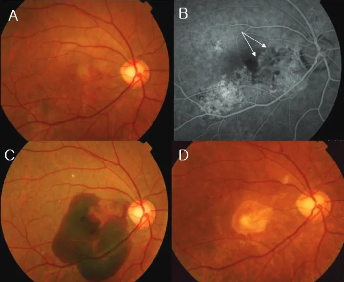
Fig. 1. Case 4, with exudative maculopathy in the right eye. (A) Fundus photograph showing subretinal blood and subretinal serous detachment. (B) Midphase fluorescein angiogram (FA) showing an irregular hyperfluorescent area corresponding to the interconnecting vascular network (arrow). (C) Fundus photograph 4 weeks after photodynamic therapy (PDT) showing a massive subretinal hemorrhage, including the posterior pole. (D) Fundus photograph after two intravitreal bevacizumab injections followed by an intravitreal injection of tissue plasminogen activator (t‐PA) and 100% C
3F
8(0.3 ml), showing chorioretinal scarring.
of PCV.
11Finally, although eyes affected with PCV have several retinal features in common with CNV of AMD, they also have some unique features such as a large (greater than 4‐disc area) serosanguinous retinopathy, orange polyp‐like structures, an absence of macular drusen, and multiple lesions scattered throughout the posterior pole including the peripapillary region.
1Since the role of VEGF in the pathogenesis of CNV was established, several studies have reported on the efficacy of anti‐VEGF therapy for the treatment of AMD‐related CNV.
15‐17