• 검색 결과가 없습니다.

Effects of Bevacizumab on Bcl-2 Expression and Apoptosis in Retinal Pigment Epithelial Cells under Oxidative Stress

N/A
N/A
Protected

Academic year: 2022

Share "Effects of Bevacizumab on Bcl-2 Expression and Apoptosis in Retinal Pigment Epithelial Cells under Oxidative Stress"

Copied!
9
0
0

로드 중.... (전체 텍스트 보기)

전체 글

(1)

© 2015 The Korean Ophthalmological Society

This is an Open Access article distributed under the terms of the Creative Commons Attribution Non-Commercial License (http://creativecommons.org/licenses /by-nc/3.0/) which permits unrestricted non-commercial use, distribution, and reproduction in any medium, provided the original work is properly cited.

Original Article

Effects of Bevacizumab on Bcl-2 Expression and Apoptosis in Retinal Pigment Epithelial Cells under Oxidative Stress

Sukjin Kim, Young Jun Kim, Na Rae Kim, Hee Seung Chin

Department of Ophthalmology and Inha Vision Science Laboratory, Inha University School of Medicine, Incheon, Korea

Purpose: To evaluate the effects of bevacizumab on expression of B-cell leukemia/lymphoma (Bcl)-2 and apop- tosis in retinal pigment epithelial (RPE) cells under oxidative stress conditions.

Methods: RPE cells were treated with H

2

O

2

(0, 100, 200, 300, and 400 µM) and bevacizumab at or above the doses normally used in clinical practice (0, 0.33, 0.67, 1.33, and 2.67 mg/mL). Cell apoptosis was measured using flow cytometry with annexin V-fluorescein isothiocyanate. The expression of Bcl-2 mRNA was deter- mined using reverse transcription polymerase chain reaction.

Results: Under low oxidative stress conditions (H

2

O

2

100 µM), cell apoptosis was not significantly different at any concentration of bevacizumab, but Bcl-2 mRNA expression decreased with increasing concentration of bevacizumab (0.33, 0.67, 1.33, and 2.67 mg/mL). Under moderate oxidative stress conditions (H

2

O

2

200 µM), Bcl-2 mRNA expression decreased with increasing concentration of bevacizumab (0.33, 0.67, 1.33, and 2.67 mg/mL), but cell apoptosis increased only at 2.67 mg/mL of bevacizumab. Under high oxidative stress (300 µM) conditions, cell apoptosis increased at high concentrations of bevacizumab (1.33 and 2.67 mg/mL), but it did not correlate with Bcl-2 expression.

Conclusions: Withdrawal of vascular endothelial growth factor can lead to RPE cell apoptosis and influences the expression of anti-apoptotic genes such as Bcl-2 under oxidative stress conditions. Since oxidative stress levels of each patient are unknown, repeated injections of intravitreal bevacizumab, as in eyes with age-relat- ed macular degeneration, might influence RPE cell survival.

Key Words: Apoptosis, Bcl-2, Bevacizumab, Retinal pigment epithelial cell

Vascular endothelial growth factor (VEGF) has been implicated as a pro-angiogenic factor that stimulates neo-

vascularization in age-related macular degeneration (AMD), and retinal pigment epithelial (RPE) is an import- ant source of VEGF [1]. VEGF expression by RPE is a fea- ture of tissues excised from human eyes showing AMD-re- lated choroidal neovascularization [2]. Oxidants have been reported to increase the deposition of oxidized proteins or other oxidized compounds in Bruch’s membrane in a pro- cess that might involve complement activation and inflam- mation, provoking VEGF-A release in patients with exuda- tive AMD [1]. Thus, VEGF-A is induced by chronic exposure to oxidative stress, suggesting a role of this stress

Received: January 17, 2015 Accepted: July 7, 2015

Corresponding Author: Hee Seung Chin, MD, PhD. Department of Oph- thalmology and Inha Vision Science Laboratory, Inha University Hospi- tal, #27 Inhang-ro, Jung-gu, Incheon 22332, Korea. Tel: 82-32-890-2408, Fax: 82-32-890-2403, E-mail: [email protected]

Parts of this work have been presented and selected as 'hot topics' at the

annual meeting of the Association for Research in Vision and Ophthal-

mology (ARVO) at Fort Lauderdale, FL, USA on May 2012.

(2)

S Kim, et al. Bevacizumab Effects on Bcl-2 and Apoptosis in RPE

in both neovascular and advanced dry AMD [3].

AMD eyes exhibited increased RPE apoptosis compared with normal eyes [3,4]. In particular, RPE apoptosis is an important feature of advanced forms of AMD; indeed, patchy loss of RPE is one of the classic features of AMD, and this process involves apoptosis of RPE cells [4]. In eyes with exudative AMD, apoptotic cells are present in choroidal neovascular membranes [5]. B-cell leukemia/

lymphoma (Bcl)-2 is a key anti-apoptotic member of the Bcl-2 family that regulates the intrinsic apoptosis pathway and is comprised of important survival factors in RPE cells [5-7]. Oxidative stress activates the intrinsic apoptosis pathway, a process mediated by enhanced mitochondrial membrane permeability and decreased Bcl-2 [8].

Anti-VEGF therapy plays a significant role in the man- agement of retinal and retinal vascular disorders and has demonstrated the best clinical outcomes of all approaches tested to date. However, VEGF-A is a known survival fac- tor of vascular endothelial cells and the developing and mature retina [9-11]. RPE tears and choroidal atrophy in specimens from patients with anti-VEGF treated AMD raise questions about the long-term safety of anti-VEGF treatment [12].

The effects of bevacizumab, which might involve cell apoptosis and regulation of anti-apoptotic protein Bcl-2 ex- pression, have not been fully investigated using RPE cells under oxidative stress. Our research therefore focused on evaluating the effects of bevacizumab on expression of Bcl-2 and apoptosis in RPE cells under various oxidative stress conditions and the possible complications of intrav- itreal bevacizumab injections in patients.

Materials and Methods

Cell culture and exposure to oxidative stress

A human retinal pigment epithelial (ARPE-19) cell line was obtained from ATCC (Manassas, VA, USA). Cells were maintained in Dulbecco’s modified Eagle’s medium/

Ham’s F-12 nutrient medium (Invitrogen-Gibco, Carlsbad, CA, USA) supplemented with 10% fetal bovine serum, penicillin, streptomycin, and amphotericin B. The ARPE- 19 cells were used for four to six passages and plated in six-well plates at 1.5 × 10

5

cells per well. They were incu- bated for 24 hours in a humidified 5% CO

2

atmosphere at

37°C after reaching approximately 70% confluence. We next washed the cells with pH 7.4 phosphate buffered sa- line (PBS). The cells were serum-starved for four hours before H

2

O

2

exposure and treated with H

2

O

2

(100 to 400 µM) for 16 hours to induce oxidative stress before they were harvested for cell death analysis.

Enzyme-linked immunosorbent assay after exposure to oxidative stress

The cells were treated with various concentrations of H

2

O

2

. The supernatants were collected at baseline (0 hours) and at 16 hours, centrifuged to remove cell debris, and stored at -70°C until enzyme-linked immunosorbent assay was performed according to the manufacturer’s instruc- tions. In this analysis, a monoclonal antibody specific for VEGF-A was pre-coated onto a microplate. Standards and samples were pipetted into the wells, and any VEGF-A present was bound by the immobilized antibody. After washing any unbound substances, an enzyme-linked poly- clonal antibody specific for VEGF-A was added to the wells. Following a wash to remove any unbound anti- body-enzyme reagent, a substrate solution was added to the wells, and color developed in proportion to the amount of VEGF-A bound in the initial step. The color develop- ment was then stopped, and the intensity of the color was measured. To determine the optical density of each well, we used a microplate reader at 450 and 540 nm. The concentrations of VEGF-A standard numbers 1 to 8 were 0, 15.6, 31.2, 62.5, 125, 250, 500, and 1,000 pg/mL of recombi- nant VEGF-A in a buffered protein base with preserva- tives, respectively. The level of VEGF-A protein was mea- sured in cell-free supernatant using a human VEGF-A Quantikin ELISA kit (catalog no. DVE00; R&D Systems, Minneapolis, MN, USA).

Bevacizumab treatment and flow cytometric analysis of apoptosis after exposure to oxidative stress

The cells were washed with PBS and incubated in se- rum-free Dulbecco’s modified Eagle’s medium in the pres- ence of H

2

O

2

(100, 200, 300, and 400 µM) for 16 hours.

Bevacizumab (0.33, 0.67, 1.33, and 2.67 mg/mL, respective-

ly) was added 2 hours before H

2

O

2

treatment. An annexin

V-fluorescein isothiocyanate (FITC) apoptosis kit (BD

Biosciences, Franklin Lakes, NJ, USA) was used to detect

(3)

phosphatidylserine externalization as an index of apopto- sis. The cells were washed and incubated for 15 minutes at room temperature in the presence of annexin V labeled with FITC and propidium iodide (PI). In total, 10,000 cells were excited at 488 nm, and emission was measured at 530 and 584 nm to assess FITC and PI fluorescence, respective- ly. The cells were analyzed with a flow cytometer (BD Biosciences). The number of gated cells was plotted on a dot plot with reference to both annexin V and PI staining.

Reverse transcription polymerase chain reaction The cells were diluted to 1 × 10

5

cell/mL and were incu- bated for 24 hours in six-well plates (Falcon, BD Biosci- ences). After washing the culture media twice with PBS, serum-free media was applied. H

2

O

2

and bevacizumab were challenged at different concentrations (0, 100, 200, 300, 400 µM and 0.33, 0.67, 1.33, 2.67 mg/mL) and incubat- ed for 16 hours. Total RNA was isolated using the total RNA purification kit (Invitrogen). Isolated RNA was quantified, and 1 µg of total RNA and 100 pmol of oligo dT were added to the reverse transcription (RT) premix (Bioneer, Seoul, Korea) to prepare 20 µL of cDNA. Pre- formed cDNA, Bcl-2 primer, and glycerol-3-phosphate de- hydrogenase (GAPDH) primer were mixed with poly- merase chain reaction (PCR) premix (Bioneer), and PCR was performed. The sequences of the primers are de- scribed in Table 1. Next, 10 µL of each amplificate were assessed using 1.5% agarose gel electrophoresis. Quantifi- cation of Bcl-2 mRNA content was performed using com- puter-assisted video densitometry (Eagle Eye II-system;

Stratagene, La Jolla, CA, USA).

Statistical analysis

Data are expressed as percentage of control or mean ± standard deviation of results in three or more independent

experiments. Statistical analysis was performed using SPSS ver. 18.0 (SPSS Inc., Chicago, IL, USA). The Krus- kal-Wallis test was used to compare the difference between control and experimental groups. A p-value ≤0.05 was considered statistically significant.

Results

Influence of bevacizumab on apoptosis of retinal pig- ment epithelial cells without H

2

O

2

The cells were treated with bevacizumab for 16 hours (0, 0.33, 0.67, 1.33, and 2.67 mg/mL). Cell apoptosis was not significantly different at control, clinical (0.33 and 0.67 mg/mL), or high (1.33 and 2.67 mg/mL) doses of bevaci- zumab without H

2

O

2

(Fig. 1).

Effects of H

2

O

2

on apoptosis of retinal pigment epithe- lial cells

The cells were cultured with H

2

O

2

for 16 hours (0, 100, 200, 300, and 400 μM). Cell apoptosis was 3.26% for RPE at 100 µM H

2

O

2

compared to 3.82% for the controls, but the difference was not significant. Cell apoptosis decreased at 200 µM H

2

O

2

(2.65%) but increased at 300 and 400 µM H

2

O

2

(10.42%, 17.99%). The means at these concentrations were significantly different than the control (p < 0.05) (Fig.

2A).

Vascular endothelial growth factor-A level under oxi- dative stress conditions

VEGF-A expression was increased after addition of 50, 100, and 200 µM H

2

O

2

to RPE cells. However, after being treated with 300 and 400 µM H

2

O

2

, VEGF-A expression decreased. Means at these concentrations were significant- ly different from the control (p < 0.05) (Fig. 2B).

Table 1. Primer used for reverse transcription polymerase chain reaction

Target gene Primer Sequence Product size (bp)

Bcl-2 Forward 5’-CTA CGA GTG GGA TGC GGG AGA TG-3’ 478

Reverse 5’-GGT TCA GGT ACT CAG TCA TCC ACA G-3’ 478

GAPDH Forward 5’-CCA CTG GCG TCT TCA CCA C-3’ 481

Reverse 5’-CCT GCT TCA CCA CCT TCT TG-3’ 481

Bcl = B-cell leukemia/lymphoma; GAPDH = glycerol-3-phosphate dehydrogenase.

(4)

S Kim, et al. Bevacizumab Effects on Bcl-2 and Apoptosis in RPE

Expression of Bcl-2 mRNA after exposure to oxidative stress

The cells were cultured with H

2

O

2

for 16 hours (0, 100, 200, and 300 μM). Expression of Bcl-2 mRNA decreased as oxidative stress increased (p < 0.05) (Fig. 2C).

Influence of bevacizumab on apoptosis of RPE cells and Bcl-2 mRNA expression under low oxidative stress

ARPE-19 cells were treated with bevacizumab (0, 0.33, 0.67, 1.33, and 2.67 mg/mL) under low oxidative stress conditions (100 µM of H

2

O

2

). Cell apoptosis was not sig- nificantly different at any concentration of bevacizumab at 100 µM H

2

O

2

. Expression of Bcl-2 mRNA decreased under low oxidative stress conditions, and the decrease was pro- portional to the increase in bevacizumab. There was a sta- tistically significant difference at all concentrations compared to the result of the control. The mRNA expres- sion of Bcl-2 was normalized to GAPDH (a housekeeping gene). Each bar shows the mean ± standard deviation of results in three or more independent experiments (p < 0.05) (Fig. 3).

Influence of bevacizumab on apoptosis of RPE cells and Bcl-2 mRNA expression under moderate oxidative stress ARPE-19 cells were treated with bevacizumab (0, 0.33, 0.67, 1.33, and 2.67 mg/mL) under moderate oxidative stress conditions (200 µM of H

2

O

2

). As mentioned previ- ously, cell apoptosis decreased at 200 µM H

2

O

2

(2.65%) compared with the control (3.82%). Cell apoptosis in- creased to 3.59%, 3.50%, and 4.13% at bevacizumab con- centrations of 0.33, 0.67, and 1.33 mg/mL, respectively, but the differences were not statistically significant. However, cell apoptosis increased significantly to 10.16% at a high dose of bevacizumab (2.67 mg/mL) under the same oxida- tive stress conditions. Expression of Bcl-2 mRNA de- creased proportionally to the increase in bevacizumab dose. There was a statistically significant difference at all concentrations (p < 0.05) (Fig. 4).

Influence of bevacizumab on apoptosis of RPE cells and Bcl-2 mRNA expression under high oxidative stress

ARPE-19 cells were treated with bevacizumab (0, 0.33, 0.67, 1.33, and 2.67 mg/mL) under high oxidative stress conditions (300 µM H

2

O

2

). As mentioned previously, cell apoptosis increased at 300 µM H

2

O

2

(10.42%) compared with the control (3.82%). Cell apoptosis increased to 12.65% and 10.97% at clinical doses of bevacizumab (0.33, 0.67 mg/mL) under high oxidative stress conditions, but there was no statistically significant difference compared to the control. Cell apoptosis did significantly increase to 19.83% and 24.85% at high doses of bevacizumab (1.33, 2.67 mg/mL) under the same oxidative stress. Expression of Bcl-2 mRNA decreased under high oxidative stress con- ditions (300 µM H

2

O

2

), but there was no trend of increase or decrease as the dose of bevacizumab increased (Fig. 5).

At 400 µM H

2

O

2,

the Bcl-2 mRNA data were not able to be interpreted. We suggest that the reverse transcription-PCR results might be a result of severe cellular apoptosis under high oxidative stress conditions.

Discussion

In the present study, we tested the possible toxicity of bevacizumab on RPE cells under various oxidative stress conditions, and we examined the expression of the an-

0 0.33 0.67 1.33

Bevacizumab concentration (mg/mL)

2.67 30

25 20 15 10 5 0

Apoptotic cells (%)

Fig. 1. Influence of bevacizumab on apoptosis of retinal pigment

epithelial cells in the absence of H

2

O

2

. The retinal pigment epi-

thelial cells were treated with bevacizumab for 16 hours (0, 0.33,

0.67, 1.33, and 2.67 mg/mL). Cellular apoptosis was not signifi-

cantly different at control, clinical (0.33 and 0.68 mg/mL) and

high (1.33 and 2.67 mg/mL) doses of bevacizumab in the absence

of H

2

O

2

(p = 0.121, 0.439, 0.221, and 0.063, respectively). Each bar

shows the mean ± standard deviation of results of three or more

independent experiments.

(5)

Korean J Ophthalmol Vol.29, No.6, 2015

ti-apoptotic protein Bcl-2 and the apoptosis of RPE cells.

We used H

2

O

2

to induce oxidative stress in order to repli- cate in vivo conditions that are implicated in the pathogen- esis of AMD and the annexin V-FITC apoptosis kit to de- tect phosphatidylserine externalization as an index of apoptosis.

Intravitreal injections of anti-VEGF agents such as beva- cizumab are widely used for the treatment of various reti- nal disorders associated with new vessels such as AMD or proliferative diabetic retinopathy in order to reduce angio- genesis [13]. Bevacizumab is a full-length, recombinant humanized immunoglobulin G antibody that binds to and

inhibits all biologically active forms of VEGF-A isoforms.

Despite ongoing clinical trials with intravitreal bevaci- zumab in many neovascular ocular disorders, its ocular safety remains an issue for research. Hypothetically, re- peated injections with bevacizumab could interfere with the neuroprotective and survival actions of VEGF under oxidative stress.

In AMD patients, several studies have reported cases of RPE tears after intravitreal injections of bevacizumab for neovascular AMD [12]. Atrophy of the choriocapillaries or loss of endothelial cell fenestrations impairs nutritional support, which can lead to functional and morphologic

0 100 200 300

H

2

O

2

concentration (μM)

400 20

15 10 5 0

Apoptotic cells (%)

*

*

**

0 50 100 200 300

H

2

O

2

concentration (μM)

400 250

200 150 100 50 0

VEGF-A (pg/mL)

0 100 200 300

H

2

O

2

concentration (μM) 1.25

1 0.75 0.5 0.25 0

Fold induction of Bcl-2 mRNA

0 100 200 300

H

2

O

2

concentration (μM)

400 30

25 20 15 10 5 0

Apoptotic cells (%)

*

*

**

0 50 100 200 300

H

2

O

2

concentration (μM)

400 250

200 150 100 50 0

VEGF-A (pg/mL)

0 100 200 300

H

2

O

2

concentration (μM) 1.25

1 0.75 0.5 0.25 0

Fold induction of Bcl-2 mRNA

Fig. 2. Apoptosis and expression of vascular endothelial growth factor (VEGF)-A and B-cell leukemia/lymphoma (Bcl)-2 after treatment of retinal pigment epithelial (RPE) cells with H

2

O

2

. (A) Effects of H

2

O

2

on apoptosis of RPE cells. The RPE cells were treated with various concentrations of H

2

O

2

for 16 hours.

Cell apoptosis was 3.26% for RPE at 100 µM H

2

O

2

compared to 3.82% for the control, but the difference was not significant. Cell apoptosis decreased at 200 µM H

2

O

2

(2.65%) but increased at 300 and 400 µM of H

2

O

2

(10.42%, 17.99%). Means were significantly different than the control (p < 0.05). Each bar shows the mean

± standard deviation of results of three or more independent experiments. The asterisk indicates a statistically significant dif- ference within the group (

*

increased,

**

decreased, p < 0.05). (B) Expression of VEGF-A after exposure to H

2

O

2

. VEGF-A excretion into the medium was measured using enzyme-linked immuno- sorbent assay. VEGF-A expression increased after addition of 50, 100, or 200 µM H

2

O

2

to RPE cells. However, after being treated with 300 and 400 µM H

2

O

2

, VEGF-A expression decreased. Data are expressed as the mean ± standard deviation of the results of three or more independent experiments. Means were statistically significant compared to the control at all concentrations of H

2

O

2

(p

< 0.05). (C) Expression of Bcl-2 mRNA after exposure to H

2

O

2

. Cells were cultured with various concentrations of H

2

O

2

for 16 hours (0, 100, 200, and 300 μM). Expression of Bcl-2 mRNA decreased as oxidative stress increased. Data are expressed as the mean ± standard deviation of the results of three or more independent experiments. Means were statistically significant compared with the control at all concentrations of H

2

O

2

(p < 0.05).

A

B

C

(6)

S Kim, et al. Bevacizumab Effects on Bcl-2 and Apoptosis in RPE

0 0 0.33 0.67

0 100

1.33 2.67 Bevacizumab (mg/mL)

H

2

O

2

(μM) 30 25 20 15 10 5 0

Apoptotic cells (%)

Bevacizumab (mg/mL) H

2

O

2

(μM) Bcl-2

GAPDH

0 0.33 0.67

100

1.33 2.67

0 0.33 0.67

100

1.33 2.67

Bevacizumab (mg/mL) H

2

O

2

(μM)

1.25 1 0.75 0.5 0.25 0

Fold induction of Bcl-2 mRNA

Fig. 3. Influence of bevacizumab on apoptosis of retinal pigment epithelial (RPE) cells and B-cell leukemia/lymphoma (Bcl)- 2 mRNA expression under low oxidative stress (100 µM H

2

O

2

).

(A) Influence of bevacizumab on apoptosis of RPE cells under low oxidative stress (100 µM H

2

O

2

). The RPE cells were treated with various concentrations of bevacizumab (0, 0.33, 0.67, 1.33, and 2.67 mg/mL) under low oxidative stress (100 µM H

2

O

2

). Cell apoptosis was not significantly different at all concentrations of bevacizumab less than 100 µM H

2

O

2

. Each bar shows the mean

± standard deviation of results of three or more independent experiments. p < 0.05 vs. control. (B,C) Expression of Bcl-2 mRNA with bevacizumab under low oxidative stress (100 µM H

2

O

2

). Expression of Bcl-2 mRNA decreased under low oxidative stress (100 µM H

2

O

2

), and this decrease was proportional to the increase in bevacizumab dose. There was a statistically significant difference at all concentrations. The mRNA expres- sion of Bcl-2 was normalized to glycerol-3-phosphate dehydro- genase (GAPDH, housekeeping gene). Each bar shows the mean

± standard deviation of results of three or more independent experiments. p < 0.05 vs. control.

Fig. 4. Influence of bevacizumab on apoptosis of retinal pigment epithelial (RPE) cells and B-cell leukemia/lymphoma (Bcl)- 2 mRNA expression under moderate oxidative stress (200 µM of H

2

O

2

). (A) Influence of bevacizumab on apoptosis of RPE cells under moderate oxidative stress (200 µM H

2

O

2

). The RPE cells were treated with various concentrations of bevacizumab (0, 0.33, 0.67, 1.33, and 2.67 mg/mL) under moderate oxidative stress (200 µM H

2

O

2

). Cell apoptosis decreased at 200 µM H

2

O

2

(2.65%) compared with the control (3.82%). Cell apoptosis did not show significant change until a bevacizumab concentration of at least 1.33 mg/mL. Apoptosis increased to 10.16% at high doses of bevacizumab (2.67 mg/mL) under the same oxidative stress conditions. Each bar shows the mean ± standard deviation of results of three or more independent experiments. The asterisk indicates a statistically significant difference within the group (

*

p < 0.05). (B,C) Expression of Bcl-2 mRNA with bevacizumab under moderate oxidative stress (200 µM H

2

O

2

). Expression of Bcl-2 mRNA decreased under moderate oxidative stress (200 µM H

2

O

2

), and that decrease was proportional to the increase in bevacizumab dose. There was a statistically significant difference at all concentrations. The mRNA expression of Bcl-2 was normal- ized to glycerol-3-phosphate dehydrogenase (GAPDH, housekeep- ing gene). Each bar shows the mean ± standard deviation of results of three or more independent experiments. p < 0.05 vs. control.

0 0 0.33 0.67

0 200

1.33 2.67 Bevacizumab (mg/mL)

H

2

O

2

(μM) 30 25 20 15 10 5 0

Apoptotic cells (%) *

0 0.33 0.67

200

1.33 2.67

Bevacizumab (mg/mL) H

2

O

2

(μM)

1.25 1 0.75 0.5 0.25 0

Fold induction of Bcl-2 mRNA

Bevacizumab (mg/mL) H

2

O

2

(μM) Bcl-2

GAPDH

0 0.33 0.67

200

1.33 2.67

A A

B B

C C

(7)

damage to the RPE and photoreceptors, with particular adverse effects if the macular lesion is affected [14]. The development of geographic atrophy contributed to visual acuity loss after repeated anti-VEGF injections [15]. In ad- dition, in a recent study in which a retinal neovasculariza- tion model was developed, it was suggested that caution is warranted in the treatment of patients with acute or severe retinal neovascularization using anti-VEGF drugs such as bevacizumab in order to prevent capillary nonperfusion and macular ischemia [16].

According to many recent reports, bevacizumab at con- centrations at or above the dose normally used in clinical practice is not toxic to RPE cells [17-20]. Several preclini- cal experimental toxicity studies have reported the histo- pathological effects of bevacizumab on retinal cells, retinal neovascular membranes, and capillaries in paraffin-em- bedded sections [16,21,22] or organotypic culture [23], as well as in ultrastructural evaluations [14,17,21]. Based on in vitro assays, bevacizumab has little toxic effect on gangli- on cells, neuroretinal cells, RPE cells, choroidal endotheli- al cells, or corneal epithelial cells [19,24]. However, there are limited previous preclinical studies associated with RPE under oxidative stress conditions [25].

In our study, RPE cell apoptosis did not show a signifi- cant difference at 100 µM H

2

O

2

but decreased at 200 µM H

2

O

2

and increased with higher oxidative stress conditions (300 and 400 µM H

2

O

2

) (Fig. 1A). It seems that RPE cells can tolerate up to 200 µM H

2

O

2

due to cell-protective mechanisms, but higher oxidative stress conditions caused in vitro mitochondrial DNA damage and promoted apop- tosis. Ballinger et al. [26] and Jin et al. [27] reported that exposure of RPE cells to concentrations of H

2

O

2

that cause in vitro mitochondrial DNA damage also promotes apop- tosis, and this correlates well with our results under high oxidative stress conditions.

In different cell types, including vascular smooth muscle cells [28], VEGF expression is triggered by oxidative stress. Other studies of VEGF expression are consistent with our results when H

2

O

2

in concentrations up to 200 µM was added to RPE cells. However, VEGF expression decreased at 300 and 400 µM H

2

O

2

, suggesting that VEGF production decreases according to the decrease in live RPE cells [29].

Oxidative stress triggers multiple signaling pathways, in- cluding some that are cytoprotective and others that con- tribute to cell damage and eventually cell death. Among these are the pathways of the Bcl-2 family proteins, which have subcellular locations on the mitochondrial outer mem- brane, nuclear envelope, and endoplasmic reticulum [30].

Expression of Bcl-2 mRNA decreased as oxidative stress increased, which was consistent with another study [27].

The retina is a highly oxygen-consumptive tissue that al- ways functions under high oxygen tension; RPE cells are therefore exposed to oxidative stress. Chronic oxidative stress can lead to impairment and death of RPE cells, im- plicating it as a risk factor of AMD. In our study, low to high oxidative stress conditions of RPE cells were impli- cated in the phase of AMD.

Oxidatively stressed RPE cells undergo apoptosis [27], and Byeon et al. [25] have reported that addition of a high concentration (2.5 mg/mL) of bevacizumab to the culture medium did not affect the survival of either control RPE cells or cells under a low level of oxidative stress (150 μM H

2

O

2

). However, under higher oxidative stress levels (200 or 300 μM H

2

O

2

), pretreatment with bevacizumab (2.5 mg/

mL) induced a significantly higher level of RPE cell death [25].

Addition of clinically applicable (0.33 and 0.67 mg/mL) and high concentrations (1.33 and 2.67 mg/mL) of bevaci-

0 0 0.33 0.67

0 300

1.33 2.67 Bevacizumab (mg/mL)

H

2

O

2

(μM) 40 35 30 25 20 15 10 5 0

Apoptotic cells (%)

*

*

Fig. 5. Influence of bevacizumab on apoptosis of retinal pig- ment epithelial cells under high oxidative stress (300 µM H

2

O

2

).

The retinal pigment epithelial cells were treated with various concentrations of bevacizumab (0, 0.33, 0.67, 1.33, and 2.67 mg/

mL) under high oxidative stress (300 µM of H

2

O

2

). As mentioned previously, cell apoptosis increased at 300 µM H

2

O

2

(10.42%) compared with the control (3.82%). Cell apoptosis increased to 12.65% and 10.97% at clinical doses of bevacizumab (0.33, 0.67 mg/mL) under high oxidative stress, but there was no statistically significant difference between the increases. Cell apoptosis significantly increased to 19.83% and 24.85% at high doses of bevacizumab (1.33, 2.67 mg/mL) under the same oxidative stress.

Each bar shows the mean ± standard deviation of results of three

or more independent experiments. The asterisk indicates a statis-

tically significant difference within the group (

*

p < 0.05).

(8)

S Kim, et al. Bevacizumab Effects on Bcl-2 and Apoptosis in RPE

zumab did not affect the survival of RPE cells under low oxidative stress (100 μM H

2

O

2

); nevertheless, Bcl-2 expres- sion decreased in a dose-dependent manner (Fig. 3). Al- though Bcl-2 is an important survival factor, it is sensitive to oxidative stress, and a decrease in Bcl-2 precedes cell apoptosis.

Under moderate oxidative stress conditions (200 μM H

2

O

2

), Bcl-2 expression decreased with increasing concen- tration of bevacizumab; however, cell apoptosis did not show significant change until 1.33 mg/mL of bevacizumab.

It then increased to 10.16% at high doses of bevacizumab (2.67 mg/mL) under the same oxidative stress (Fig. 4).

Under high oxidative stress conditions (300 μM H

2

O

2

), RPE cell apoptosis increased at high bevacizumab concen- trations (1.33 and 2.67 mg/mL), but this was not correlated with Bcl-2 expression (Fig. 5). This might be due to multi- ple survival factors in RPE cells or experimental error due to low Bcl-2 expression.

In our experiment, RPE apoptosis did not increase with increased concentration of bevacizumab in the absence of H

2

O

2

; however, as the oxidative stress level increased, apoptosis of RPE cells increased. The higher was the oxi- dative stress, the lower was the concentration of bevaci- zumab that induced cellular apoptosis. Although cellular apoptosis at clinically applicable levels of bevacizumab (0.33 and 0.67 mg/mL) did not show significant increase, care should be taken since Bcl-2 expression showed a sig- nificant decrease even at lower levels of oxidative stress.

To our knowledge, we have shown for the first time that bevacizumab influences Bcl-2 expression and apoptosis of RPE cells under oxidative stress. Our study has some lim- itations. First, our conclusions are drawn from in vitro studies, which are not the best approximation of in vivo conditions. Moreover, some previous studies have shown that non-confluent ARPE-19 cells behave quite differently in terms of oxidative stress compared to confluent mono- layers [31]. Further studies are needed to examine these findings in porcine retina-RPE-choroid cultures or on mouse retinae. Second, it is difficult to determine the de- gree of H

2

O

2

simulating oxidative stress conditions of hu- man eyes. Third, we could not identify the additional path- way mechanisms of Bcl-2 expression. Further studies are needed to examine the intracellular pathway or cumulative effects at repeated doses. Fourth, we could not determine the additional mechanism of action of bevacizumab in our study. Bevacizumab might bind RPE cell surface receptors

in addition to known VEGF receptors.

In our experiment, we demonstrated the possibility that addition of the VEGF inhibitor bevacizumab might block the protective effect of VEGF in our in vitro model under high oxidative stress conditions mimicking those in AMD eyes. Under increased oxidative stress conditions, high doses of bevacizumab might influence RPE cell survival.

Furthermore, bevacizumab might affect the expression of anti-apoptotic genes such as Bcl-2 under all oxidative con- ditions. Since the oxidative stress levels of each patient are unknown, repeated injections of intravitreal bevacizumab, as in eyes with AMD, might influence RPE cell survival.

Therefore close monitoring is needed in patients who receive repeated injections of intravitreal bevacizumab.

Conflict of Interest

No potential conflict of interest relevant to this article was reported.

Acknowledgements

This work was supported by an Inha University research grant.

References

1. Kliffen M, Sharma HS, Mooy CM, et al. Increased expres- sion of angiogenic growth factors in age-related maculopa- thy. Br J Ophthalmol 1997;81:154-62.

2. Otani A, Takagi H, Oh H, et al. Vascular endothelial growth factor family and receptor expression in human choroidal neovascular membranes. Microvasc Res 2002;64:

162-9.

3. Liang FQ, Godley BF. Oxidative stress-induced mitochon- drial DNA damage in human retinal pigment epithelial cells: a possible mechanism for RPE aging and age-related macular degeneration. Exp Eye Res 2003;76:397-403.

4. Dunaief JL, Dentchev T, Ying GS, Milam AH. The role of apoptosis in age-related macular degeneration. Arch Oph- thalmol 2002;120:1435-42.

5. Hinton DR, He S, Lopez PF. Apoptosis in surgically ex-

cised choroidal neovascular membranes in age-related

(9)

macular degeneration. Arch Ophthalmol 1998;116:203-9.

6. Zhang N, Peairs JJ, Yang P, et al. The importance of Bcl-xL in the survival of human RPE cells. Invest Ophthalmol Vis Sci 2007;48:3846-53.

7. Godley BF, Jin GF, Guo YS, Hurst JS. Bcl-2 overexpression increases survival in human retinal pigment epithelial cells exposed to H(2)O(2). Exp Eye Res 2002;74:663-9.

8. Sorenson CM. Bcl-2 family members and disease. Biochim Biophys Acta 2004;1644:169-77.

9. Alon T, Hemo I, Itin A, et al. Vascular endothelial growth factor acts as a survival factor for newly formed retinal vessels and has implications for retinopathy of prematurity.

Nat Med 1995;1:1024-8.

10. Gerber HP, Dixit V, Ferrara N. Vascular endothelial growth factor induces expression of the antiapoptotic proteins Bcl- 2 and A1 in vascular endothelial cells. J Biol Chem 1998;

273:13313-6.

11. Gora-Kupilas K, Josko J. The neuroprotective function of vascular endothelial growth factor (VEGF). Folia Neuro- pathol 2005;43:31-9.

12. Gelisken F, Ziemssen F, Voelker M, et al. Retinal pigment epithelial tears after single administration of intravitreal bevacizumab for neovascular age-related macular degener- ation. Eye (Lond) 2009;23:694-702.

13. CATT Research Group, Martin DF, Maguire MG, et al.

Ranibizumab and bevacizumab for neovascular age-related macular degeneration. N Engl J Med 2011;364:1897-908.

14. Peters S, Heiduschka P, Julien S, et al. Ultrastructural find- ings in the primate eye after intravitreal injection of beva- cizumab. Am J Ophthalmol 2007;143:995-1002.

15. Ying GS, Kim BJ, Maguire MG, et al. Sustained visual acuity loss in the comparison of age-related macular de- generation treatments trials. JAMA Ophthalmol 2014;132:

915-21.

16. Ameri H, Chader GJ, Kim JG, et al. The effects of intravit- reous bevacizumab on retinal neovascular membrane and normal capillaries in rabbits. Invest Ophthalmol Vis Sci 2007;48:5708-15.

17. Inan UU, Avci B, Kusbeci T, et al. Preclinical safety evalu- ation of intravitreal injection of full-length humanized vas- cular endothelial growth factor antibody in rabbit eyes. In- vest Ophthalmol Vis Sci 2007;48:1773-81.

18. Manzano RP, Peyman GA, Khan P, Kivilcim M. Testing intravitreal toxicity of bevacizumab (Avastin). Retina 2006;26:257-61.

19. Luthra S, Narayanan R, Marques LE, et al. Evaluation of

in vitro effects of bevacizumab (Avastin) on retinal pig- ment epithelial, neurosensory retinal, and microvascular endothelial cells. Retina 2006;26:512-8.

20. Brar VS, Sharma RK, Murthy RK, Chalam KV. Evaluation of differential toxicity of varying doses of bevacizumab on retinal ganglion cells, retinal pigment epithelial cells, and vascular endothelial growth factor-enriched choroidal en- dothelial cells. J Ocul Pharmacol Ther 2009;25:507-11.

21. Heiduschka P, Fietz H, Hofmeister S, et al. Penetration of bevacizumab through the retina after intravitreal injection in the monkey. Invest Ophthalmol Vis Sci 2007;48:2814-23.

22. Shahar J, Avery RL, Heilweil G, et al. Electrophysiologic and retinal penetration studies following intravitreal injec- tion of bevacizumab (Avastin). Retina 2006;26:262-9.

23. Kaempf S, Johnen S, Salz AK, et al. Effects of bevacizum- ab (Avastin) on retinal cells in organotypic culture. Invest Ophthalmol Vis Sci 2008;49:3164-71.

24. Luke M, Warga M, Ziemssen F, et al. Effects of bevaci- zumab on retinal function in isolated vertebrate retina. Br J Ophthalmol 2006;90:1178-82.

25. Byeon SH, Lee SC, Choi SH, et al. Vascular endothelial growth factor as an autocrine survival factor for retinal pigment epithelial cells under oxidative stress via the VEGF-R2/PI3K/Akt. Invest Ophthalmol Vis Sci 2010;51:

1190-7.

26. Ballinger SW, Van Houten B, Jin GF, et al. Hydrogen per- oxide causes significant mitochondrial DNA damage in hu- man RPE cells. Exp Eye Res 1999;68:765-72.

27. Jin GF, Hurst JS, Godley BF. Hydrogen peroxide stimulates apoptosis in cultured human retinal pigment epithelial cel- ls. Curr Eye Res 2001;22:165-73.

28. Castilla MA, Caramelo C, Gazapo RM, et al. Role of vascu- lar endothelial growth factor (VEGF) in endothelial cell protection against cytotoxic agents. Life Sci 2000;67:1003-13.

29. Kannan R, Zhang N, Sreekumar PG, et al. Stimulation of apical and basolateral VEGF-A and VEGF-C secretion by oxidative stress in polarized retinal pigment epithelial cells.

Mol Vis 2006;12:1649-59.

30. Krajewski S, Tanaka S, Takayama S, et al. Investigation of the subcellular distribution of the bcl-2 oncoprotein: resi- dence in the nuclear envelope, endoplasmic reticulum, and outer mitochondrial membranes. Cancer Res 1993;53:4701-14.

31. Rabin DM, Rabin RL, Blenkinsop TA, et al. Chronic oxida-

tive stress upregulates Drusen-related protein expression in

adult human RPE stem cell-derived RPE cells: a novel cul-

ture model for dry AMD. Aging (Albany NY) 2013;5:51-66.

수치

Table 1. Primer used for reverse transcription polymerase chain reaction
Fig. 1. Influence of bevacizumab on apoptosis of retinal pigment  epithelial cells in the absence of H 2 O 2
Fig. 2. Apoptosis and expression of vascular endothelial growth  factor (VEGF)-A and B-cell leukemia/lymphoma (Bcl)-2 after  treatment of retinal pigment epithelial (RPE) cells with H 2 O 2
Fig. 3. Influence of bevacizumab on apoptosis of retinal pigment  epithelial (RPE) cells and B-cell leukemia/lymphoma  (Bcl)-2 mRNA expression under low oxidative stress (100 µM H 2 O 2 )
+2

참조

관련 문서

Inhibitory effect of Quercetin and Aronia extract on the MITF, Tyrosinase, TRP-1, TRP-2 and Actin in expression B16F10 cells.. Inhibitory effect of C3G on the Tyrosinase,

1) Effects of methanol extracts of Capsella bursa-pastoris on cyclooxygenase-2 (COX-2) and Inducible Nitric oxide synthase (iNOS) expression in human prostate cancer cell

Inhibitory effects of classified methanol extracts of Smilax china L on the COX-2 and iNOS expression of human colorectal cancer cell lines... Inhibitory

“The economic order of the Republic of Korea shall be based on a respect for the freedom and creative initiative of individuals in economic affairs.” The State may only

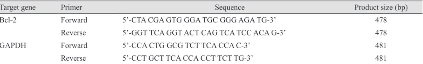
In this study, hydrogen peroxide(H 2 O 2 ) was used for producing low molecular weight sodium alginates(LMWSAs) under e-beam irradiation or controlled

4 &gt; Effects of Taro on COX-2 expression and iNOS expression(hot water) in human thyroid cancer cells. The cells were pretreated for 48hours with either

And Western Blotting was used to see the effects of Cnidium officinale MAKINO extracts on inducible Nitric Oxide Synthase (iNOS) and cyclooxygenase-2 (COX-2) expression..

This research was supported by Korea Atomic Energy Research Institute (NTIS-1711139325) and National Research Foundation of Korea (NRF) Grant funded by the Korean