Radiological Manifestations of Childhood Fractures
소아 골절의 영상의학적 소견
Jae-Yeon Hwang, MD*
Department of Radiology, Research Institute for Convergence of Biomedical Science and Technology, Pusan National University Yangsan Hospital, College of Medicine, Pusan National University, Yangsan, Korea
Musculoskeletal injury is the most common cause of children visiting the department of emer- gency medicine. Since the bone is still developing, pediatric patients have characteristic radio- logical manifestations, including plastic deformation, greenstick fractures, and buckle (or torus) fractures. Furthermore, growth arrest can occur in those with physeal fractures. Various mecha- nisms are responsible for pediatric musculoskeletal injury since children have different ranges of activities, depending on their age, such as birth injury and fall and traffic accidents. Some fractures have characteristic locations and radiological manifestations. In this review, we will discuss various radiological manifestations of fractures involving both upper and lower limbs in pediatric patients.
Index terms Child; Radiology; Trauma; Fractures, Bone
서론
근골격계의 외상은 소아가 응급실로 내원하는 가장 흔한 원인 중의 하나이다. 국내에서는 2015년 현재 연간 약 100만 명의 15세 이하 소아가 근골격계 외상으로 응급실을 방문하고 있 는 것으로 보고된 바 있으며, 유병률은 2006년부터 2014년까지 꾸준히 증가하는 것으로 보고 되었다(1). 남아에서 더 흔하게 발생하며, 4세 이하 소아의 근골격계 외상의 빈도는 그 이상 연 령의 소아보다 약 1.5배 정도 높았다. 또한 상지의 손상이 하지의 손상보다 더 많이 발생하였 으며, 계절적으로는 외부 활동이 많은 봄과 여름에 더 높은 빈도로 발생하였다.
아직 발달 중에 있는 소아의 근골격계 외상은 성인과는 다른 임상적 및 영상의학적 특성을 보이며, 나이에 따라서 외상의 위치, 손상의 범위 및 양상이 다양하다. 소아는 골절의 치유와 뼈의 재형성 과정이 성인보다 빠르며, 이 과정에서 외상 후 변형이 성인보다 잘 교정되는 특성 이 있다. 또한 뼈의 성장 및 골화가 진행중이며 뼈와 연부조직의 특성이 성인과 다르기 때문에 골절 및 합병증의 양상도 성인과는 구분된다(2). 특히 소아는 성장판의 손상이 일어날 수 있기
Received March 23, 2020 Revised June 2, 2020 Accepted June 23, 2020
*Corresponding author Jae-Yeon Hwang, MD Department of Radiology, Pusan National University Yangsan Hospital,
20 Geumo-ro, Mulgeum-eup, Yangsan 50612, Korea.
Tel 82-55-360-1840 Fax 82-55-360-1848
E-mail jyhwang79@gmail.com This is an Open Access article distributed under the terms of the Creative Commons Attribu- tion Non-Commercial License (https://creativecommons.org/
licenses/by-nc/4.0) which permits unrestricted non-commercial use, distribution, and reproduc- tion in any medium, provided the original work is properly cited.
ORCID iD Jae-Yeon Hwang https://
orcid.org/0000-0003-2777-3444
https://doi.org/10.3348/jksr.2020.81.4.806 807 때문에 뼈 성장의 장애가 합병증으로 동반될 수 있다. 따라서 영상의학과 의사는 소아에서 발생할 수 있는 근골격계 외상의 다양한 영상의학적 소견을 이해해야 하며, 성장 장애나 변형과 같은 합병 증을 예측할 수 있어야 한다. 본 종설에서는 소아 근골격계 외상 중 사지 골절을 중심으로 영상의학 적 소견을 고찰하고자 한다.
불완전 골절(Incomplete Fracture)
소아의 뼈는 성인보다 부드럽고 탄성이 있기 때문에, 피질골의 완전한 결손을 동반하지 않는 불완 전 골절(incomplete fracture)이 발생할 수 있다. 불완전 골절에는 소성변형(plastic deformity), 생 나무 골절(greenstick fracture), 죔쇠 골절/융기 골절(buckle fractur or torus fracture) 등이 있다.
소성변형(Plastic Deformation)
뼈가 가지고 있는 탄성 한계보다 약한 외력이 가해졌을 때에는 뼈의 가역적인 변형이 일어난다.
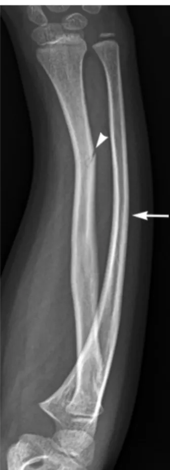
하지만 이 탄성 한계보다 큰 힘이 가해졌을 경우에는 힘이 가해진 반대측(tensile side, convex side) 에 현미경적 손상이 발생하게 되고, 비가역적인 변화가 발생한다(3). 이를 소성변형(plastic defor- mation)이라고 하며, 일반적인 골절과 달리 피질골의 파괴는 영상에서 보이지 않는다. 소성변형은 아래팔이나 하지의 긴 뼈에서 가장 흔하게 볼 수 있다. 일반 촬영에서 뼈의 변형이 보이지만, 골절선 은 보이지 않는다(Fig. 1).
Fig. 1. Plastic deformation and greenstick fracture.
The plain radiograph of the forearm shows plastic deformation in the ulna (arrow) and greenstick fracture in the distal radius (arrowhead). Note the angulation of the distal radius.
생나무 골절(Greenstick Fracture)
생나무 골절(greenstick fracture)은 소성변형을 일으키는 힘보다 더 강한 외력이 가해졌을 때 발 생하며, 외력에 의해 뼈가 구부러지고 힘이 가해진 반대측에 골절이 생긴 것을 의미한다. 힘이 가해 진 측(concave side)의 골절은 발생하지 않지만, 미세 손상과 소성변형은 동반한다. 일반 촬영에서 피질의 한쪽면에서만 골절이 보이며 각변형(angulation)을 동반한다(Fig. 1).
죔쇠 골절(Buckle Fracture)/융기 골절(Torus Fracture)
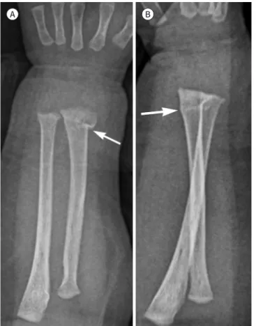
긴 뼈에 축 방향으로 외력이 가해지는 경우, 피질골의 결손(disruption)이 발생하지 않은 상태에 서 외력의 수직 방향으로 피질골의 융기가 발생할 수 있는데, 이를 죔쇠 골절(buckle fracture), 혹은 융기 골절(torus fracture)이라 부른다. 요골 원위부의 골간단(metaphysis)과 골간(diaphysis)의 경계부에서 가장 흔하게 볼 수 있다(Fig. 2).
성장판 손상
소아 골절의 약 18%가 성장판 손상을 동반하는 것으로 보고되고 있는데(4). 성장판 손상은 뼈가 빨리 자라는(growth spurt) 시기에 잘 발생한다. 성장판은 뼈의 다른 부위나 인대, 힘줄보다 물리 적인 힘에 취약하기 때문에 연부조직보다 먼저 손상된다. 성인보다 두껍고 질긴 뼈막은 성장판을 둘 러싸면서 성장판을 보호하는 역할을 한다. 다양한 종류의 힘이 성장판 손상을 일으킬 수 있지만 성
A B
Fig. 2. Buckle fracture.
A, B. Anteroposterior (A) and lateral (B) views show a buckle fracture in the metaphysis of the distal radius (arrows).
https://doi.org/10.3348/jksr.2020.81.4.806 809 장판은 비틀림(torsion)이나 엇갈림(shear) 손상에 취약하며 축 방향의 으깸(crush)이나 견인(trac- tion)에는 상대적으로 강한 것으로 알려져 있다(5). 성장판은 조직학적으로 골단에서 골간단 방향으 로 germinal zone, zone of differentiation, zone of proliferation, zone of hypertrophy, zone of provisional calcification으로 이루어져 있다(Supplementary Fig. 1 in the online-only Data Supplement). 이 중 zone of hypertrophy와 zone of provisional calcification이 외력에 가장 취약한 부분으로 알려져 있다. 석회화가 이루어진 골간단에 가까운 부분은 상대적으로 손상에 강하 기 때문에 성장판 손상은 zone of hypertrophy 혹은 zone of hypertrophy와 zone of provisional calcification의 경계 부위에서 가장 잘 발생한다.
성장판 손상은 뼈 성장 장애의 예후에 따라 분류한 Salter-Harris 분류가 가장 널리 이용되고 있다 (Fig. 3). 그러나 손상의 예후를 예측할 때에는 성장판 손상의 분류뿐만이 아니라 손상 부위, 성장판 의 해부학적인 구조, 혈관 분포 등도 고려해야 한다(6).
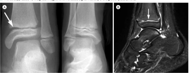
제1형 Salter-Harris 골절은 성장판에 골절이 생겨 골간단과 골단이 분리되는 손상이며, 손가락이 나 발가락뼈에서 가장 흔하게 발생한다(7). 일반 촬영에서는 전위(displace) 된 골단을 발견함으로써 진단할 수 있으나 골단이 전위되지 않은 경우에는 성장판의 확장이나 단순한 연부조직 부종만 보일 수도 있다(Fig. 4).
Normal
Type III
Type I
Type VI
Type II
Type V Fig. 3. Salter-Harris classification of physeal injuries.
제2형 Salter-Harris 골절은 골절선이 성장판과 골간단에서 발생한 손상이며, 가장 흔하게 발생하 는 유형이다. 요골 원위부, 경골과 비골의 원위부, 손가락이나 발가락뼈에서 잘 발생한다(Fig. 5). 일 반 촬영에서 골간단의 골절이 작을 경우 진단이 어려울 수 있는데, 세모꼴의 골간단 골절파편(“cor- ner sign”)이 보이면 제2형 Salter-Harris 골절로 진단할 수 있다. 제1형 및 제2형 Salter-Harris 골절 은 골단에 가까운 germinal zone의 연골세포와 혈관의 손상을 동반하지 않아 예후가 좋은 편이다.
제3형과 4형 Salter-Harris 골절은 골단에 골절이 발생한 경우로, germinal zone의 연골 세포와
Fig. 4. Type 1 Salter-Harris injury.
A. The plain radiograph shows irregular widening (arrow) of the growth plate of the right distal tibia (left image). The right image represents a normal growth plate.
B. The fat-suppressed T2-weighted image shows increased signal intensity of the growth plate (arrow).
A B
A B C
Fig. 5. Type 2 Salter-Harris injury.
A. The plain radiograph shows widening of the anterior portion of the growth plate of the distal tibia, accompanied by fractures in the me- taphysis.
B. The sagittal fat-suppressed T2-weighted image shows hyperintensity of the growth plate (arrow) and a fracture line in the metaphysis.
C. The sagittal T2-weighted image shows entrapment of the disrupted periosteum within the physeal fracture (arrowhead).
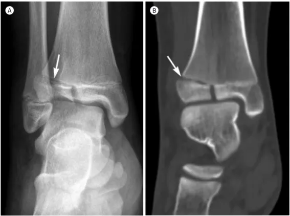
https://doi.org/10.3348/jksr.2020.81.4.806 811 혈관의 손상을 동반하기 때문에 제1형과 2형 골절에 비해서 예후가 나쁘다. 제3형 Salter-Harris 골 절은 성장판과 골단에 골절이 발생하나 골간단의 골절은 동반하지 않는다(Fig. 6). 예후는 제4형 Salter-Harris 골절보다 양호하다. 제4형 Salter-Harris 골절은 골절이 골단의 관절면에서 시작하 여 성장판을 가로지르고 골간단까지 연장되며, 상완골과 경골 원위부에서 잘 발생한다(Fig. 7). 제5 형 Salter-Harris 골절은 성장판에 압박이 가해져 발생하며, 예후는 가장 좋지 않다. 가장 드문 형태 이나 성장판 이 발생한 다른 형태의 Salter-Harris 골절에서도 제5형 골절이 동반되었을 것으로 추 측하기도 한다(2). 대퇴골 원위부나 경골 근위부에서 가장 흔하게 발생하며, 골간의 골절과 동반되 는 경우가 많다. 초기 영상 검사에서는 정상이거나 성장판의 협착(narrowing) 및 불규칙한 경계 등 의 소견이 보일 수 있다(Fig. 8). 발견이 매우 어려워 손상 기전을 염두에 두고 정상 측과 비교를 하 면서 영상을 판독하는 것이 중요하며, 추적 영상 검사에서 뒤늦게 발견되는 경우가 흔하다.
MRI의 액체민감영상(fluid-sensitive sequences)에서 성장판의 골절은 성장판의 확장 및 증가된 신호강도를 보이며(Figs. 4B, 5B), 조영증강 영상에서 골절 부위의 성장판은 조영증강이 되지 않는 다. 3차원 경사에코영상에서는 성장판의 고신호강도가 소실되어 보일 수도 있다(8). 특히 골절 부위 에 뼈막이나 인대, 힘줄, 근육과 같은 연부조직이 포착(entrapment) 된 경우 성장판 조기유합(pre- mature physeal closure)과 같은 후유증의 발생 위험이 높기 때문에 주의 깊은 평가가 필요하다(9, 10). 정복 후 추적 영상 검사에서 성장판의 넓이가 3 mm 이상으로 확장되었을 경우에는 골절 부위 에 연부조직이 포착되었을 가능성을 의심해야 하며(9, 11), 골절 부위에 포착된 뼈막은 MRI에서 저
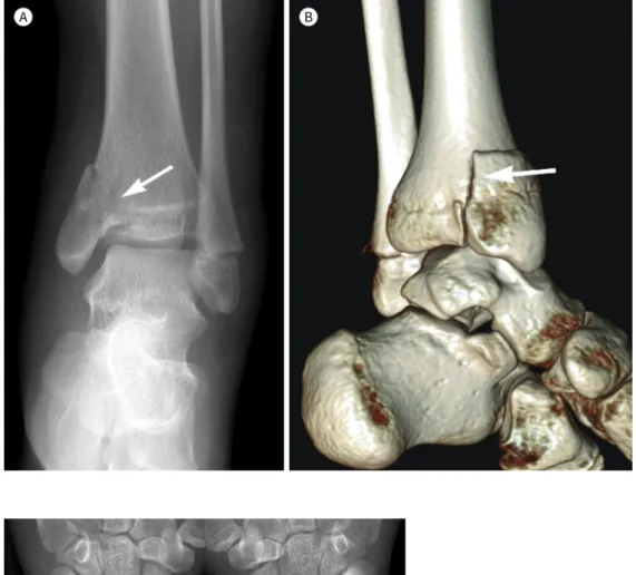
A B
Fig. 6. Tillaux fracture (type 3 Salter-Harris injury) in a 10-year-old child.
A, B. The plain radiograph (mortise view) (A) and coronal reformatted CT image (B) show vertical fracture of the epiphysis and widening of the growth plate (arrows) of the distal tibia.
신호강도로 보인다(Fig. 5C).
성장판 조기유합은 성장판 골절의 약 15%에서 발생하며, 손상 후 대략 3개월 정도에 발생하는 것 으로 알려져 있다(12). 치유기에 성장판을 가로지르는 뼈다리(bone bridge)를 형성할 수 있고, 성장 판의 조기유합으로 인하여 뼈의 성장 장애를 유발할 수도 있다(13). 일반 촬영에서 성장판의 조기유 합이 발생한 부분은 방사선투과성(radiolucent)의 성장판 음영이 소실된다(Fig. 9A). 성장판의 중심 부분에서 조기유합이 진행된 경우 일반 촬영에서 성장판의 모양은 컵 모양(cupped appearance)으
Fig. 7. Type 4 Salter-Harris injury.
A, B. The plain radiograph (A) and volume rendering CT image (B) show a fracture involving the epiphysis and metaphysis crossing the growth plate in the distal tibia (arrows).
A B
Fig. 8. Type 5 Salter-Harris injury.
The plain radiograph (left image) shows obliteration and marginal irregularity of the growth plate in the distal radi- us (arrow). Note the associated buck- ling of the cortex (arrowhead). The right image reveals a normal left dis- tal radius.
https://doi.org/10.3348/jksr.2020.81.4.806 813 로 변형되어 유합이 발생된 부위의 골단과 성장판이 골간단 부위로 돌출되는 모양(Supplementary Fig. 2 in the online-only Data Supplement)을 보이며(12, 14), 사지 길이의 차이를 동반하는 경우 가 많다. 성장판의 주변부에서 유합이 발생한 경우에는 뼈 성장 속도의 차이로 인하여 각변형을 일 으키게 된다.
뼈다리는 MRI에서 저신호강도 혹은 골수와 동일한 신호강도를 보인다(Fig. 9B). 특히 3차원 지 방억제 양자밀도강조영상이나 지방억제 T2 강조영상과 같은 연골이나 액체에 민감한 영상에서 뼈 다리가 생성된 부위는 저신호강도로 보이며 성장판의 고신호강도가 소실된다. 다평면 영상 재구성 과 최대신호강조영상 기법 등으로 성장판 조기유합의 위치와 범위를 자세히 평가할 수 있다 (Supplementary Fig. 3 in the online-only Data Supplement) (12, 15).
상지 골절
상완골(Humerus) 골절
소아에서 상완골(humerus) 근위부 골절의 양상은 나이에 따라 다양하게 나타나는데, 5세 이하에 서는 제1형 Salter-Harris 골절이, 5세에서 10세에는 골간단의 융기 골절이, 10세 이상에서는 제2형 Salter-Harris 골절이 자주 발생한다. 어린 소아에서는 관절을 감싸고 있는 단단한 연부조직으로 인
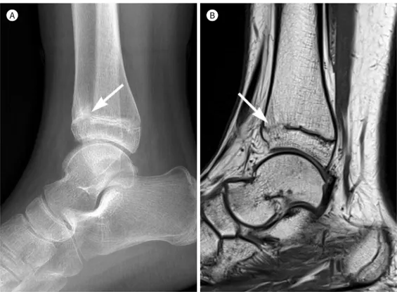
Fig. 9. Premature physeal closure of the peripheral portion of the growth plate.
A. The plain radiograph shows focal sclerotic changes in the anterior portion of the growth plate of the dis- tal tibia (arrow).
B. The sagittal T1-weighted image reveals a bone bridge in the anterior portion of the growth plate of the distal tibia, isointense with the bone marrow (arrow).
A B
하여 성장판 손상이 발생하여도 오목위팔관절(glenohumeral joint)의 탈구가 동반되는 경우는 드 물다.
신생아에서는 분만 외상으로 인해 근위부 상완골에 골절이 발생할 수 있다. 특히 제1형 Salter- Harris 골절이 발생한 경우, 신생아에서는 아직 골단의 골화 중심이 일반 촬영에서 보이지 않기 때 문에 일반 촬영에서 단순한 오목위팔관절의 탈구로 오인할 수 있어 주의가 필요하다. 신생아에서는 초음파가 진단에 유용하며, 성장판과 골간단의 골절, 전위, 연부조직 부종 등의 소견을 확인할 수 있다(Fig. 10).
팔꿈치 골절
정상 팔꿈치의 영상 소견
팔꿈치는 상완골, 요골, 척골 등으로 이루어져 있으며, 성장함에 따라 일반 촬영에서 6개의 골화 중심(ossification center)들이 순차적으로 나타난다. 일반 촬영에서 골화중심들이 보이기 시작하
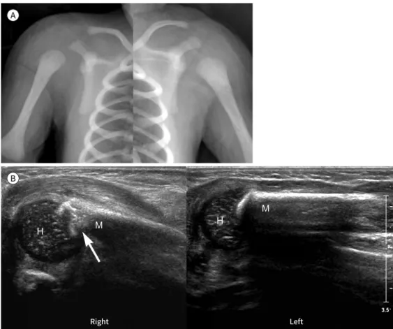
Fig. 10. Type 1 Salter-Harris injury in a neonate as a birth injury.
A. The plain radiograph shows lateral displacement of the right humerus (left image). The right image re- veals a normal left humerus.
B. Ultrasonography shows a fracture involving the growth plate and metaphysis of the right proximal hu- merus (arrow). Note the displacement of the humeral head (H) from the metaphysis (M) and soft tissue swelling, including the deltoid muscle.
Right Left
A
B
https://doi.org/10.3348/jksr.2020.81.4.806 815 는 연령은 개인마다 차이가 있을 수 있으나, 나타나는 순서는 거의 일정하다. 이 골화중심을 간혹 골 절 파편으로 오인할 수 있어 주의가 필요하다(Supplementary Fig. 4 in the online-only Data Supplement). 어린 소아의 팔꿈치를 이루는 뼈는 아직 골화가 되지 않은 부분이 많기 때문에 일반 촬영에서 보이지 않는 연골의 범위를 예측할 수 있어야 골절의 범위, 분류, 및 성장판과 연골 손상 여부를 정확히 평가할 수 있다.
미세한 융기 골절이나 생나무 골절 등은 일반 촬영에서 잘 보이지 않을 수 있기 때문에 팔꿉관절 액(elbow joint effusion) 증가 여부를 평가하는 것이 진단에 도움이 된다. 정상 팔꿉관절의 측면 촬 영에서는 상완골 원위부의 앞쪽으로 오목한 모양(sliverlike, concave) 혹은 삼각형 모양의 앞지방 패드(anterior fat pad)를 볼 수 있다. 그러나 팔꿉관절액이 증가한 경우 앞지방패드가 볼록한 모양 을 보이면서 융기되는 소견(sail sign)이 나타날 수 있다. 한편 상완골 원위부 뒤쪽의 지방패드는 팔 꿈치오목(olecranon fossa)에 가려져 정상적으로는 보이지 않으나, 팔꿉관절액이 증가한 경우에 는 보일 수 있다(Fig. 11).
팔꿈치를 이루는 뼈의 정렬은 전방상완골피질선(anterior humeral line)과 요골상완골선(radio- capitellar line)으로 평가할 수 있다(Supplementary Fig. 5 in the online-only Data Supplement).
전방상완골피질선은 상완골의 앞쪽 피질을 따라 그은 선이며, 이는 소두(capitellum)의 중간 1/3 지 점을 지나야 하는데, 4세 이하에서는 이보다 약간 앞쪽으로 지날 수도 있다. 요골상완골선은 요골의 중심에서 긴축을 따라 그린 선이며, 전후면 촬영과 측면 촬영 영상에서 소두를 지나야 한다. 전방상 완골피질선은 과상 골절(supracondylar fracture)의 진단에 유용하며, 요골상완골선은 요골상완 골탈구(radiocapitellar dislocation), 외과 골절(lateral condylar fracture), 요골 두(radial head)
A B
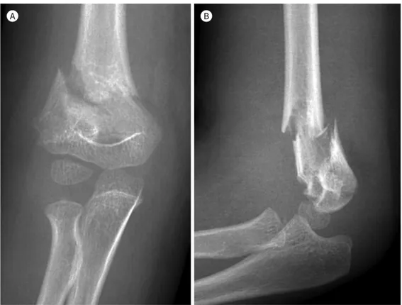
Fig. 11. Type 1 supracondylar fracture.
A. The anteroposterior view shows a transverse fracture (arrow) in the distal humerus.
B. The lateral view (left image) reveals elevation of the anterior fat pad (sail sign) and appearance of the posterior fat pad (arrowheads). Normally (right image), the anterior fat pad shows a concave or triangular shape (curved arrow), and the posterior fat pad should not be seen.
골절 등의 진단에 유용하다.
과상 골절(Supracondylar Fracture)
과상 골절(supracondylar fracture)은 소아의 상완골 원위부 골절 중 가장 흔하게 발생하는 형태 이다. 대부분의 과상 골절은 손과 팔이 펴진 채로 낙상을 당할 때 발생하는데(FOOSH-fall onto outstretched hand) (16), 상완골 뒤쪽이 척골의 팔꿈치머리(olecranon)에 의해서 충격을 받게 되 면서 골절이 발생한다. 성장판 손상을 수반하는 경우는 드물지만, 동반된 아래팔 골절 여부를 주의 깊게 살펴봐야 한다. 골절은 일반적으로 수정된 Gartland 분류에 의해서 4가지 형태(Figs. 11, 12, Supplementary Figs. 6, 7 in the online-only Data Supplement)로 Table 1과 같이 구분할 수 있다(17-20). 팔꿉관절의 탈구를 동반하는 경우는 드물다. 골절이 심하지 않고 전위가 없는 경우 일 반 촬영에서 진단이 어려울 수 있으며, 전방상완골피질선이나 팔꿈치 지방패드의 변화를 주의 깊게 살펴보는 것이 진단에 도움이 된다(Fig. 11, Supplementary Fig. 6 in the online-only Data Supplement).
외과 골절(Lateral Condylar Fracture)
외과 골절(lateral condylar fracture)은 소아에서 두 번째로 흔한 상완골 원위부 골절이며(21), 아래팔이 회외전(supination) 되어 있고, 팔꿈치가 펴진(extension) 상태에서 내반력(varus stress)
A B
Fig. 12. Type 3 supracondylar fracture.
A, B. Anteroposterior (A) and lateral view (B) images reveal fractures in both anterior and posterior aspects of the distal humerus, accompanied by mild displacement.
https://doi.org/10.3348/jksr.2020.81.4.806 817 이 가해졌을 때 발생한다. 외과 골절은 골절의 범위와 외측과(lateral condyle)의 전위 여부에 따라 서 구분한 Jakob 분류(Fig. 13, Supplementary Figs. 8, 9 in the online-only Data Supplement) 를 일반적으로 사용하며 Table 2와 같이 구분한다(2, 22, 23). 소두의 유합이 완전히 이루어지지 않 은 소아에서는 상완골 골간단과 소두 사이에서 골간단과 평행한 방향의 골절을 볼 수 있다. 골절조 각이 상완골의 후외측(posterolateral)에 위치하기 때문에 내측경사촬영에서(internal oblique view) 골절을 더 명확히 볼 수 있다(Fig. 13B) (21).
Fig. 13. Type 1 lateral condylar fracture.
A. The anteroposterior view shows a fracture of the lateral condyle of the distal humerus (arrow), with no dis- placement.
B. The fracture (arrow) is more evident in the internal oblique view.
A B
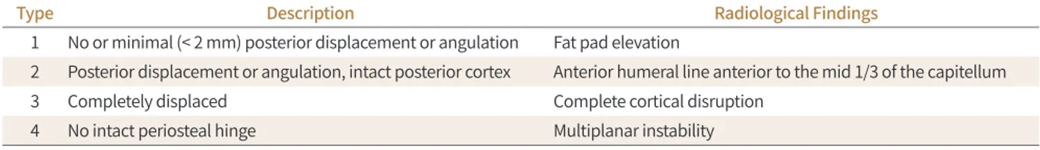
Table 1. Modified Gartland Classification of Supracondylar Fracture (18, 19)
Type Description Radiological Findings
1 No or minimal (< 2 mm) posterior displacement or angulation Fat pad elevation
2 Posterior displacement or angulation, intact posterior cortex Anterior humeral line anterior to the mid 1/3 of the capitellum
3 Completely displaced Complete cortical disruption
4 No intact periosteal hinge Multiplanar instability
Table 2. Jakob Classification of Lateral Condylar Fracture (22, 23)
Type Description Radiological Findings
1 Incomplete Displaced < 2 mm, intact articular cartilage Fracture line enters between the capitellum and trochlear 2 Complete Displaced ≥ 2 mm, intact articular surface Fracture line extends to the articular surface
3 Complete, displaced Displaced ≥ 2 mm, displaced articular surface Completely displaced lateral condyle
내상과 골절(Medial Epicondylar Fracture)
상완골 내상과(medical epicondyle)는 아래팔의 굽힘근육(flexor muscle)들과 내측측부인대 (ulnar collateral ligament)의 부착부위이다. 골절은 주로 팔꿈치가 과신전된(hyperextension) 상태에서 외반력(valgus stress)이 작용했을 때 발생한다. 낙상과 같은 직접적인 손상(Fig. 14)이나 공 던지기와 같은 반복적인 팔꿉관절의 사용으로 인해서 견열 골절(avulsion fracture)이 발생할 수 있다(Little leaguer’s elbow) (Supplementary Fig. 10 in the online-only Data Supplement). 팔 꿉관절의 탈구를 자주 동반하기 때문에, 영상에서 팔꿉관절의 탈구가 발견된 경우에는 내상과 골절 여부를 신중히 평가하여야 한다. 척골신경의 손상을 수반할 수도 있다(24).
일반 촬영에서 전위된 골절 조각이나 내상과 뼈돌기(apophysis)를 발견함으로써 진단할 수 있으 나(Fig. 14), 전위가 적은 경우에는 발견이 어려울 수도 있어 정상 측과 비교하면서 판독하는 것이 중요하다(Supplementary Fig. 10 in the online-only Data Supplement). 또한 팔꿉관절 탈구의 정복 후 영상에서 내상과가 정상적인 위치에 보이지 않는 경우에는 골절 조각이나 내상과 뼈돌기가 팔꿉관절 내에 포착되어 있는지 살펴볼 필요가 있다. MRI는 내측측부인대나 척골신경 손상 여부, 팔꿉관절 외측에서 동반된 상완골이나 요골의 뼈연골손상(osteochondral injury), 골수 부종 등의 평가에 유용하다.
요골 두(Radial Head) 및 경부(Radial Neck) 골절
소아에서 요골 두(head)나 경부(neck)의 골절은 주로 융기 골절 혹은, 제1형이나 제2형 Salter- Harris 골절의 형태로 발생하며, 요골 근위부의 각변형이나 전위를 동반할 수 있다(Fig. 15). 요골상
Fig. 14. Medial epicondyle avulsion fracture in a 6-year-old child after a fall.
A, B. The anteroposterior view (A) and lateral view (B) show avulsion and displacement of the apophysis of the medial epicondyle (arrows). Note the disruption of the radiocapitellar line that suggest dislocation of the radiocapitellar joint.
A B
https://doi.org/10.3348/jksr.2020.81.4.806 819 완골관절의 탈구를 동반하는 경우는 흔하지 않지만, 팔꿈치머리(olecranon)나 내상과 견열 골절 등을 동반할 수 있다.
팔꿈치머리(Olecranon) 골절
팔꿈치머리 골절은 견열 골절, 가로 골절(transverse fracture), 경사 골절(oblique frcture), 분쇄 골절(comminuted fracture) 등 다양한 형태로 발생할 수 있으며, 측면 촬영이 골절의 평가에 가장 유용하다. 소아에서 팔꿈치머리 단독 골절은 드물기 때문에 동반 손상을 평가하는 것이 중요하며, 약 1/3에서 요골 두나 내상과, 갈고리돌기(coronoid process)의 골절을 동반한다(25). 소아에서는 정상적인 팔꿈치머리의 골화중심을 골절과 혼동할 수 있으므로 주의가 필요하다. 골절은 보통 골화 중심보다 얇고, 경계가 불규칙하며, 팔꿈치머리의 성장판 방향과 평행하고, 피질골을 보이지 않는 다(Fig. 16).
아래팔 골절
소아의 아래팔 골절은 과상 골절과 마찬가지로 FOOSH 손상에 의해서 발생하는 경우가 많으며, 요골과 척골 원위부의 골절이 가장 흔하다. 요골 원위부는 소아에서 죔쇠 골절이 가장 자주 발견되 는 부위이기도 하다. 요골 골간과 척골 골간의 골절은 동시에 발생하는 경우가 많으며, 요골 원위부 의 성장판 손상은 제2형 Salter-Harris 골절이 가장 흔하다. 요골 원위부의 작은 죔쇠 골절은 일반 촬영에서 잘 보이지 않을 수 있으므로 여러 방향에서 촬영한 영상을 주의 깊게 평가하는 것이 중요 한데, 미세한 각변형이나 피질골의 융기가 보인다면 죔쇠 골절을 의심할 수 있다(Fig. 2).
Fig. 15. Radial head fracture.
The anteroposterior view shows the type 2 Salter-Harris injury involving the proximal radius (arrow). Note the angulation and dislocation of the radial head.
아래팔 골절은 탈구를 동반할 수 있기 때문에 팔꿈치와 손목 관절의 영상을 함께 평가해야 한다.
Monteggia 손상은 척골 근위부의 골절과 요골상완골관절의 탈구를 동반한 경우를 말하며 소아에 서 척골 근위부의 단독 골절은 드물기 때문에, 척골 근위부 골절이 발견되면 Monteggia 손상의 가 능성을 염두에 두어야 한다(Fig. 17). Galeazzi 손상은 요골 원위부 골절에 원위요척관절(distal ra- dioulnar joint)의 탈구가 동반된 손상을 말하며, 요골 원위부 골절이 있는 경우에는 원위요척관절 의 탈구 여부를 함께 평가해야 한다(Fig. 18). 척골경상돌기(ulnar styloid process)의 골절, 요골의 5 mm 이상 단축 (shortening), 2 mm 이상의 원위요척관절의 확장(widening) 등의 소견들이 원 위요척관절 탈구의 진단에 도움이 된다. 소아에서는 Monteggia 손상보다 Galeazzi 손상이 더 흔하 게 발견된다.
주상골(Scaphoid) 골절
주상골(scaphoid)은 소아의 손목뼈 중 가장 흔하게 손상되는 뼈이며, 삼각골(triquetrum) 및 대 능형골(trapezium) 등이 주상골 다음으로 흔하게 손상된다(2). 과거에는 주상골 원위극(distal pole) 의 골절이 가장 흔한 것으로 보고되었으나, 최근에는 신체질량지수(body mass index)의 증가와 운동 손상의 증가로 주상골 허리(scaphoid waist)의 골절이 흔한 것으로 보고되고 있다(26, 27).
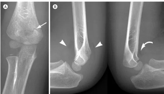
골절의 평가에 가장 좋은 영상은 손목의 척골 편위(unlar deviation) 상태에서 시행한 주상골 촬 영(scaphoid view)이다. 초기 영상에서는 골절이 명확히 보이지 않을 수 있어 약 10일에서 14일 후 골절의 재평가가 필요할 수도 있다. 급성 시기에는 일반 촬영보다 CT나 MRI가 골절의 발견에 더 민감하다(Fig. 19) (28, 29). 일반 촬영에서 주상골과 월상골(lunate)의 간격이 3 mm 이상인 경우에 Fig. 16. Olecranon fracture.
A. The lateral view shows a fracture in the metaphyseal-equivalent zone of the olecranon (arrow). The frac- ture shows a thin, sliver-like appearance and an irregular border with lack of cortication.
B. The lateral view of the normal ossification center of the olecranon. Note the smooth border and cortica- tion of the apophysis.
A B
https://doi.org/10.3348/jksr.2020.81.4.806 821 는 주상월상인대(scapholunate ligament)의 손상을 의심해야 하며, 이는 주상월상골 진행성 붕괴 (scapholunate advanced collapse, SLAC)와 같은 합병증과 연관이 있다. 주상골 동맥은 원위극에 서 근위극(proximal pole)의 방향으로 주행하기 때문에, 근위극의 골괴사(osteonecrosis)나 불유 합(non-union)과 같은 합병증이 발생할 수 있다. 골괴사가 진행된 경우에는 주상골의 경화성 변화 및 연골하 뼈의 낭성 변화, 붕괴(collapse) 등의 소견들을 볼 수 있다(Supplementary Fig. 11 in the online-only Data Supplement).
손가락 골절
손뼈의 골절은 9세에서 14세에 가장 흔하게 발생한다(30). 성장판 손상의 동반이 흔한 편이지만, 뼈 성장의 장애로 진행하는 경우는 상대적으로 드물다. 원위부 손가락뼈사이관절(distal interpha- langeal joint)의 과굴곡(hyperflexion)으로 인해 끝마디뼈(distal phalanx) 기저부의 후방에 발생 한 제3형 Salter-Harris 견열 골절을 Mallet finger라고 부른다(Supplementary Fig. 12 in the online-only Data Supplement). 반대로 손가락의 과신전에 의해서 생긴 끝마디뼈나 중간마디뼈 (middle phalanx)의 손가락굽힘근힘줄(flexor digitorum tendon) 부착 부위인 손바닥판(volar plate)에도 제3형 Salter-Harris 손상이 발생할 수 있다(Supplementary Fig. 13 in the online-only Data Supplement). 끝마디뼈의 골절은 어린 소아에서 흔하게 발생할 수 있는데, 문틈과 같은 좁은 부위에 손가락이 끼어서 발생하는 압궤손상(crushing injury)에 의한 경우가 많다(Supplementary Fig. 14 in the online-only Data Supplement). 사춘기에는 다섯 번째 중수골(metacarpal bone) Fig. 17. Monteggia fracture.
The anteroposterior view shows a fracture involving the diaphysis of the proximal ulna (arrow) and dislocation of the radiocapitellar joint (arrowhead).
머리의 골절이 흔하게 발생하며(Boxer’s fracture) 골절 원위부는 손바닥쪽으로 각변형을 일으키는 경우가 많다(Supplementary Fig. 15 in the online-only Data Supplement).
하지 골절
대퇴골두골단분리증(Slipped Capital Femoral Epiphysis)
대퇴골두골단분리증(slipped capital femoral epiphysis)은 대퇴골두골단(capital femoral epiph-
Fig. 18. Galeazzi fracture.
The anteroposterior view shows a fracture involving the diaphysis of the distal radius (arrow) and dislocation of the distal radioulnar and ulnocarpal joints (arrowhead).
Fig. 19. Scaphoid fracture.
A, B. The scaphoid view (A) and volume-rendering CT (B) images show a fracture involving the scaphoid waist (arrows). The scapholunate in- terval is normal (< 3 mm).
C. The coronal fat-suppressed T2-weighted image reveals a fracture (arrow) and combined bone marrow edema involving the scaphoid bone.
Note the normal scapholunate ligament (arrowhead).
A B C
https://doi.org/10.3348/jksr.2020.81.4.806 823 ysis)이 성장판에서 분리되어 후내측(posteromedial)으로 전위되는데, 사춘기 시기의 남아에서 호 발한다. 사춘기 시기의 뼈 성장과 연관이 있으나, 외상이나 내분비 혹은 염증성 질환과도 연관이 있 다. 높은 신체질량지수는 대퇴골두골단분리증의 위험 요인 중에 하나이며, 양측성 질환의 빈도는 약 60%로 알려져 있다(31). 외상에 의한 제1형 Salter-Harris 골절에 동반된 대퇴골두골단분리와 구분 하기도 한다(32). 환자가 목발과 같은 보조 기구 없이 몸무게를 지탱할 수 있는지의 여부에 따라서 안정성(stable slip)과 불안정성(unstable slip) 질환으로 구분한다(33).
대퇴골두골단분리를 평가하기 위해서는 대퇴관절 전후면 영상과 개구리다리자세 측면 영상(frog leg lateral view)를 함께 평가하는 것이 중요하다. 대퇴관절 전후면 영상에서 대퇴골 경부(femoral neck)의 피질을 따라 그린 선을 Klein 선(Klein’s line)이라고 부르며, 정상적으로 이 Klein 선은 대 퇴골두골단의 바깥쪽을 지난다. 만일 대퇴골두와 Klein 선이 만나지 않는 경우에는 대퇴골두의 내 측 전위를 진단할 수 있으나(Supplementary Fig. 16 in the online-only Data Supplement), 대 퇴골두골단의 내측 전위의 정도가 적을 경우에는 전후면 영상에서 진단이 어려울 수 있다. 대퇴골 두골단은 후내측으로 전위되기 때문에 전후면 영상에서 대퇴골단의 내측 전위가 잘 보이지 않는 경 우에는 개구리다리자세 측면 영상이 대퇴골두의 후측 전위를 평가하는 데 유용하다(Fig. 20A, B).
대퇴골두골단의 전위가 일어나기 전(pre-slip phase)에는 대퇴골두 성장판의 확장 및 불규칙한 경 계면, 골단의 방사선투과성 변화 등의 소견만이 보일 수 있어, 일반 촬영에서 이상 소견이 명확히 보 이지 않는 경우 MRI를 고려할 수 있다(Fig. 20C).
경골 근위부(Proximal Tibia) 및 경골거친면(Tibial Tuberosity) 골절
측면에서 봤을 때 경골 근위부의 성장판은 뒤집힌 L자의 형태를 이루고 있으며, 경골 근위부 골단 과 경골거친면(tibial tuberosity)은 약 15~17세 경에 유합된다. 따라서 소아에서 경골 근위부 골단 과 경골거친면까지 확장되는 골절(Supplementary Fig. 17 in the online-only Data Supplement) 은 성장판 손상을 주의 깊게 평가해야 한다(2). 경골거친면의 골절은 반복적인 도약에 의한 손상이 흔하다. 오스굿-슐라터병(Osgood-Schlatter disease)과 경골거친면 골절의 감별이 필요한데, 전자 는 무릎힘줄(patellar tendon) 부착부위의 반복적인 장력(tension)으로 인해 경골거친면의 염증과 박리가 서서히 발생하는 질환인 반면, 후자는 외력에 의해서 급성으로 발생한 성장판의 골절인 점이 다르다.
골절의 위치와 형태에 따라서 제2형(Supplementary Fig. 17 in the online-only Data Supplement) 혹은 제3형(Fig. 21) Salter-Harris 골절이 발생할 수 있으며, CT나 MRI와 같은 단면 영상이 골절의 형태와 범위의 평가에 유용하다. 특히 성장판, 관절 연골, 동반된 연부조직의 손상 여부 평가가 필요할 경우에는 MRI가 유용하다.
경골(Tibia)과 비골(Fibula)의 골간 골절
경골 골간은 소아 하지 골절이 가장 흔하게 일어나는 부위이다. 대개 직접적인 외력에 의해서 발 생하며, 비골 골간의 골절을 자주 동반한다(34). 특히 약 9개월에서 3세 사이의 유아에서 발생하는 경골 골간의 나선형 골절을 유아기 골절(toddler fracture)이라고 부른다. 전형적으로 약한 비틀림
이나 낙상에 의해서 발생하는 경골의 골절을 유아기골절로 일컬어 왔으나, 최근에는 유아기에 생긴 다리나 발의 골절을 모두 유아기 골절 스펙트럼으로 부르기도 한다. 유아기 골절 스펙트럼에서는 경 골의 골절이 가장 흔하고 비골이나 입방골(cuboid)의 골절도 발생할 수 있다(2, 35-38).
유아기 골절은 일반 촬영에서 하지 긴뼈(long bone)의 골간단이나 골간에서 사선형이나 나선형 의 골절선을 보이며, 대개 골절의 전위는 동반하지 않는다(Supplementary Fig. 18 in the online- only Data Supplement). 초기 영상에서 골절선이 명확히 보이지 않을 수도 있으며. 2~3주 후에 시
Fig. 20. Slipped capital femoral epiphysis.
A. The anteroposterior view shows widening of the growth plate (ar- row) of the right femoral head.
B. The frog lateral view more clearly depicts posterior displacement of the right capital femoral epiphysis.
C. The coronal fat-suppressed T2-weighted image reveals widening and high signal intensity of the growth plate and bone marrow edema of the head and neck of the right proximal femur. Note the effusion in the right hip joint.
A
B
C
https://doi.org/10.3348/jksr.2020.81.4.806 825 행한 추적 영상에서 골절선 주변의 경화성 변화 및 뼈막주위의 뼈형성(periosteal new bone for- mation) 등으로 발견되기도 한다. 전후면 촬영, 측면 촬영, 경사면 촬영 중 한 방향의 영상에서만 골 절선이 보일 수 있어 주의가 필요하다.
경골 원위부 틸로우 골절(Tillaux Fracture) 및 삼면 골절(Triplane Fracture)
경골 원위부의 성장판은 약 10세 전후로 유합되기 시작하는데, 내측에서 외측 방향으로 유합이 진 행되어 성장판의 전외측(anterolateral)이 가장 늦게 유합된다. 따라서 성장판이 유합되는 시기에 경골 원위부에 외력이 가해졌을 경우 성장판 유합의 정도에 따라서 다양한 양상의 골절이 발생할 수 있으며, 주로 외회전 방향의 외력에 의해서 틸로우 골절(Tillaux fracture)과 삼면 골절(triplane fracture)이 발생할 수 있다.
틸로우 골절은 경골 원위부 성장판의 내측이 유합되고 외측은 유합되지 않은 상태에서, 전방경비 인대(anterior tibiofibular ligament)에 외회전(external rotation) 방향의 외력이 가해져 경골 원 위부 골단의 전외측에 발생한 견열 골절을 말한다. 성장판과 경골 골단에서 골절이 발생하므로 제3 형 Salter-Harris 골절로 분류할 수 있다(Fig. 6).
삼면 골절은 틸로우 골절에서 경골 골간단에 관상면(coronal plane) 방향의 골절이 추가된 형태 로 전형적으로 세 가지 방향의 골절—1) 경골 원위부 골단의 시상면(sagittal plane) 골절, 2) 성장판
A B
Fig. 21. Tibial tuberosity avulsion fracture.
A, B. The plain radiograph (A) and sagittal reformatted CT (B) image show an avulsion fracture of the tibial tuberosity extending to the proximal tibial epiphysis (arrow). The fracture can be regarded as the type 3 Salter-Harris fracture.
의 횡단 골절(transverse fracture), 3) 경골 원위부 골간단의 관상면 골절—이 발생한다(Fig. 22). 경 골 원위부 골단과 성장판, 그리고 골간단의 골절이 동반됨에도 불구하고, 제2형 및 제3형 Salter- Harris 골절이 함께 발생한 것으로 간주하지만, 골절의 부위가 2개로 나뉘는 경우에는 제4형 Salter- Harris 골절로 분류할 수 있다(32).
일반 촬영에서는 복잡한 골절선의 구분이 잘되지 않아 정확한 골절의 범위를 평가하기가 어려울 수 있으며, 이러한 경우 CT나 MRI와 같은 단층 영상이 골절의 범위와 형태, 관절면 침범 여부, 성장 판과 연부조직 손상의 평가에 유용하다.
거골(Talus) 골절
거골(talus)의 골절은 소아에서는 흔하지 않으나 유아기 골절(toddler fracture)이 발견되는 부위 중에 하나이다. 견열 골절이나 골연골 골절, 거골의 목이나 몸통 등에서 골절이 발생할 수 있으며 (Supplementary Fig. 19A in the online-only Data Supplement), 어린 소아에서는 죔쇠 골절이 발견되기도 한다. 치유기에 근위극에서 골괴사가 발생할 수 있는데, 약 2개월 뒤 추적 영상에서 거 골원개(talar dome)의 연골하 방사선투과성음영(subchondral radiolucency)이 보이면(Hawkins sign) 골괴사 발생의 위험이 낮은 것으로 알려져 있다(Supplementary Fig. 19B in the online- only Data Supplement) (39).
A B
Fig. 22. Triplane fracture involving the distal tibia in a 10-year-old child.
A. The mortise view shows a fracture in the distal tibial epiphysis in the sagittal plane (arrow) and widening of the growth plate in the lateral aspect.
B. The lateral view reveals a fracture involving the metaphysis of the distal tibia (arrow) in the coronal plane.
https://doi.org/10.3348/jksr.2020.81.4.806 827 입방골(Cuboid) 골절
입방골(cuboid) 골절은 과굴곡(hyperflexion) 된 발에 수직 방향의 힘이 가해졌을 때 중족골 (metatarsal bones) 종골(calcaneus) 사이에서 입방골이 충격(impact)을 받아 발생하는 경우가 많다(40). 유아기 골절이 발생할 수 있는 부위이나, 골절의 발견이 어려워 간과하기 쉽다(41, 42). 따 라서 바깥쪽 발의 통증이 있고 몸무게를 지탱할 수 없는 환아에서 입방골 골절을 의심하는 것이 중 요하다. 전위가 없는 골절인 경우 초기 영상에서 진단이 어려울 수 있으며, 치유기에 골절 주변부의 경화성 변화나 뼈모양의 변형 등으로 발견되기도 한다. 어린 소아에서 입방뼈의 모양은 다양하게 보일 수 있으므로 정상 측과 비교하는 것이 중요하다(Supplementary Fig. 20 in the online-only Data Supplement).
중족골 골절
소아의 중족골(metatarsus)과 발가락뼈의 골절은 발의 다른 부위 골절보다 비교적 흔하며, 발등 쪽에 무거운 물체가 떨어지면서 발생하는 직접적인 외력에 의해서 발생하는 경우가 많다. 중족골 기저부에서 골절이 발생한 경우에는 발목발허리관절의 손상(Lisfranc injury)의 가능성을 고려해 야 한다.
소아에서 첫 번째 중족골 기저부에 축방향의 외력에 의해서 죔쇠 골절이 발생할 수 있는데, 2단 침대에서 딱딱한 마룻바닥을 향해 뛰어내릴 때 자주 발생한다고 하여 2단 침대 골절(bunk bed fracture)로 불리기도 한다. 일반 촬영에서는 첫 번째 중족골 기저부의 피질의 융기 및 각변형을 볼 수 있다(Fig. 23).
5번째 중족골거친면(5th metatarsal tuberosity)의 견열 골절은 발의 과도한 내번(inversion)이 나 내전(adduction)에 의해서 발생하며 짧은종아리근(Peroneus brevis)이나 새끼벌림근(Adduc- tor digiti minimi), 또는 발바닥널힘줄(plantar aponeurosis)에 의해 힘줄 부착부위가 당겨져 발 생하는 것으로 알려져 있다(43, 44). 골절은 중족골의 횡축 방향으로 발생한다(Fig. 24). 간혹 5번째
Fig. 23. Buckle fracture involving the 1st metatarsal base (bunk bed fracture).
Note the buckling of the 1st metatarsal base (arrow).
중족골거친면의 정상적인 견인골단(apophysis)을 견열 골절로 오인할 수 있으나, 정상적인 견인골 단은 중족골의 장축과 평행한 모양으로 보이기 때문에 감별이 가능하다(Supplementary Fig. 21 in the online-only Data Supplement). 그러나 아직 유합되지 않은 견인골단에 박리가 발생한 경 우에는 진단이 어려울 수 있는데, 정상 측과 비교하여 병변 측 견인골단이 근위부로 전위되거나 중 족골거친면 성장판이 확장된 소견 등이 보이면 진단할 수 있다.
반복적인 내번에 의해서 5번째 중족골거친면에서 1.5 cm 이내의 골간에 발생한 횡단 골절을 Jones 골절이라고 하며(Supplementary Fig. 22 in the online-only Data Supplement), 5번째 중족골 거친면 견열 골절과 감별을 요한다. 소아에서는 Jones 골절보다는 5번째 중족골거친면 견열 골절이 더 흔하게 발생한다.
결론
외상으로 인한 근골격계의 손상은 성장하는 소아에 있어 다양한 후유증과 성장 장애를 유발할 수 있으므로 정확한 영상의 평가가 환자의 치료에 중요한 역할을 한다. 소아 골절은 성인과는 다른 영 상의학적 특성을 보이기 때문에 영상 소견에 익숙하지 않는 경우 진단에 어려움을 겪을 수도 있다.
특히 소성변형, 죔쇠 골절, 생나무 골절 등은 소아에서만 특징적으로 보이므로 이러한 영상 소견을 숙지하고 있어야 조기 진단을 놓치는 실수를 줄일 수 있다. 또한 성장판 근처의 골절은 항상 성장판 손상을 염두에 두고 영상을 평가해야 하며, 치유기의 영상에서는 뼈다리의 형성이나 뼈의 변형 및 뼈 성장 장애 등에 주의를 기울여야 한다.
소아 골절의 대부분은 일반 촬영으로 진단이 가능하지만 골절이 명확히 보이지 않는 경우에는 여 러 방향에서 시행한 영상을 조합하여 판독하여야 한다. 골단의 골화가 완전히 이루어지지 않고 성 장판이 유합되기 전에는 손상과 정상의 구분이 어려울 수 있으므로, 일반 촬영에서 보이지 않는 연 골의 범위를 염두에 두면서 정상 측과 비교하며 영상을 평가하는 것이 중요하다. CT는 미세 골절의
Fig. 24. Avulsion fracture involving the 5th metatarsal tuberosity.
The oblique view reveals a transverse fracture involv- ing the 5th metatarsal tuberosity (arrow).
https://doi.org/10.3348/jksr.2020.81.4.806 829 발견이나 골절의 범위를 평가하는데 유용할 수 있으나, 일반 촬영에 비해 방사선 피폭의 위험이 높 기 때문에 필요한 경우에만 선택적으로 시행하여야 한다. MRI는 연골이나 관절 손상, 연부조직의 손상, 미세 골절, 성장판 손상 등의 평가가 필요할 때 유용하게 이용할 수 있으나, 검사 시간이 길고 어린 소아에서는 진정이 필요할 수도 있다는 점에서 제한이 있다. 초음파는 아직 널리 이용되고 있 지 않으나, 신생아 골절이나 관절의 손상, 연부조직이나 혈관 손상 평가 등에 선택적으로 이용할 수 있다. 영상의학과 의사는 소아 골절의 다양한 임상적, 영상의학적 특성을 이해하고 영상을 평가할 수 있어야 하며, 소아의 특수성을 고려하여 상황에 맞는 적절한 영상 검사를 제안할 수 있어야 하겠다.
Supplementary Materials
The online-only Data Supplement is available with this article at http://dx.doi.org/10.3348/
jksr.2020.81.4.806.
Conflicts of Interest
The author has no potential conflicts of interest to disclose.
Acknowledgments
This study was supported by a 2020 research grant from Pusan National University Yangsan Hospital.
REFERENCES
1. Kang MS, Kim HS. Characteristics and trends of traumatic injuries in children visiting emergency depart- ments in South Korea: a retrospective serial cross-sectional study using both nationwide-sample and sin- gle-institutional data. PLoS One 2019;14:e0220798
2. Coley BD. Caffey’s pediatric diagnostic imaging e-book. Amsterdam: Elsevier Health Sciences 2013 3. Borden S 4th. Roentgen recognition of acute plastic bowing of the forearm in children. Am J Roentgenol
Radium Ther Nucl Med 1975;125:524-530
4. Mizuta T, Benson WM, Foster BK, Paterson DC, Morris LL. Statistical analysis of the incidence of physeal inju- ries. J Pediatr Orthop 1987;7:518-523
5. Dodwell ER, Kelley SP. Physeal fractures: basic science, assessment and acute management. Orthop Trauma 2011;25:377-391
6. Salter RB, Harris WR. Injuries involving the epiphyseal plate. JBJS 1963;45:587-622 7. Rogers LF, Poznanski AK. Imaging of epiphyseal injuries. Radiology 1994;191:297-308
8. Jaramillo D, Kammen BF, Shapiro F. Cartilaginous path of physeal fracture-separations: evaluation with MR imaging--an experimental study with histologic correlation in rabbits. Radiology 2000;215:504-511
9. Barmada A, Gaynor T, Mubarak SJ. Premature physeal closure following distal tibia physeal fractures: a new radiographic predictor. J Pediatr Orthop 2003;23:733-739
10. Wirth T, Byers S, Byard RW, Hopwood JJ, Foster BK. The implantation of cartilaginous and periosteal tissue into growth plate defects. Int Orthop 1994;18:220-228
11. Nguyen JC, Markhardt BK, Merrow AC, Dwek JR. Imaging of pediatric growth plate disturbances. Radio- graphics 2017;37:1791-1812
12. Ecklund K, Jaramillo D. Patterns of premature physeal arrest: MR imaging of 111 children. AJR Am J Roent- genol 2002;178:967-972
13. Boutis K. The emergency evaluation and management of pediatric extremity fractures. Emerg Med Clin North Am 2020;38:31-59
14. Hensinger RN. Complications of fractures in children. In Green NE, Swiontkowski M, eds. Skeletal trauma in children. Philadelphia: Saunders Elsevier 2008:159-205
15. Sailhan F, Chotel F, Guibal AL, Gollogly S, Adam P, Bérard J, et al. Three-dimensional MR imaging in the as- sessment of physeal growth arrest. Eur Radiol 2004;14:1600-1608
16. Lins RE, Simovitch RW, Waters PM. Pediatric elbow trauma. Orthop Clin North Am 1999;30:119-132
17. Wilkins KE. Fracture and dislocation of the elbow region. Fracture in Children 1984:363-576
18. De Boeck H, De Smet P, Penders W, De Rydt D. Supracondylar elbow fractures with impaction of the medial condyle in children. J Pediatr Orthop 1995;15:444-448
19. Alton TB, Werner SE, Gee AO. Classifications in brief: the Gartland classification of supracondylar humerus fractures. Clin Orthop Relat Res 2015;473:738-741
20. Leitch KK, Kay RM, Femino JD, Tolo VT, Storer SK, Skaggs DL. Treatment of multidirectionally unstable su- pracondylar humeral fractures in children. A modified Gartland type-IV fracture. J Bone Joint Surg Am 2006;
88:980-985
21. Tejwani N, Phillips D, Goldstein RY. Management of lateral humeral condylar fracture in children. J Am Acad Orthop Surg 2011;19:350-358
22. Weiss JM, Graves S, Yang S, Mendelsohn E, Kay RM, Skaggs DL. A new classification system predictive of complications in surgically treated pediatric humeral lateral condyle fractures. J Pediatr Orthop 2009;29:
602-605
23. Jakob R, Fowles JV, Rang M, Kassab MT. Observations concerning fractures of the lateral humeral condyle in children. J Bone Joint Surg Br 1975;57:430-436
24. Eiff MP, Hatch RL. Fracture management for primary care updated edition e-book. Amsterdam: Elsevier Health Sciences 2017
25. Landin LA, Danielsson LG. Elbow fractures in children. An epidemiological analysis of 589 cases. Acta Orthop Scand 1986;57:309-312
26. Larson AN, Stans AA, Shaughnessy WJ, Dekutoski MB, Quinn MJ, McIntosh AL. Motocross morbidity: eco- nomic cost and injury distribution in children. J Pediatr Orthop 2009;29:847-850
27. Skaggs DL, Loro ML, Pitukcheewanont P, Tolo V, Gilsanz V. Increased body weight and decreased radial cross- sectional dimensions in girls with forearm fractures. J Bone Miner Res 2001;16:1337-1342
28. Bhat M, McCarthy M, Davis TR, Oni JA, Dawson S. MRI and plain radiography in the assessment of displaced fractures of the waist of the carpal scaphoid. J Bone Joint Surg Br 2004;86:705-713
29. Lozano-Calderón S, Blazar P, Zurakowski D, Lee SG, Ring D. Diagnosis of scaphoid fracture displacement with radiography and computed tomography. J Bone Joint Surg Am 2006;88:2695-2703
30. Young K, Greenwood A, MacQuillan A, Lee S, Wilson S. Paediatric hand fractures. J Hand Surg Eur Vol 2013;
38:898-902
31. DeLee JC, Drez D, Stanitski CL. Orthopaedic sports medicine: principles and practice. Yonkers: Saunders 1994 32. Azar FM, Canale ST, Beaty JH. Campbell’s operative orthopaedics e-book. Amsterdam: Elsevier Health Sci-
ences 2016
33. Loder RT, Richards BS, Shapiro PS, Reznick LR, Aronson DD. Acute slipped capital femoral epiphysis: the im- portance of physeal stability. J Bone Joint Surg Am 1993;75:1134-1140
34. Patel NK, Horstman J, Kuester V, Sambandam S, Mounasamy V. Pediatric tibial shaft fractures. Indian J Or- thop 2018 ;52:522-528
35. Blumberg K, Patterson RJ. The toddler’s cuboid fracture. Radiology 1991;179:93-94 36. Donnelly LF. Toddler’s fracture of the fibula. AJR Am J Roentgenol 2000;175:922
37. Laliotis N, Pennie BH, Carty H, Klenerman L. Toddler’s fracture of the calcaneum. Injury 1993;24:169-170 38. Swischuk LE, John SD, Tschoepe EJ. Upper tibial hyperextension fractures in infants: another occult toddler’s
fracture. Pediatr Radiol 1999;29:6-9
39. Kay RM, Tang CW. Pediatric foot fractures: evaluation and treatment. J Am Acad Orthop Surg 2001;9:308-319 40. George MP, Bixby S. Frequently missed fractures in pediatric trauma: a pictorial review of plain film radiog-
raphy. Radiol Clin North Am 2019;57:843-855
41. Simonian PT, Vahey JW, Rosenbaum DM, Mosca VS, Staheli LT. Fracture of the cuboid in children. A source of leg symptoms. J Bone Joint Surg Br 1995;77:104-106
42. Englaro EE, Gelfand MJ, Paltiel HJ. Bone scintigraphy in preschool children with lower extremity pain of un- known origin. J Nucl Med 1992;33:351-354
43. Ribbans WJ, Natarajan R, Alavala S. Pediatric foot fractures. Clin Orthop Relat Res 2005;432:107-115 44. Richli WR, Rosenthal DI. Avulsion fracture of the fifth metatarsal: experimental study of pathomechanics.
AJR Am J Roentgenol 1984;143:889-891
https://doi.org/10.3348/jksr.2020.81.4.806 831
소아 골절의 영상의학적 소견
황재연*
근골격계의 외상은 소아가 응급실로 내원하는 가장 흔한 원인 중에 하나이다. 소아에서 뼈는 아직 완전히 골화가 되지 않았기 때문에 불완전 골절(소성변형, 생나무 골절, 죔쇠 골절)과 같 이 성인과는 다른 영상의학적 특성을 보이기도 하며, 성장판의 손상으로 인해 성장 장애를 유발하기도 한다. 소아는 연령에 따라서 활동 범위나 활동 양상이 다르기 때문에 분만 손상 에서부터 낙상, 교통사고에 이르기까지 다양한 기전에 의해서 골절이 발생하며, 각각의 손상 기전에 따라 특징적인 골절의 발생 부위 및 골절의 형태를 보이기도 한다. 본 종설에서는 소 아의 다양한 근골격계 외상 중 소아에서 흔하게 볼 수 있는 사지 골절의 영상의학적 특성에 대해서 고찰해보고자 한다.
부산대학교 의과대학 양산부산대학교병원 영상의학과, 의생명융합연구소