ISSN 0378-6471 (Print)⋅ISSN 2092-9374 (Online)
http://dx.doi.org/10.3341/jkos.2016.57.1.6
Original Article
코눈물관폐쇄 수술 환자에서 눈물주머니누석의 임상병리학적 연구
Clinicopathological Study of Lacrimal Sac Dacryolith in Patients Treated for Nasolacrimal Duct Obstruction
최승우1⋅박종서1⋅권아영2⋅유혜린1
Seung Woo Choi, MD1, Jong Seo Park, MD1, A Young Kwon, MD2, Helen Lew, MD1
차의과학대학교 분당차병원 안과학교실1, 차의과학대학교 분당차병원 병리학교실2
Department of Ophthalmology, CHA Bundang Medical Center, CHA University School of Medicine1, Seongnam, Korea Department of Pathology, CHA Bundang Medical Center, CHA University School of Medicine2, Seongnam, Korea
Purpose: To evaluate the correlation between clinical manifestation of patients with nasolacrimal duct obstruction and patho- logical characteristics of lacrimal sac and dacryolith found during endoscopic dacryocystorhinostomy.
Methods: This retrospective study included 158 patients (189 eyes) who received endoscopic dacryocystorhinostomy. We defined lacrimal dacryolith by gross discovery in the lacrimal sac during surgery or findings during pathological examination as dacryolith or calcification, including size and distribution in pathology slides. We correlated the relationship between the patients’ clinical manifestations, surgical results, lacrimal sac's pathological findings including calcification, inflammation and fibrosis, and patho- logical findings of lacrimal sac dacryolith.
Results: Dacryolith was found in 61 eyes (32.3%) and among them, grossly found in 13 eyes (6.9%). Dacryolith's filling defect on dacryocystography was found in 17 eyes (9.0%) and based on grossly found dacryolith, dacryocystography's sensitivity and specif- icity were 58.8% and 1.7%, respectively. The average size of dacryolith was 0.3 ± 0.8 mm2 with an average distribution of 20.1 ± 17.9%. Distribution of dacryolith and the proportions of inflammatory cells and fibrosis in the lacrimal sac were negatively correlated (p < 0.05). The size of dacryolith and the proportions of fibrosis in the lacrimal sac were also negatively correlated (p = 0.008). In cases where the proportion of calcification in pathology slides was over 50%, the duration of symptoms in the calcification-dominant group was shorter than in other groups (p = 0.006). The success rates of endoscopic dacryocystorhinostomy with dacryolith and without dacryolith were 91.8% and 80.5%, respectively (p = 0.046).
Conclusions: The patients with lacrimal sac dacryolith in nasolacrimal duct obstruction showed shorter duration of symptoms, lower fibrosis of lacrimal sac, and higher surgical success rates than the other cases. Therefore, additional research may be necessary to determine the mechanism of dacryolith formation and early treatment in nasolacrimal duct obstruction with lacrimal sac dacryolith.
J Korean Ophthalmol Soc 2016;57(1):6-13
Key Words: Dacryolith, Endoscopic dacryocystorhinostomy, Lacrimal sac, Nasolacrimal duct obstruction, Pathology
■Received: 2015. 6. 12. ■ Revised: 2015. 8. 24.
■Accepted: 2015. 10. 15.
■Address reprint requests to Helen Lew, MD
Department of Ophthalmology, CHA Bundang Medical Center,
#59 Yatap-ro, Bundang-gu, Seongnam 13496, Korea Tel: 82-31-780-5330, Fax: 82-31-780-5333 E-mail: [email protected]
* This study was presented as a narration at the 113th Annual Meeting of the Korean Ophthalmological Society 2015.
ⓒ2016 The Korean Ophthalmological Society
This is an Open Access article distributed under the terms of the Creative Commons Attribution Non-Commercial License (http://creativecommons.org/licenses/by-nc/3.0/) which permits unrestricted non-commercial use, distribution, and reproduction in any medium, provided the original work is properly cited.
누석(dacryolith, lacrimal stone)은 눈물기관에서 발생하 는 결석을 모두 칭하는 용어로 눈물주머니, 코눈물관, 눈물 소관, 눈물샘 등 눈물기관의 석회화를 일컫는다.1-3 눈물샘에 생기는 누석의 경우 속눈썹이나 감염, 그리고 누선소관염 (lacrimal gland ductulitis)과 관련이 있는 것으로 알려져 있고4 눈물소관(canaliculus)에 생기는 누석의 경우 Actinomyces나 Canalicular diverticulum과 연관된 것으로 보고되고 있다5.
Figure 1. Histopathologic findings of calcification in lacrimal
sac dacryolith. (A) Lacrimal sac epithelium. (B) Submucosa layer. (C) Calcification (Hematoxylin & Eosin stain, ×100).눈물주머니 누석(lacrimal sac dacryolith)은 눈물주머니코안 연결술 중 5.7-18%의 경우에서 발견되는 것으로 보고되었
는데1,3,6-10 최근 원발성 후천성 코눈물관 폐쇄(primary ac-
quired nasolacrimal duct obstruction)와 관련된 것으로 알려 져 있으나11 눈물주머니에서 발생하는 누석에 대한 병리학 적 정보나 자세한 발생기전 등에 대해서는 잘 알려져 있지 않다.
눈물주머니 조직검사는 눈물주머니코안연결술 중 코눈 물관 폐쇄의 원인을 알고 종양과 같은 악성기저질환을 배 제하기 위해 통상적으로 시행하게 된다.12,13 눈물주머니코 안연결술 시 눈물주머니의 병리소견으로 눈물주머니 상피 조직의 염증침윤, 섬유화, 술잔세포 소실, 편평상피로의 화 생을 관찰할 수 있다고 알려져 있으나,14 눈물주머니 누석 에 대한 병리학적 판독은 누석, 석회화 등 여러 가지이며 이에 대한 의견일치가 없는 상황이다.
이에 본 연구의 저자들은 수술 중 육안적으로 눈물주머니 에서 누석이 발견되거나 병리검사상 누석이나 석회화가 발 견되는 경우를 눈물주머니 누석이라고 정의하고 내시경적 눈물주머니코안연결술(endoscopic dacryocystorhinostomy) 후 눈물주머니 조직에서 병리소견을 통하여 눈물주머니 누 석의 빈도, 양상을 조사하고 눈물주머니 누석의 병리학적 특성과 코눈물관 폐쇄 환자들의 임상양상과의 상관관계를 비교 · 분석하여 눈물주머니 누석이 코눈물관 폐쇄와 어떤 연관성이 있는지 파악하고자 한다.
대상과 방법
2009년 8월부터 2015년 1월까지 본원에서 원발성 후천 성 코눈물관 폐쇄로 진단 받고 내시경적 눈물주머니코안 연결술을 시행한 환자 158명의 환자 189안을 대상으로 후 향적으로 조사하였다. 수술 전 세극등 검사, 눈물소관 관 류술, 전안부 빛간섭 단층촬영(anteiror segment optical co- herence tomography, OCT) (Spectralis HRA-OCT, Heidelberg Engineering, Heidelberg, Germany)을 이용한 아래눈물띠높 이(lower tear meniscus height) 측정 검사 및 눈물주머니 조 영술(dacryocystography)을 실시하였다.
눈물소관 관류술은 주사침을 눈물점을 통해 눈물소관을 따라 4 mm 정도 넣은 뒤 생리 식염수를 주입하고 환자의 느낌과 검사자의 역류 관찰 소견을 근거로 분류하였다. 환 자가 식염수의 관류를 느끼고 역류가 전혀 없는 경우를 ‘완 전 통과’, 환자가 식염수의 관류를 느끼지만 식염수가 역류 되는 경우를 ‘부분 통과’, 그리고 환자가 식염수의 관류를 전혀 느끼지 못하고 식염수 역류 소견 또한 관찰할 수 있는 경우를 ‘통과 못함’으로 정의하고 각각 1-3점으로 점수화하
였다.
OCT를 이용한 아래눈물띠 높이를 측정할 때의 검사실 온도와 습도는 23 ± 2°C와 40 ± 5%로 유지되었으며, 불을 끈 후 정면 주시 상태에서 검사를 시행하였다. 모든 환자들 은 2-3차례 자연스럽게 깜박인 다음 OCT 상의 고정 타깃 을 똑바로 바라보는 상태에서 각막의 6시 방향을 수직으로 지나는 6 mm 선으로 눈물띠를 촬영하였다. 아래눈물띠 높 이는 아래 눈꺼풀 가장자리와 각막 사이에 생기는 삼각형 모양 영역에서의 높이로 정의하였다.15
눈물주머니 조영술은 주사기에 조영제(BONOREX 300 Inj., Central Medical Service Co., Seoul, Korea)를 채우고 세척용 주사침으로 눈물점을 통해 조영제를 주사한 뒤 Caldwell view로 눈물길을 촬영하는 방법으로 시행되었 다.16 눈물주머니 조영술상 눈물주머니 부분에서 충만결손 (filling defect)이 나타나는 경우, 이를 눈물주머니 누석으로 분류하였다.
모든 대상 환자에서 내시경적 눈물주머니코안연결술을 시행하였으며, 최대한 충분한 크기로 눈물주머니의 생검을 실시한 뒤, 고정액에 담긴 상태로 병리과에 제출하였고, 눈 물주머니 누석이 발견된 경우 또한 고정액에 담긴 상태로 병리과에 제출하였다. 제출된 모든 표본(눈물주머니 조직 생검 평균 크기 0.47 ± 0.53 mm2)은 헤마톡실린-에오신 염 색을 실시하여 표본으로 제작하였으며, 병리 표본은 한 명 의 병리과 의사에 의해 판독되었다.
본 연구에서 눈물주머니 누석은 눈물주머니 조직 내에서 발생한 미세한 석회화 및 누석이 크기가 커지면서 육안적 으로 외부에서 발견되는 것으로 생각하고 코눈물관폐쇄에 서 발생하는 여러 병리학적 소견의 같은 연장선상에 있다
Figure 3. The major pathologic findings of the lacrimal sac. (A) Calcification-dominant lacrimal sac (Hematoxylin & Eosin stain,
×100). (B) Inflammation-dominant lacrimal sac (Hematoxylin & Eosin stain, ×100). (C) Fibrosis-dominant lacrimal sac (Hematoxylin & Eosin stain, ×100).
Figure 4. Histopathologic analysis of lacrimal sac based on the severity of fibrosis and inflammation. (A) Indicator for measurement
of tissue’s occupying ratio (arrow, 150 μm-sized, HE stain, ×100). (B) Inflammatory portion of lacrimal sac (surrounded by dash- ed-line, HE stain, ×100; Black solid line = 150 μm). (C) Fibrotic portion of lacrimal sac (surrounded by dashed-line, HE stain,×100; Black solid line = 150 μm). HE = Hematoxylin & Eosin.
Figure 2. Measurement of size for lacrimal sac dacryolith.
Size (mm2) = longest length (A) × shortest length (B) of da- cryolith (Hematoxylin & Eosin stain, ×100).
는 가설을 설정하였다. 이에 따라 눈물주머니 누석은 수술 중 육안적으로 눈물주머니에서 누석이 발견되거나 병리검 사상 누석이나 석회화가 발견되는 경우(Fig. 1)로 정의하고 다음과 같은 병리학적 세부 정의로서 평가하였다.
누석의 크기(size)는 병리 슬라이드상에서 누석들의 장경 과 단경의 곱의 총합으로서 정의하였다(Fig. 2). 누석의 분
포(distribution)는 병리 슬라이드의 조직샘플 크기, 누석이 차지하는 크기의 비율로서 정의하였다.
눈물주머니의 대표 병리 소견을 특징적 소견에 따라 세 그룹 으로 분류하였으며, 병리 슬라이드상에서 석회화(calcification) 가 차지하는 비율이 50% 이상인 경우에는 석회화 우세 그 룹, 염증세포들이 차지하는 비율이 50% 이상인 경우에는 염 증 우세 그룹, 그리고 섬유화가 차지하는 비율이 50% 이상 인 경우에는 섬유화 우세 그룹으로 정의하였다(Fig. 3). 또한 눈물주머니 조직의 염증과 섬유화 정도는 눈물주머니에서 염증세포와 섬유화가 차지하는 비율로 평가하였으며, 비율 은 현미경상의 indicator (40배율에서 350 μm, 100배율에서 150 μm)를 이용하여 염증세포와 섬유화의 면적, 병리 슬라 이드상의 샘플 전체의 면적을 구하여 계산하였다(Fig. 4).
내시경적 눈물주머니코안연결술의 성공은 눈물흘림의 증상이 없고 눈물소관관류술에서 완전통과하며 내시경 소 견에서 수술로 형성된 개구부가 잘 열려 있는 경우로 정의 하였고, 수술의 실패는 눈물흘림 증세가 재발하거나 눈물 소관 관류술에서 통과하더라도 수술로 형성한 개구부가 좁 아져 추가적인 다이오드 레이저 치료가 필요하거나 재수술 (revision)을 한 경우로 정의하였다.
A B C
A B C
Table 1. Demographic characteristics of the patients treated with endoscopic dacryocystorhinostomy
Dacryolith (+) Dacryolith (-) Total p-value*
Cases (%) 61 (32.3) 128 (67.7) 189
Sex (male:female) 11:50 26:102 37:152 0.76
Age (years) 58.0 ± 14.2 57.2 ± 13.3 57.6 ± 13.8 0.74
Duration of tearing (months) 72.0 ± 73.4 79.3 ± 94.7 75.9 ± 85.3 0.62
Values are presented as mean ± SD unless otherwise indicated.
*By student t-test.
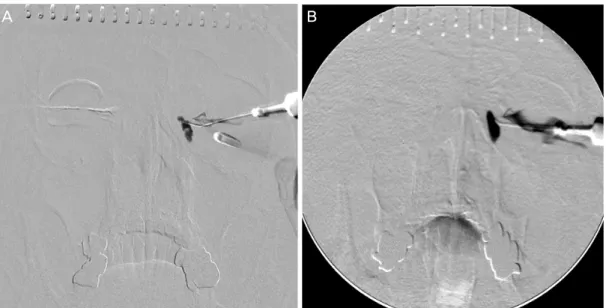
Figure 5. The results of dacryocystography in patients with dacryolith. (A) Filling defect of lacrimal sac in dacryocystography’s
finding. (B) Nasolacrimal duct stenosis without filling defect of the lacrimal sac.Table 2. Dacryocystographic findings related with lacrimal
sac dacryolith grossly found in operationDCG filling defect* Presence (%) Absence (%) Grossly found dacryolith in
operation
Presence (n = 13) 10 (76.9) 3 (23.1) Absence (n = 176) 7 (4.0) 169 (96.0)
Total (n = 189) 17 (9.0) 172 (91.0)
DCG = dacryocystography.
*Pearson χ2 78.694 (p = 0.00).
위 분석 방법을 토대로 눈물주머니 누석이 발견된 환자 들에서 눈물주머니의 대표 병리 소견에 따라 분류한 그룹 간 눈물흘림 증상 지속기간, 눈물소관 관류술 결과, OCT를 이용한 아래눈물띠 높이 측정 결과, 눈물주머니 조영술 결 과를 비교 · 분석하고 눈물주머니 누석이 발견된 누석군과 눈물주머니 누석이 발견되지 않은 비누석군의 성공률을 비 교하였다. 통계학적인 처리는 SPSS 21.0 버전(SPSS Inc., Chicago, IL, USA)을 이용한 t-test, Mann-Whiteny U-test, Chi-square test, Pearson’s correlation 분석을 사용하였다.
결 과
내시경적 눈물주머니코안연결술을 시행한 대상 환자들 의 평균 연령은 57.6 ± 13.8세였고 총 189안 중 61안 (32.28%)에서 병리검사상 누석이 발견되었으며, 누석군과 비누석군 각각 58.0 ± 14.2세와 57.2 ± 13.3세였다(p=0.74, Table 1). 누석군 중 13안(21.31%)은 수술 중 육안상 눈물 주머니 누석으로 관찰되었다. 눈물주머니 조영술에서 눈물 주머니 누석의 충만결손(Fig. 5)과 내시경적 눈물주머니코 안연결술로 확인한 눈물주머니 누석의 존재 여부를 교차분
석한 결과, 누석군에서의 눈물주머니 조영술 충만결손이 발견된 경우가 그렇지 않은 경우에 비해 유의하게 높게 나 타났으며(p<0.05), 육안상 발견된 누석 기준으로 누석군의 76.9%에서 눈물주머니 조영술상 충만결손을 보였고, 수술 중 육안상 발견된 누석을 기준으로 눈물주머니 조영술의 눈물주머니 누석 진단에 대한 민감도(sensitivity)는 58.8%, 특이도(specificity)는 1.7%였다(Table 2).
수술한 전체 189안 중 염증 우세 그룹(36안)에서 누석이 발견된 경우는 9안(25.0%)이었고, 섬유화 우세 그룹(144안) 에서 누석이 발견된 경우는 43안(29.9%)이었으며 통계적으
A B
Table 4. Clinical characteristics according to the major pathologic findings of the lacrimal sac
Major pathologic findings Duration of tearing(months)
History of eye discharge
(number of eyes) Syringing score* LTMH (μm)†
Calcification dominant (n = 9) 31.2 ± 39.3‡ 4 1.9 ± 0.9 484.5 ± 255.5
Non-calcification dominant (n = 52) 79.9 ± 76.1‡ 23 2.1 ± 0.8 578.9 ± 420.6
Inflammation dominant (n = 9) 67.7 ± 80.6 5 1.9 ± 0.9 578.5 ± 153.4
Fibrosis dominant (n = 43) 81.9 ± 76.1 18 2.2 ± 0.8 579.0 ± 438.8
Values are presented as mean ± SD unless otherwise indicated.
LTMH = lower tear meniscus height.
*The results of canaliculus irrigation test. 1 for complete passage, 2 for partial passage and 3 for no passage; †Lower tear meniscus height meas- ured with anteiror segment optical coherence tomography) (Spectralis optical coherence tomography [OCT], Heidelberg Engineering, Heidelberg, Germany); ‡p = 0.006, Mann-Whitney test.
Figure 6. The results of correlation analysis between pathologic properties of dacryolith and the lacrimal sac.
Table 3. The distribution of dacryolith according to pathologic
findings of the lacrimal sac in the total patientsPathologic findings of lacrimal sac
Dacryolith Presence (%) Absence (%) Calcification dominant (n = 9) 9 (100.0) 0 (0.0) Inflammation dominant (n = 36) 9 (25.0)* 27 (75.0)* Fibrosis dominant (n = 144) 43 (29.9)* 101 (70.1)*
Total (n = 189) 61 (32.3) 128 (67.7)
*Pearson χ2 0.331 (p = 0.57).
로 유의한 차이는 없었다(p=0.57) (Table 3). 눈물주머니 누 석의 평균 크기는 0.3 ± 0.8 mm2이고 평균 분포는 20.1 ± 17.9%였다. 눈물주머니 병리소견은 석회화가 우세한 경우 는 9안, 나머지는 52안이었으며, 두 군 간의 눈물흘림 증상 지속기간은 각각 31.2 ± 39.3개월과 79.9 ± 76.1개월로 유 의한 차이를 보였다(p=0.006) (Table 4). 추가적으로 눈곱
발생 증상을 호소한 경우는 석회화 우세 그룹에서 4안 (44.4%), 염증 우세 그룹에서 5안(55.6%) 그리고 섬유화 우 세 그룹에서 18안(41.9%)으로 각 군 비교 시 통계적으로 차이는 없었다(p=0.72).
눈물주머니 누석과 눈물주머니의 병리학적 결과를 상관 분석한 결과 눈물주머니 누석의 분포와 눈물주머니의 염증 세포 비율 및 섬유화 비율은 음의 상관관계를 보였다(각각 p=0.000, p=0.010). 또한 눈물주머니 누석의 크기와 눈물주 머니의 섬유화 비율은 음의 상관관계를 보였다(p=0.008) (Fig. 6).
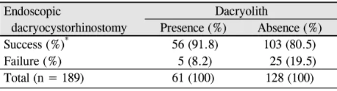
그 외에 눈물주머니 누석의 병리학적 특성과 눈물소관 관류술 및 OCT를 이용한 아래눈물띠높이를 포함한 임상양 상 간의 유의한 결과는 없었다(Table 5). 수술성공률은 누 석군에서 91.8%, 비누석군에서 80.5%로, 통계적으로 유의 한 차이를 보였다(p=0.046, Table 6).
R2 = 0.051
Table 5. Correlation analysis between pathologic properties of lacrimal sac dacryolith and lacrimal sac and clinical findings
(n = 61) Lacrimal sac dacryolith Duration of tearing* Syringing score*† LTMH*‡Size (mm2) 0.3 ± 0.8 0.07 (0.59) -0.18 (0.17) -0.17 (0.39)
Distribution (%) 20.1 ± 17.9 -0.01 (0.97) -0.11 (0.40) -0.15 (0.44)
Values are presented as mean ± SD unless otherwise indicated.
LTMH = lower tear meniscus height.
*Pearson coefficient of correlation (p-value); †The results of canaliculus irrigation test. 1 for complete passage, 2 for partial passage and 3 for no passage; ‡Lower tear meniscus height measured with anteiror segment optical coherence tomography (Spectralis OCT, Heidelberg Engineering, Heidelberg, Germany).
Table 6. The success rate of endoscopic dacryocystorhinostomy
according to lacrimal sac dacryolithEndoscopic
dacryocystorhinostomy
Dacryolith
Presence (%) Absence (%)
Success (%)* 56 (91.8) 103 (80.5)
Failure (%) 5 (8.2) 25 (19.5)
Total (n = 189) 61 (100) 128 (100)
*Pearson χ2 3.98 (p = 0.046).
고 찰
누석은 코눈물관 폐쇄의 한 원인으로 눈물주머니 조영술 을 이용한 수술 전 진단에서 발견되는 경우가 있으나 일반 적으로 수술 전 진단은 비교적 어려우며 보통 수술 중에 발 견됨으로써 진단되는 경우가 대부분으로, 본 연구에서는 눈물주머니 누석의 발견 빈도가 32.2%로 이전 연구들에 비 해 좀 더 높게 나타났다. 이는 본 연구에서 수술 중 육안적 으로 눈물주머니에서 누석이 발견된 경우뿐만 아니라 병리 검사상 누석이나 석회화가 발견되는 경우 또한 눈물주머니 누석으로 정의하였기 때문으로 사료된다.
눈물주머니 누석의 구성성분에 대한 연구를 보면 Orhan et al17은 누석의 대부분이 유기질성분(organic composition)이며 그 중심부는 작은 엽(lobe)으로 이루어진 비세포성 무정형구 조(acellular amorphous structure)라고 보고하였다. Iliadelis et al18은 유기질성분은 98.85%, 무기질성분(inorganic compo- sition)은 1.15%를 차지하며 무기질성분은 Ca2+ 0.79%, Na+ 0.21%, Mg2+ 0.11%, K+ 0.04%의 비율이라고 보고하였다.
Rhim et al19은 국내에서 시행한 누석의 성분분석상 누석의 구성을 유기질성분은 97.91%, 무기질성분 2.09%로 분석하 였으며 무기질성분은 Ca2+ 0.82%, Na+ 0.76%, Mg2+ 0.27%, K+ 0.24%로 세분화되어 있음을 보고하였다.
본 연구에서는 눈물주머니 누석의 크기와 분포가 높을수 록 눈물주머니의 섬유화 비율이 낮아지는 것을 관찰할 수 있었다. 코눈물관 폐쇄 환자에서는 상피하층의 섬유모세포 의 증가와 콜라겐 섬유 다발의 증가로 인한 섬유화가 관찰 되며, 눈물주머니와 코눈물관 점막의 상피층과 상피하층에
염증세포들이 침윤된다.20,21 Lee et al14은 코눈물관 폐쇄 환 자에서의 눈물주머니의 병리학적 소견으로 눈물주머니 상 피조직의 염증 침윤, 섬유화, 술잔세포 소실, 편평상피로의 화생을 보고하였으며 그중에서도 섬유화가 특징적인 폐쇄 의 병리라고 하였고, 눈물흘림 환자에서의 피부경유 눈물 주머니코안연결술(external dacryocystorhinostomy) 후 눈물 주머니의 조직병리학적 특징을 연구한 Salour et al22은 만 성 염증이 95.5%의 눈물흘림 환자에서 발견되었고 섬유화 가 3.8%의 환자에서 발견되었음을 보고하였다. Ozer et al23 은 만성눈물주머니염증(chronic dacryocystitis)으로 인해 피 부경유 눈물주머니코안연결술을 시행한 환자들에서 얻은 눈물주머니 조직을 분석하였으며, 심한 섬유화(severe fib- rosis)가 44%의 조직에서 발견되었음을 보고하였다.
기존의 연구를 보면 눈물주머니 내 섬유화는 만성적인 염증에 의해 발생하는 것으로 생각할 수 있으며, 본 연구의 결과상 눈물주머니 누석이 많고 클수록 섬유화의 비율이 낮으므로, 눈물주머니 누석의 발생은 만성적인 코눈물관 폐쇄보다 급성이나 아급성으로 발생한 코눈물관 폐쇄에서 더 높게 나타난다고 생각할 수 있다. 또한 본 연구에서는 병리학적 슬라이드 분석상 염증세포가 우세하거나 섬유화 조직이 우세한 눈물주머니 누석을 가진 환자군보다 석회화 가 우세한 눈물주머니 누석을 가진 환자군에서 그렇지 않 은 군에 비해 눈물흘림 증상 지속기간이 짧게 나타났는데, 이 사실 또한 눈물주머니 누석이 만성보다는 급성이나 아 급성으로 발생한 코눈물관 폐쇄와 더 연관성을 가짐을 뒷 받침해 준다. Repp et al5은 눈물주머니 코안연결술 중 눈물 주머니 누석이 발견된 환자군에서 눈물흘림 기간이 더 짧 음을 보고하였으며, 이는 본 연구와 비슷한 양상을 보인다.
눈물주머니 조영술은 관류용 도관으로 조영제를 아래 눈 물소관으로 주입하고 Caldwell view와 측면 사진을 촬영하 는 방법으로 눈물길을 직접적으로 조영하여 코눈물길 폐쇄 에서 협착이나 폐쇄의 정확한 위치와 그 정도를 확인하고 치료 방향을 결정하는 데 큰 도움을 주며 눈물샛길이나 종 양, 누석의 유무를 직접적으로 확인할 수 있다는 장점을 가 지고 있다.24-26 하지만 본 연구에서는 눈물주머니 누석이
발견된 누석군의 16.4%만이 눈물주머니 조영술에서 눈물 주머니 누석의 음영이 발견되었는데, 이는 수술 전 눈물주 머니 조영술이 눈물주머니 누석을 발견해 내는 데 있어서 한계가 있음을 의미하는 것으로 생각된다. 하지만 수술 중 육안상으로 누석이 발견된 13예를 보면 10예(76.9%)에서 눈물주머니 조영술상 충만결손이 확인되었으며, 다만 눈물 주머니 안에 있는 눈물주머니 누석뿐만 아니라 눈물주머니 자체의 병리조직에서 발견한 석회화까지 눈물주머니 누석 으로 정의한 본 연구의 특성에 의해 눈물주머니 조영술의 눈물주머니 누석 발견 확률이 낮게 측정되었을 가능성도 고려해야 할 것으로 생각된다.
Marcet et al27이 이전 문헌을 고찰한 바에 따르면 내시경 적 눈물주머니코안연결술의 평균 수술성공률은 91.33%였 다. 본 연구에서 내시경적 눈물주머니코안연결술의 전체 수술성공률은 84.13%였으며, 눈물주머니 누석이 발견된 누 석군에서 91.80%, 그렇지 않은 비누석군에서 80.47%로, 눈 물주머니 누석이 발견된 환자군에서 유의하게 수술성공률 이 높았다(p<0.05). 이는 누석이 발견된 경우의 수술성공률 이 그렇지 않은 경우보다 더 높다고(각각 100%와 91.1%, p<0.001) 보고한 Komínek et al11의 연구 결과와 같은 결과 이다. 본 연구에서는 눈물주머니 누석의 크기와 분포가 높 을수록 눈물주머니의 섬유화 비율이 낮아지는 것을 관찰하 였으며 이는 누석의 발견이 눈물주머니코안연결술의 높은 수술성공률을 예상하는 데 있어서 긍정적인 영향을 주는 인자라고 생각할 수 있겠다.
본 연구는 대상과 방법에 기술한 바와 같이 눈물주머니 누석의 정의 자체를 수술 중 육안적으로 눈물주머니에서 누석이 발견되거나 병리검사상 누석이나 석회화가 발견되 는 경우로 한정지어 연구하였다는 한계가 있고, 또한 피부 경유 눈물주머니코안연결술이 아닌 내시경적 누낭비강문 합수술로서 수술적 치료를 하였기 때문에 눈물주머니의 일 부 조직만을 채취해서 확인할 수밖에 없다는 한계가 있으 나, 많은 수의 환자들을 대상으로 연구한 만큼 의미있는 결 과에 부합할 것으로 사료된다.
본 연구는 내시경적 눈물주머니코안연결술 후 눈물주머 니 병리검사상 누석이나 석회화가 발견되는 경우를 눈물주 머니 누석으로 정의하고 눈물주머니 누석과 눈물주머니의 병리학적 결과 및 임상양상과의 연관성을 분석하였으며 눈 물주머니 누석이 증상 지속기간과 수술성공률과 연관성이 있음을 확인할 수 있었다. 아직까지 눈물주머니 누석의 발 생 기전이 정확히 알려져 있지 않아 누석이 코눈물관 폐쇄 에 미치는 정확한 인과관계를 알 수 없지만, 누석이 있는 환자의 눈물주머니 병리소견이 일반적인 코눈물관 폐쇄 환 자의 대표적인 섬유화보다는 염증과 석회화가 특징적인 것
과 유병기간이 짧고 수술 후의 결과가 우수한 임상양상을 고려할 때 코눈물관 폐쇄 질환의 조기 현상이거나 다른 질 환으로 이해되고 치료가 계획되어야 할 것으로 생각된다. 향후 누석의 발생원인과 생성 기전에 대한 연구가 진행되 어 코눈물관 폐쇄의 치료 방향을 결정하는 데 도움이 되길 바란다.
REFERENCES
1) Berlin AJ, Rath R, Rich L. Lacrimal system dacryoliths. Ophthalmic Surg 1980;11:435-6.
2) Rosen WJ, Rose GE. Intranasal passage of dacryoliths. Br J Ophthalmol 2000;84:799-800.
3) Yazici B, Hammad AM, Meyer DR. Lacrimal sac dacryoliths: pre- dictive factors and clinical characteristics. Ophthalmology 2001;
108:1308-12.
4) Halborg J, Prause JU, Toft PB, et al. Stones in the lacrimal gland: a rare condition. Acta Ophthalmol 2009;87:672-5.
5) Repp DJ, Burkat CN, Lucarelli MJ. Lacrimal excretory system concretions: canalicular and lacrimal sac. Ophthalmology 2009;
116:2230-5.
6) Herzig S, Hurwitz JJ. Lacrimal sac calculi. Can J Ophthalmol 1979;14:17-20.
7) Hawes MJ. The dacryolithiasis syndrome. Ophthal Plast Reconstr Surg 1988;4:87-90.
8) Wilkins RB, Pressly JP. Diagnosis and incidence of lacrimal calculi. Ophthalmic Surg 1980;11:787-9.
9) Jones LT. Tear-sac foreign bodies. Am J Ophthalmol 1965;60:
111-3.
10) Andreou P, Rose GE. Clinical presentation of patients with dacryolithiasis. Ophthalmology 2002;109:1573-4.
11) Komínek P, Červenka S, Zeleník K, et al. Lacrimal sac dacryolith (76 cases): a predictive factor for successful endonasal dacryocys- torhinostomy? Eur Arch Otorhinolaryngol 2014;271:1595-9.
12) Anderson NG, Wojno TH, Grossniklaus HE. Clinicopathologic findings from lacrimal sac biopsy specimens obtained during dacryocystorhinostomy. Ophthal Plast Reconstr Surg 2003;19:
173-6.
13) Tucker N, Chow D, Stockl F, et al. Clinically suspected primary ac- quired nasolacrimal duct obstruction: clinicopathologic review of 150 patients. Ophthalmology 1997;104:1882-6.
14) Lee SH, Lew H, Sim JY. Pathologic features of lacrimal sac muco- sa in primary acquired nasolacrimal duct obstruction patients. J Korean Ophthalmol Soc 2004;45:173-9.
15) Park DI, Lew H, Lee SY. Tear meniscus measurement in nasolacri- mal duct obstruction patients with Fourier-domain optical coher- ence tomography: novel three-point capture method. Acta Ophthalmol 2012;90:783-7.
16) Kim CH, Lew H, Yun YS. Correspondence among the canaliculus irrigation test, dacryocystography and Jones test in the epiphora patients. J Korean Ophthalmol Soc 2007;48:1017-22.
17) Orhan M, Onerci M, Dayanir V, et al. Lacrimal sac dacryolith: a study with atomic absorption spectrophotometry and scanning electron microscopy. Eur J Ophthalmol 1996;6:478-80.
18) Iliadelis E, Karabatakis V, Sofoniou M. Dacryoliths in chronic da-
= 국문초록 =
코눈물관폐쇄 수술 환자에서 눈물주머니누석의 임상병리학적 연구
목적: 내시경적 눈물주머니코안연결술 후 발견된 눈물주머니 누석 및 눈물주머니의 병리학적 특성과 코눈물관폐쇄 환자들의 임상양 상과의 상관관계를 알아보고자 한다.
대상과 방법: 2009년 8월부터 2015년 1월까지 원발성 후천성 코눈물관폐쇄로 내시경적 눈물주머니코안연결술을 시행한 158명 189안 을 후향적 조사하였다. 수술 중 육안적으로 눈물주머니에서 누석이 발견되거나 병리검사상 발견된 누석이나 석회화를 눈물주머니 누석으로 정의하였다. 눈물주머니 석회화, 염증, 섬유화를 평가하고 환자 임상양상과 수술 결과, 슬라이드에서의 누석 크기 및 분포와 병리소견과의 연관관계를 분석하였다.
결과: 눈물주머니 누석은 61안(32.3%), 그중 육안상 13안(6.9%)에서 발견되었다. 17안(9.0%)에서 눈물주머니 조영술상 음영결손이 보 였고 육안상 발견된 누석 기준 민감도 58.8%, 특이도 1.7%였다. 누석 크기 0.3 ± 0.8 mm2, 분포 20.1 ± 17.9%, 누석분포와 눈물주머 니 염증, 섬유화는 음의 상관관계를, 누석 크기와 눈물주머니 섬유화는 음의 상관관계를 보였다(p<0.05). 병리슬라이드상 석회화가 차지하는 비율이 50% 이상인 석회화 우세 그룹에서 그렇지 않은 그룹보다 증상 기간이 더 짧았다(p<0.05). 수술성공률은 누석군 91.8%, 비누석군 80.5%였다(p<0.05).
결론: 코눈물관폐쇄 환자들 중 눈물주머니 누석을 보인 경우 증상 기간이 짧고 눈물주머니 섬유화가 적었으며 수술성공률이 높았다.
따라서 코눈물관 폐쇄 중 누석을 동반한 경우에서의 누석 생성 기전과 조기치료에 대한 추가적인 연구가 필요할 것으로 생각된다.
<대한안과학회지 2016;57(1):6-13>
cryocystitis and their composition (spectrophotometric analysis).
Eur J Ophthalmol 1999;9:266-8.
19) Rhim WI, Lew H, Yun YS, Ahn HJ. A case of morphological and chemical analysis of recurrent dacryoliths after dacryocystorhinostomy.
J Korean Ophthalmol Soc 2002;43:1128-32.
20) Lee-Wing MW, Ashenhurst ME. Clinicopathologic analysis of 166 patients with primary acquired nasolacrimal duct obstruction.
Ophthalmology 2001;108:2038-40.
21) Paulsen FP, Thale AB, Maune S, Tillmann BN. New insights into the pathophysiology of primary acquired dacryostenosis. Oph- thalmology 2001;108:2329-36.
22) Salour H, Hatami MM, Parvin M, et al. Clinicopathological study of lacrimal sac specimens obtained during DCR. Orbit 2010;
29:250-3.
23) Ozer O, Eskiizmir G, Unlü H, et al. Chronic inflammation: a poor prognostic factor for endoscopic dacryocystorhinostomy. Eur Arch Otorhinolaryngol 2012;269:839-45.
24) Nixon J, Birchall IW, Virjee J. The role of dacryocystography in the management of patients with epiphora. Br J Radiol 1990;
63:337-9.
25) Milder B, Demorest BH. Dacryocystography. I. The normal lac- rimal apparatus. AMA Arch Ophthalmol 1954;51:180-95.
26) Ewing AE. Roentgen ray demonstration of the lacrimal abscess cavity. Am J Ophthalmol 1909;24:1-4.
27) Marcet MM, Kuk AK, Phelps PO. Evidence-based review of surgi- cal practices in endoscopic endonasal dacryocystorhinostomy for primary acquired nasolacrimal duct obstruction and other new indications. Curr Opin Ophthalmol 2014;25:443-8.