Effects of Acupuncture at SP
6on Reflux Esophagitis in Rats
※Yun Kyu Lee1, Sung Soo Rho2 and Jae Soo Kim1,*
1Department of Acupuncture & Moxibustion, Meridian & Acupoint, College of Korean Medicine, Deagu Haany University
2Department of Herbology, College of Korean Medicine, Deagu Haany University
[Abstract]
Objectives : The purpose of this study was to evaluate whether acupuncture at SP6 attenuates esophageal inflammation on refluxed-induced esophagitis.
Methods : Acupuncture at SP6 was stimulated by acupuncture torsion technique for 30 seconds four times every hour after an operation induced reflux esophagitis(RE), and its effects were assessed in comparison with RE rats without acupuncture, and normal rats.
Results : SP6 acupuncture stimulation markedly ameliorated mucosal damage in the histological evaluation. Reflux-induced esophagitis rats exhibited the down-regulation of antioxidant-related protein expression levels such as heme oxygenase-1(HO-1) in the esophagitis; however, the associated levels with SP6 acupuncture stimulation were significantly higher than those in RE rats without acupuncture stimulation. Moreover, SP6 acupuncture stimulation significantly reduced the expression of inflammatory proteins through mitogen-activated protein kinase(MAPK)- related signaling pathways. The increased protein expressions of inflammatory mediators, cyclooxygenase-2(COX-2) and inducible nitric oxide synthase(iNOS), by nuclear factor-kappa B(NF-kB) activation were significantly suppressed through SP6 acupuncture stimulation.
Conclusions : Our findings support the therapeutic evidence for SP6 acupuncture stimulation alleviating the development of esophagitis via regulating inflammation through the activation of the antioxidant pathway.
Key words : SP6;
Gastroesophageal reflux disease(GERD);
Reflux esophagitis;
Anti-inflammatory;
Antioxidation
Received : 2015. 08. 07.
Revised : 2015. 08. 23.
Accepted : 2015. 08. 24.
On-line : 2015. 09. 20.
※ This research was supported by a grant from Daegu Haany University Ky․lin Foundation in 2012
✱ Corresponding author : Department of Acupuncture & Moxibution, Meridian & Acupoint, College of Korean Medicine, Daegu Hanny University, 136, Sincheondong-ro, Suseong-gu, Daegu, 42158, Republic of Korea
Tel : +82-53-770-2112 E-mail : [email protected]
This is an Open-Access article distributed under the terms of the Creative Commons Attribution Non-Commercial License (http://creativecommons.org/licenses/by-nc/3.0) which permits unrestricted non-commercial use, distribution, and reproduction in any medium, provided the original work is properly cited.
The Acupuncture is the Journal of Korean Acupuncture & Moxibustion Medicine Society. (http://www.TheAcupuncture.org) Copyright © 2014 KAMMS. Korean Acupuncture & Moxibustion Medicine Society. All rights reserved.
Ⅰ. Introduction
Gastroesophageal reflux disease(GERD) is characterized by excessive reflux of gastric content(acid, pepsin, etc) into the esophagus causing symptoms of heartburn and acid regurgitation, and mucosal inflammation and damage. GERD is a common and costly disease;
however, despite great achievements in the under- standing of the pathophysiology and treatment of the disease, the incidence of GERD seems to be rising worldwide lately1). Approximately 10~20 % of people in the Western world have GERD. GERD significantly impacts patient quality of life and may lead to long-term complications, such as Barrett’s esophagus and esophageal adenocarcinoma. Current therapy of GERD relies predominantly on the use of acid- suppressant medications, such as proton pump inhibitors, but response to these medications is less than optimal. For this reason, novel treatments remain desirable, and to develop them, a clearer understanding of the molecular mechanisms responsible for damage of the esophageal epithelium is needed2).
Acupuncture, a therapeutic modality with few or no adverse effects, has been used in the treatment of several diseases for at least 5,200 years in china. The widespread application of acupuncture includes the treatment of infections, inflammatory diseases like rheumatoid arthritis, autonomic dysfunction, neurological diseases, cardiovascular diseases, pulmonary diseases, drug abuse, psychological disorders and many other illnesses3). In the case of inflammation, the effects of acupuncture could be reduced clinical symptoms like burning pain, redness, swelling, changing temperature and loss of function. Acupuncture is accomplished by the insertion the tips of thin, stainless steel needle on specific points(called acupoints) and induces marked changes close to the needle in all the different tissues that are penetrated4,5).
SP6 is the commonly used acupoints in many disorders including gynecologic, genitourinary, allergic, insomnia, immunological and psychosomatic diseases
and pain control6-8). In addition, the SP6 treatment also exerted the anti-inflammatory effect in a model of carrageenan-induced peritonitis and the antioxidative effect in a mouse model of Parkinson's disease9,10).
During the latest decades, a considerable number of studies have been performed on acupuncture for the treatment of gastrointestinal disorders and underlying mechanisms. However, SP6 has yet to be reported the protective effect in GERD. Therefore, this study was designed to clarify the anti-inflammatory effect of the acupuncture at SP6 on reflux-induced esophagitis rats.
II. Materials and Methods
A. Materials
The protease inhibitor mixture solution and ethyl- enediaminetetraacetic acid(EDTA) were purchased from Wako Pure Chemical Industries, Ltd.(Osaka, Japan).
Phenylmethylsulfonyl fluoride(PMSF) and b-actin were purchased from Sigma Chemical Co(St Louis, MO, USA). The pierce bicinchoninic acid(BCA) protein assay kit was obtained from Thermo Scientific(Rockford, IL, USA). ECL Western Blotting Detection Reagents and pure nitrocellulose membranes were supplied by GE healthcare(Piscataway, NJ, USA). Rabbit polyclonal antibodies against heme oxygenase-1(HO-1), phosphor- p38(p-p38), and phosphor-extracellular signal-regulated kinase 1/2(p-ERK1/2), nuclear factor-kB(NF-kB);
mouse monoclonal antibodies against cyclooxygenase-2 (COX-2), inducible nitric oxide synthase(iNOS), histone and b-actin were purchased from Santa Cruz Biotechnology, Inc(Santa Cruz, CA, USA). Rabbit anti-goat, goat anti-rabbit, and goat anti-mouse immunoglobulin G(IgG) horseradish peroxidase(HRP)- conjugated secondary antibodies were acquired from Santa Cruz Biotechnology, Inc(Santa Cruz, CA, USA).
All other chemicals and reagents were purchased from Sigma Chemical Co(St Louis, MO, USA).
B. Experimental animals and acupuncture treatment
Animal experiments were carried out according to the “guidelines for animal experimentation” approved by ethics committee of the Daegu Haany University (IRB : DHU2012-004). Six- weeks-old male Sprague- Dawley rats were purchased from Samtako(Osan, Korea). Rats were maintained under a 12 hrs light/
dark cycle, housed at a controlled temperature(24 ± 1℃), and humidity(about 55 %), and kept in raised mesh-bottom cages to prevent coprophagy. After adaptation(1 week), the rats were divided into three groups of equal number (n=6, each), avoiding any inter-group differences in body weight. The rats were fasted for 24 hrs prior to surgical procedures, but were provided free access to water. The rats were anaesthetized with an injection of zoletil 0.75 ㎎/㎏ (Virbac S. A. France). A midline laparotomy was performed to expose the stomach, and then both the pylorus and the transitional junction between the forestomach and the corpus were first exposed and later ligated with a 2-0 silk thread but without a plyoric ring, employing the method originally proposed by Omura11). The vagus nerves were left intact. (1) normal group(N), (2) reflux esophagitis(RE) control group without acupuncture stimulation(Veh), (3) RE group with SP6 acupuncture stimulation(SP6).
SP6 group was received acupuncture at Both SP6, and stimulated by acupuncture torsion techinque for 30 secs four times every hour after the operation.
Disposable acupuncture needles(0.7 ㎜, 160 ㎛ in diameter) were inserted into the acupoints. N group was not received any treatment, and Veh group was also not received any treatment after operation.
Experimental groups were sacrificed 6 hrs after operation. The entire esophagus was removed immediately and examined for gross mucosal injury. The esophageal tissue was immediately frozen in liquid nitrogen and blood samples were collected by vena cava puncture from anesthetized rats. Subsequently, the esophagus and serum were kept at –80℃ until analysis.
C. Esophageal lesion score
The rat esophagus was cut with scissor in the longitudinal direction from the gastroesophageal junction to the pharynx after sacrifice. The inner mucous was washed away with 0.9 % NaCl and laid out on paper. Thereafter, the dissected esophagus photographed with an optical microscope(Olympus BX51, Tokyo, Japan) and analyzed using the i-solution lite software program. The gross mucosal damage ratio was calculated as follows: the gross mucosal damage ratio(%) = [width of area with esophageal mucosal damage(mm2)/width of total area of esophagus(mm2)] × 100.
D. Histopathological studies
6 hrs after the operations of pylorus and forestomach ligation, the junction area from the esophagus to the cardia(about 5 ㎝) and a part of the fundus tissue were separated and fixed in 10 % neutral buffered formalin, after paraffin embedding, 3 ㎛ serial sections were prepared and stained with hematoxylin and eosin. Thickness of mucosa, submucosa in the esophagus, and full thickness of esophagus were measured in each prepared specimens using an optical microscope(Olympus BX51, Tokyo, Japan) as
㎜/crossly trimmed tissues. Protecting percentages of mucosa(%) =(Length of lesions on the crossly trimmed esophageal mucosa/total length of crossly trimmed esophageal mucosa) × 100.
E. Measurement of gastric secretions
After sacrifice, the stomach of each rat was washed with 1 ㎖ 0.9 % NaCl(pH 7.4) with a 1,000 ㎕ micropipette and the gastric contents were collected.
In addition, the volume of gastric juice was examined.
The pH of collected gastric juice was measured using a pH meter(EcoMet; iSTEK Co, Seoul, Korea).
F. Preparation of nuclear and post-nuclear fractions
Nuclear protein extraction was performed according to the method of Komatsu12). In brief, lung tissues were homogenized with ice-cold lysis buffer containing 5 mM Tris-HCl(pH 7.5), 2 mM MgCl2, 15 mM CaCl2, and 1.5 Msucrose, and then 0.1 M DTT and proteaseinhibitormixturesolution were added. After centrifugation(10,500×g for 20 mins at 4°C), the pellet was suspended with extraction buffer containing 20 mM 2-[4-(2-hydroxyethyl)-1-piperazyl] ethanesulfonic acid(pH 7.9), 1.5 mM MgCl2, 0.42 M NaCl, 0.2 mM EDTA, and 25 %(v/v)glycerol, and then 0.1 M DTT and protease inhibitor mixture solution were added. The mixture was placed on ice for 30 mins. The nuclear fraction was prepared by centrifugation at 20,500×g for 5 mins at 4 °C. The post-nuclear fraction was extracted from the lung of each mouse, as described below. In brief, lung tissue was homogenized with ice-cold lysis buffer(pH 7.4) containing 137 mM NaCl, 20 mM Tris-HCl, 1 % Tween 20, 10 % glycerol, 1 mM PMSF, and protease inhibitor mixture solution.
The homogenate was then centrifuged at 2,000 × g for 10 mins at 4°C. The protein concentration in each fraction was determined using a Bio-Rad protein kit(Bio-Rad Laboratories, Hercules, CA, USA).
G. Immunoblotting analyses
For the determination of NF-kBp65 and histone, 10 ㎎ of protein from each nuclear fraction was electrophoresed through 12 % sodium dodecylsulfate polyacrylamide gel(SDS-PAGE). Separated proteins were transferred to a nitrocellulose membrane, blocked with 5 %(w/v) skim milk solution for 1 h, and then incubated with primary antibodies to NF-kBp65 and histone overnight at 4°C. After the blots were washed, they were incubated with anti-rabbit or anti-mouse IgG HRP-conjugated secondary antibody for 1 h at room temperature. Also, 10~15 ㎎ of protein of each post-nuclear fraction of COX-2, iNOS, HO-1, p-p38, p-ERK1/2, and b-actin was
electrophoresed through 8~15 % SDS-PAGE. Each antigen-antibody complex was visualized using ECL Western Blotting Detection Reagents and detected by chemiluminescence with Sensi-Q 2000(Lugen sci, Gyeonggi-do, Korea). Band densities were determined using ATTO Densitograph Software(ATTO Corporation, Tokyo, Japan) and quantified as the ratio to histone or b-actin. The protein levels of groups are expressed relative to those of normal mice.
H. Statistical analysis
Data are expressed as means ± SEM. Significance was assessed by one-way analysis of variance(ANOVA) followed by Dunnett's multiple comparison test(SPSS 11.5.1 for Windows, 2002, SPSS Inc, USA). Values of p<0.05 were considered significant.
III. Results
A. Gross mucosal damage in the esophagus
Fig. 1 shows the results of the morphological examination of esophagus(Fig. 1). Morphological changes such as hyperemia and multiple erosions were observed in reflux esophagitis rats. The damage of normal rats was not apparent. The acupuncture stimulation of SP6 acupoint led to a marked decrease of gross mucosal damage.
B. Histopathological changes in the esophagus
We examined entire sizes of mucosa on histological images, calculated protecting percentages of mucosa, and measured thickness of mucosa(Table 1, Fig. 2). As shown in Fig. 2, esophagus lesions of normal group are not shown. Lesions on RE control group without acupuncture stimulation were significantly increased
A
B
Fig. 1. Gross evaluation of the esophageal mucosal damage
A : Representative microphotographs of the esophagus.
Esophageal lesion observed in RE rats was ameliorated by the acupuncture stimulation at SP6.
B : Gross mucosal injury ratio at the end of experiment.
The gross mucosal injury was increased in RE rats compared with normal rats, but SP6 acupuncture stimulation led to a significant decrease.
N : normal rats.
Veh : RE rats without acupuncture stimulation.
SP6 : RE rats and SP6 acupuncture stimulation.
Values are the means ± SEM.
* : p<0.01, ** : p<0.001.
v. RE rats without acupuncture stimulation values.
n=6 in each group.
Group Protecting percentage of mucosa(%)
Thickness o of mucosa(㎛) N 99.6 ± 0.5*** 259.2 ± 31.2***
RE Veh 25.9 ± 7.1 22.2 ± 19.0 SP6 53.0 ± 14.2* 151.5 ± 38.2**
N : normal rats.
Veh : RE rats without acupuncture stimulation.
SP6 : RE rats with SP6 acupuncture stimulation.
Values are the means ± SEM.
* : p<0.05, ** : p<0.01, *** : p<0.001.
vs. RE rat without acupuncture stimulation values, n=6 in each group.
Table 1. Effect of SP6 Acupuncture Treatment on Esophageal Histomorphometry
Fig. 2. Histophasological changes in esophagus tissue
RE control group examined the removed mucosa, edematous submucosa by inflammation(yellow arrow), and infiltration of inflammatory cells in muscularis externa(red arrow).
Whereas, SP6 group, still occurred submucosa edema(black arrow), protected the esophageal mucosa damage and had little the infiltration of inflammatory cells in ME.
A B : esophagus tissue stained with H&E on normal rat.
C D : esophagus tissue stained with H&E on reflux esophagitis rats.
E F : esophagus tissue stained with H&E on reflux esophagitis rats treated with SP6.
A C and D are captured images with isolution software with 200 magnification on Olympus microscope.
B D and F are images captured with isolution software with 400 magnification.
compared to normal group. However, SP6 group was significantly decreased. Esophagus thicknesses in RE control group were increased compared to normal group because of edema, but the esophagus thicknesses of SP6 group were significantly decreased compared to RE control group. Mucosal thicknesses in RE control group were significantly decreased compared to normal group, but mucosal thickness of SP6 group were significantly increased compared to RE control group.
The normal esophageal mucosa exhibited a thin epithelial layer with squamous cells and included mucosa(M), submucosa(SM), and muscularis externa(ME).
The reflux-induced esophagitis lead to esophageal
mucosa damage due to back diffusion of gastric acid.
Accordingly, RE control group examined the removed mucosa, edematous submucosa by inflammation(yellow arrow), and infiltration of inflammatory cells in muscularis externa(red arrow). Whereas, SP6 group, still occurred submucosa edema(black arrow), protected the esophageal mucosa damage and had little the infiltration of inflammatory cells in ME. Table 1 shows the protecting percentages and thickness of mucosa.
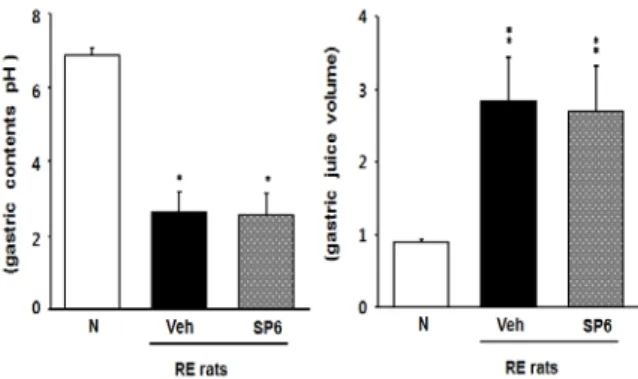
C. Gastric pH and volume in the gastric contents
The reflux-induced esophagitis rats displayed a marked decrease in gastric pH(Fig. 3). However, gastric pH was not changed by SP6 acupuncture stimulation.
Fig. 3. Effect of gastric contents pH and gastric volume
Gastric pH and volume analyses were performed as described in Materials and Methods.
N : normal rats.
Veh : RE rats without acupuncture stimulation.
SP6 : RE rats with SP6 acupuncture stimulation.
Values are the means ± SEM.
* : p<0.01. ** : p<0.001.
vs. normal rat values.
n=6 in each group.
D. Oxidative stress-related protein expression in the esophagus
Compared to normal rats, esophageal HO-1 protein expression was significantly decreased in RE rats
Fig. 4. Esophageal HO-1 protein expression
Immunoblotting analyses were performed as described in materials and methods.
N : normal rats.
Veh : RE rats without acupuncture stimulation.
SP6 : RE rats with SP6 acupuncture stimulation.
Values are the means ± SEM.
* : p<0.01.
vs. RE rat without acupuncture stimulation values.
n=6 in each group.
without acupuncture stimulation(Fig. 4). However, SP6 acupuncture stimulation adversely regulated cytosolic HO-1 expression in the esophagus of reflux-induced esophagitis rats.
E. MAPK-related protein
expressions in the esophagus
MAPK-related protein expression was augmented in the esophagus of RE rats without acupuncture stimulation compared to the normal rats but SP6
acupuncture stimulation decreased the expressions of p-p38 and p-ERK1/2(Fig. 5).
F. Inflammation-related protein expressions in the esophagus
The protein level of NF-kBp65 was enhanced in the esophagus of RE rats without acupuncture stimulation, whereas these elevated levels were significantly reduced in SP6 acupuncture stimulation rats(Fig. 6).
Especially, NF-kBp65 level was lowered nearly to
(A) (B) (C)
Fig. 6. Esophageal NF-kBp65(A), COX-2(B), and iNOS(C) protein expressions.
Immunoblotting analyses were performed as described in materials and methods.
N : normal rats. Veh : RE rats without acupuncture stimulation. SP6 : RE rats with SP6 acupuncture stimulation.
Values are the means ± SEM.
* : p<0.01. ** : p<0.001.
vs. RE rat without acupuncture stimulation values.
n=6 in each group.
(A) (B)
Fig. 5. Esophageal p-p38(A) and p-ERK(B) protein expressions.
p-p38 : phosphor-p38.
p-ERK : phosphorylated extracellular signal-regulated kinase.
Immunoblotting analyses were performed as described in materials and methods.
N : normal rats.
Veh : RE rats without acupuncture stimulation.
SP6 : RE rats with SP6 acupuncture stimulation.
Values are the means ± SEM.
* : p<0.05. ** : p<0.01. *** : p<0.001.
vs. RE rat without acupuncture stimulation values.
n=6 in each group.
that of normal rats by SP6 acupuncture stimulation.
The expression levels of COX-2 and iNOS were also enhanced in the esophagus of RE rats without acupuncture stimulation, with the results presented in Fig. 6.
These increased protein expressions were significantly attenuated by SP6 acupuncture stimulation.
Ⅳ. Discussion
Gastroesophageal reflux disease(GERD) is defined as symptoms or mucosal damage produced by the abnormal reflux of gastric contents into the esophagus. Typical symptoms of GERD are heartburn and acid regurgitation which have high specificity but low sensitivity. The range of GERD prevalence estimates an approximately 10~20 % in Europe and the USA, and of less than 5 % in Asia. This high prevalence of GERD in combination with the high cost of acid lowering medications results in the significant socioeconomic burden associated with the disease and has a negative impact on the quality of life13,14). PPIs, the suppression agent of gastric acid secretion, were one of the most commonly prescribed medications by primary physicians and are frequently used over the long term. However, although PPI therapy is effective in most patients with GERD, approximately 20~30 % continue to experience reflux symptoms despite PPI treatment15). The safety of these drugs and their potential adverse effects is of great importance to public health. In addition, several case reports suggested that acid suppressive drugs may increase the occurrence of gastric polyps or cancer16). Alternative approaches to management are required
in GERD treatment.
Acupuncture is one of major components of traditional Chinese medicine(TCM), which is a distinctive heritage of Chinese culture-related medicine when compared with the conventional Western medicine. Acupoints(or acupuncture points) are special nodes(or outlets) on the meridians, where ‘Qi’(a kind of air) enters, exits, meets and accumulates. There are 14 major meridians corresponding to internal organs, along which there are a total of 361 acupoints17). SP6, the junction point of the liver, spleen, and kidney meridians, are located on the medial border of the tibia18). SP6 is frequently used to study acupuncture effects on various physiological regulatory mechanisms and control systems changes, including gastrointestinal disorders and reproductive conditions, such as labor induction and pain relief during labor19,20). However, the mechanisms underlying the effects of SP6 acu- puncture treatment have yet to be investigated in an experimental model of reflux esophagitis. Therefore, the present study has been investigated to enhance esophageal inflammation using experimental reflux esophagitis model.
The pathogenesis of reflux esophagitis which is considered as the early stage of GERD is complex, resulting from an imbalance between aggressive factors damaging the esophagus and a number of the natural defense mechanisms. The esophageal mucosa is in a state of continuous exposure to potentially damaging endogenous and exogenous factors. The development of reflux esophagitis(RE) on a cellular level is due to hydrogen ion diffusion into the mucosa, leading to tissue acidification and necrotic damage. The basic level of esophageal defense against acid-pepsin damage consists of the anti-reflux mechanisms such as the luminal acid clearance and removal of the esophageal contents and neutralization of luminal acidity21). Major aspect in esophageal injury and esophageal blood flow during development of reflux esophagitis was characterized by the appearance of local mucosal edema and a small number of focal hemorrhagic erosions. Accordingly, histopathological change of esophagus revealed increasing of thickness, damage to the mucosa, and
hemorrhages in esophagus tissues. In addition, reflux-induced esophagitis increase both gastric volume, acid output and also decrease gastric pH22-24). In this study, RE rats group without acupuncture stimulation decreased markedly the gastric pH and increased the gastric volume like another study. However, SP6 acupuncture stimulation does not affect the regulation of gastric pH.
Nevertheless the apparatus of esophageal macroscopic lesions reduced markedly. Maybe, these results have supposed to protect the esophageal mucosal damage improve different factor without regulating gastric pH25).
Recent studies have been shown that gastro- esophageal reflux enhances the production of oxygen-derived free radicals, which subsequently led to esophageal mucosal damage26,27). Oxygen free radicals in excessively high amounts are all very reactive chemically and can impose a detrimental influence on living organisms by provoking “oxidative stress” that can be capable of damaging cellular DNA, protein, and organelles28). Furthermore, oxidative stress seems to be an important mediator in generation of esophageal mucosal injury. In recent studies it has been shown that mucosal damage in reflux oesophagitis is mediated primarily by oxygen derived free radicals29,30). Administration of various free radical scavengers has been founded to prevent esophageal mucosal damage. Various exogeneous scavengers are available that can selectively block oxygen-derived free radicals26). Especially, as a major cellular defense mechanism against oxidative stress, the Nrf2/Keap1 pathway regulates expression of enzymes involved in detoxification and anti-oxidative stress response31). In the presence of reactive oxygen species(ROS), Nrf2 is released from Keap1 and then translocates into the nucleus, activating the trans- cription of target genes, including HO-1. Nrf2/HO-1 antioxidant pathway plays a vital role in the im- provement of tissue damage as well as esophageal epithelial cells32,33). In our results, reflux esophagitis rats showed decreased HO-1 protein expression in esophageal tissues compared with normal rats;
however, SP6 acupuncture stimulation effectively
alleviates oxidative stress and results in the up-regulation of HO-1.
Besides, the overexpression of ROS in gastric epithelium has been linked to gastric carcinogenesis (as well as inflammation). High levels of ROS activates MAPK including p38 and ERK1/2. The MAPK cascades on p38 and ERK play important roles in the regulation of intracellular metabolism and gene expression including disease, apoptosis, and cellular responses to external stresses34). That is, phosphorylations of p38 and ERK1/2 lead to NF-kB translocation in the nucleus. The NF-kB is the main regulator of inducible expression of inflammatory genes. Activated ERK1/2 induces the dissociaton of IkBa to NF-kB, therefore allowing nuclear translocation and DNA-binding of NF-kB, and p38 induces the expression of p65 and p5035). In this study, increased expressions of ERK1/2 and p38 in the reflux esophagitis rats were decreased by SP6
acupuncture stimulation. The results from the present study show that SP6 acupuncture stimulation blocked NF-kB activation in the esophageal tissue. NF-κB is one of the cross-talk points of multiple signal transduction pathways, playing a key role in the regulation of immune and inflammatory responses. In particular, NF-κB is known to regulate transcription and expression of many genes such as COX-2 and iNOS36,37). COX-2 is barely detected in normal tissues, but is readily expressed in response to inflammatory cytokines, bacterial lipopolysaccharide, mitogens and reactive oxygen intermediates. Following inflammatory stimuli, excess NO by iNOS and proinflammatory prostaglandins by COX-2 have been reported to induce noxious effects in the esophagus.
These inflammatory proteins was augmented under inflammatory conditions, such as reflux esophagitis and barrett esophagus38,39). In the present study, SP6
acupuncture stimulation in the reflux esophagitis model significantly decreased up-regulation of inflammatory mediators(COX-2 and iNOS). That is, SP6 acupuncture stimulation ameliorated inflammation with esophageal mucosal injury on experimental reflux esophagitis in rats.
Ⅴ. Conclusion
The acupuncture stimulation of SP6 effectively ameliorates the inflammatory damage of esophageal mucosa through the activation of HO-1 antioxidant pathway.
Ⅵ. References
1. Herbella FA, Neto SP, Santoro IL, Figueiredo LC.
Gastroesophageal reflux disease and non-esophageal cancer. World J Gastroenterol. 2015 ; 21(3) : 815-9.
2. Fang Y, Chen H, Hu Y et al. Gastroesophageal reflux activates the NF-κB pathway and impairs esophageal barrier function in mice. Am J Physiol Gastrointest Liver Physiol. 2013 ; 305(1) : G58-65.
3. Zijlstra FJ, van den Berg-de Lange I, Huygen FJ, Klein J. Anti-inflammatory actions of acupuncture.
Mediators Inflamm. 2003 ; 12(2) : 59-69.
4. Kaptchuk TJ. Acupuncture: theory, efficacy, and practice. Ann Intern Med. 2002 ; 136(5) : 374-83.
5. Yin J, Chen JD. Gastrointestinal motility disorders and acupuncture. Auton Neurosci. 2010 ; 157(1-2) : 31-7.
6. Pavão TS, Vianna P, Pillat MM, Machado AB, Bauer ME. Acupuncture is effective to attenuate stress and stimulate lymphocyte proliferation in the elderly. Neurosci Lett. 2010 ; 484(1) : 47-50.
7. Chen MN, Chien LW, Liu CF. Acupuncture or acu- pressure at the Sanyinjiao(SP6) acupoint for the treatment of primary dysmenorrhea: a meta- analysis. Evid Based Complement Alternat Med.
2013 ; 2013 : 493038.
8. Oliveira R, Prado WA. Anti-hyperalgesic effect of electroacupuncture in a model of post-incisional pain in rats. Braz J Med Biol Res. 2000 ; 33(8) : 957-60.
9. da Silva MD, Guginski G, Werner MF, Baggio CH, Marcon R, Santos AR. Involvemen to fInterleukin-10
in the Anti-Inflammatory effect of Sanyinjiao (SP6) acupuncture in a mouse model of peritonitis.
Evid Based Complement Alternat Med. 2011 ; 2011 : 217946.
10. Wang H, Pan Y, Xue B et al. The antioxidative effect of electro-acupuncture in a mouse model of Parkinson's disease. PLoS One. 2011 ; 6(5) : e19790.
11. Omura N, Kashiwagi H, Chen G, Suzuki Y, Yano F, Aoki T. Establishment of surgically induced chronic acid reflux esophagitis in rats. Scand J Gastroenterol. 1999 ; 34(10) : 948-53.
12. Komatsu S. Extraction of nuclear proteins. Methods Mol Biol. 2007 ; 355 : 73-7.
13. Badillo R, Francis D. Diagnosis and treatment of gastroesophageal reflux disease. World J Gastrointest Pharmacol Ther. 2014 ; 5(3) : 105-12.
14. El-Serag HB, Sweet S, Winchester CC, Dent J.
Update on the epidemiology of gastro- oesophageal reflux disease: a systematic review.
Gut. 2014 ; 63(6) : 871-80.
15. Miner PB Jr, Silberg DG, Ruth M, Miller F, Pandolfino J. Dose-dependent effects of lesogaberan on reflux measures in patients with refractory gastroesophageal reflux disease: a randomized, placebo-controlled study. BMC Gastroenterol.
2014 ; 14(1) : 188.
16. Ahn JS, Eom CS, Jeon CY, Park SM. Acid suppressive drugs and gastric cancer: a meta-analysis of observational studies. World J Gastroenterol. 2013 ; 19(16) : 2560-8.
17. Ouyang H, Chen JD. Therapeutic roles of acu- puncture in functional gastrointestinal disorders.
Aliment Pharmacol Ther. 2004 ; 20(8) : 831-41.
18. Santos EL, Dias BH, Andrade AC et al. Effects of acupuncture and electroacupuncture on estradiol- induced inflammation and oxidative stress in health rodents. Acta Cir Bras. 2013 ; 28(8) : 582-8.
19. Senna-Fernandes V, Franca DL, de Souza D et al.
Acupuncture at "Zusanli"(St.36) and "Sanyinjiao"
(SP.6) points on the gastrointestinal tract: a study of the bioavailability of(99m) Tc-sodium
pertechnetate in rats. Evid Based Complement Alternat Med. 2011 ; 2011 : 823941.
20. Yesilcicek Calik K, Komurcu N. Effects of SP6
acupuncture point stimulation on labor pain and duration of labor. Iran Red Crescent Med J. 2014 ; 16(10) : e16461.
21. Pawlik MW, Kwiecien S, Pajdo R et al.
Esophagoprotective activity of angiotensin-(1-7) in experimental model of acute reflux esophagitis.
Evidence for the role of nitric oxide, sensory nerves, hypoxia-inducible factor-1alpha and proin- flammatory cytokines. J Physiol Pharmacol. 2014 ; 65(6) : 809-22.
22. Min YS, Lee SE, Hong ST et al. The inhibitory effect of quercetin-3-o-β-d-glucuronopyranoside on gastritis and reflux esophagitis in rats.
Korean J Physiol Pharmacol. 2009 ; 13(4) : 295-300.
23. Im WJ, Nam Y, Park SY, Sohn UD. Gastro- protective effect of the three glucuronopyranoside flavonoids in rats. Korean J Physiol Pharmacol.
2013 ; 17(5) : 411-5.
24. Abdel-Aziz H, Zaki HF, Neuhuber W, Kelber O, Weiser D, Khayyal MT. Effect of an herbal preparation, STW 5, in an acute model of reflux oesophagitis in rats. J Pharmacol Sci. 2010 ; 113(2) : 134-42.
25. Ku SK, Kim JS, Seo YB et al. Effect of curculigo orchioides on reflux esophagitis by suppressing proinflammatory cytokines. Am J Chin Med. 2012 ; 40(6) : 1241-55.
26. Lee JS, Oh TY, Ahn BO et al. Involvement of oxidative stress in experimentally induced reflux esophagitis and Barrett's esophagus: clue for the chemoprevention of esophageal carcinoma by antioxidants. Mutat Res. 2001 ; 480-481 : 189-200.
27. Kim YJ, Kim EH, Hahm KB. Oxidative stress in inflammation-based gastrointestinal tract diseases:
challenges and opportunities. J Gastroenterol Hepatol. 2012 ; 27(6) : 1004-10.
28. Hartman KG, Bortner JD, Falk GW et al. Modeling inflammation and oxidative stress in gastrointestinal disease development using novel organotypic
culture systems. Stem Cell Res Ther. 2013 ; 4 Suppl 1 : S5.
29. Oh TY, Lee JS, Ahn BO et al. Oxidative stress is more important than acid in the pathogenesis of reflux oesophagitis in rats. Gut. 2001 ; 49(3) : 364-71.
30. Oh TY, Lee JS, Ahn BO et al. Oxidative damages are critical in pathogenesis of reflux esophagitis:
implication of antioxidants in its treatment. Free Radic Biol Med. 2001 ; 30(8) : 905-15.
31. Chen H, Hu Y, Fang Y et al. Nrf2 deficiency impairs the barrier function of mouse oesophageal epithelium. Gut. 2014 ; 63(5) : 711-9.
32. Song HJ, Shin CY, Oh TY, Min YS, Park ES, Sohn UD. Eupatilin with heme oxygenase-1-inducing ability protects cultured feline esophageal epithelial cells from cell damage caused by indomethacin. Biol Pharm Bull. 2009 ; 32(4) : 589-96.
33. He M, Pan H, Chang RC, So KF, Brecha NC, Pu M. Activation of the Nrf2/HO-1 antioxidant pathway contributes to the protective effects of lycium barbarum polysaccharides in the rodent retina after ischemia-reperfusion-induced damage. PLoS One. 2014 ; 9(1) : e84800.
34. Liu MW, Su MX, Zhang W et al. Protective effect of Xuebijing injection on paraquat-induced pul- monary injury via down-regulating the expres-
sion of p38 MAPK in rats. BMC Complement Altern Med. 2014 ; 14(1) : 498.
35. Seo JH, Lim JW, Kim H. Differential role of ERK and p38 on NF-κB activation in helicobacter pylori-infected gastric epithelial cells. J Cancer Prev. 2013 ; 18(4) : 346-50.
36. Shin JS, Noh YS, Lee YS et al. Arvelexin from brassica rapa suppresses NF-κB-regulated pro- inflammatory gene expression by inhibiting acti- vation of IκB kinase. Br J Pharmacol. 2011 ; 164(1) : 145-58.
37. Park CH, Lee SL, Okamoto T, Tanaka T, Yokozawa T. Rokumi-jio-gan-containing prescriptions attenu- ate oxidative stress, inflammation, and apoptosis in the remnant kidney. Evid Based Complement Alternat Med. 2012 ; 2012 : 587902.
38. Surh Y, Na HK, Lee JY, Keum YS. Molecular mechanisms underlying anti-tumor promoting activities of heat-processed Panax ginseng C.A.
Meyer. J Korean Med Sci. 2001 ; 16Suppl : S38-41.
39. Ferguson HR, Wild CP, Anderson LA et al.
Cyclooxygenase-2 and inducible nitric oxide synthase gene polymorphisms and risk of reflux esophagitis, Barrett's esophagus, and esophageal adenocarcinoma. Cancer Epidemiol Biomarkers Prev. 2008 ; 17(3) : 727-31.