https://doi.org/10.14734/PN.2017.28.2.59 pISSN 2508-4887•eISSN 2508-4895
In Ji Hwang, MD1, Hyun Ok Lee, MD1, Ha Jin Oh, MD1, Eun Song Song, MD1,2, Young Youn Choi, MD1,2
1Department of Pediatrics, Chonnam National University Hospital, 2Department of Pediatrics, Chonnam National University Medical School, Gwangju, Korea
Objective: The aims of this study were to compare clinical characteristics and outcome of neo
nates with congenital myotonic dystrophy (CMD) according to the number of cytosine, thymine, guanine (CTG) copies and to analyze the relating factors for survival.
Methods: The patients were divided into two groups; less than or equal to 1,000 CTG copies (group A) and above 1,000 CTG copies (group B). This study compared the maternal CTG copies, obstetric characteristics, and patients’ clinical characteristics, morbidity, hospital course, and long term outcome between group A and B, and also analyzed the relating factors for survival.
Results: The twentythree patients were confirmed by gene analysis in the neonatal period. Nine patients (39.1%) were included in group A and fourteen patients (60.9%) in group B. There was no correlation between the number of CTG copies of the mothers and their babies. There were no significant differences in maternal obstetric characteristics, patient’s clinical findings, morbidities, hospital course and mortality between group A and B. Seven patients died before discharge and six patients among 16 who survived died after discharge. Analyzing the relating factors for survival, Apgar score at 1 and 5 minute were significantly higher in patients who survived than those who expired (P=0.0001, P=0.01, respectively). All survived patients showed developmental delay and 7 patients (58.3%) failed to thrive.
Conclusion: There was no correlation between the number of CTG copies of the mothers and their babies. There were no statistical differences in maternal obstetric characteristics, patient’s clinical findings, morbidities, hospital course, and mortality between the two groups. Apgar score at 1 minute and 5 minute were the relating factors for survival.
Key Words: Myotonic dystrophy, CTG trinucleotide copies number, Outcomes
서론
선천 근긴장 퇴행위축(congenital myotonic dystrophy, CMD)은 1960년 Vanier1에 의해 처음 기술되었으며, 발생빈도는 3,500-16,000명당 한 명으로 알려져 있다.2-4 대부분 상염 색체 우성으로 유전되며, 19번 염색체의 q13.3 위치에 있는 dystrophiamyotonica protein kinase (DMPK) 유전자 안에 있는 cytosine, thymine, guanine (CTG) 삼핵산 반복의 비정상 적인 확장에 의해 발현된다.5,6 근긴장 퇴행위축은 발병 시기에 따라 선천형, 유년기형, 성인 형, 지연형 등으로 분류되며 임상양상도 다양하다.7
CMD는 골격근뿐만 아니라 심장, 소화기관, 눈 및 중추신경계 등 여러 기관을 침범한다.8 신생아는 신체검진상 둔한 얼굴 표정, 높은 구개열, 텐트형 윗입술, 벌어진 입, 내반슬, 내반 Received: 7 February 2017
Revised: 18 May 2017 Accepted: 19 June 2017 Correspondence to Eun Song Song, MD
Department of Pediatrics, Chonnam National University Medical School, 160 Baekseoro, Donggu, Gwangju 61469, Korea
Tel: +82622206641 Fax: +82622226103 E-mail: [email protected]
Copyright© 2017 by The Korean Society of Perinatology
This is an Open Access article distributed under the terms of the Creative Com
mons Attribution NonCommercial License (http://creativecommons.org/
license/bync/4.0/), which permits unrestricted noncommercial use, distribution, and reproduction in any
Comparison of Two Congenital Myoto
nic Dystrophy Groups According to the
Number of CTG Trinucleotide Copies on
Clinical Characteristics and Outcomes
다.7,8
CTG 삼핵산의 반복은 정상인에게서도 발견되는데, 반복수 에 따라 5-35반복수는 정상, 36-49는 전돌연변이(premuta- tion) 그리고 50 이상은 환아로 분류하며, 환아인 경우 이를 더 세분하여, 50-100반복수는 경증형, 101-1,000은 고전형, 1,001 이상은 선천형으로 구분하기도 한다.7,9 그러나 CTG 삼 핵산 1,000반복수 미만인 경우에도 10-18% 정도에서 선천형 임상양상을 보였으며,2,3,10 반대로 CTG 삼핵산 반복수가 1,000 을 넘는 경우도 신생아기에 치료가 필요 없을 정도로 심하지 않 은 환아도 보고되어,3 CTG 삼핵산 반복수에 따른 분류와 중증 도 관련 여부에 대해서는 논란의 여지가 많다.
국내에서는 2006년 이후 분자유전학 검사로 확진된 1예씩의 증례보고만 있고,11-13 CTG 삼핵산 반복수에 대한 연구는 없기 에, 본 연구는 단일 기관에서 경험한 23명의 환아들을 대상으로 CTG 삼핵산 반복수에 따른 임상양상과 이환, 사망률 및 예후를 비교하고, 입원 기간 동안 생존과 관련된 인자에 대해 알아보고 자 하였다.
대상 및 방법
1. 대상
2003년 1월부터 2015년 12월까지 13년간 전남대학교병원 신생아집중치료실에 입원한 환아 중 임상소견으로 의심되어 유 전자 검사로 확진된 CMD 환아 23명과 그 어머니를 대상으로 하였다. 확진 환아 23명을 CTG 삼핵산 반복수에 따라 1,000 이 하군(A군, 9명)과 1,000 초과군(B군, 14명) 두 군으로 분류하였 다. 또한 확진된 경우 유전양상을 보기 위해 어머니의 유전자 검 사를 권유하였으며, 이 중 14명(증례 17과 20은 남매)에서 실시 되었다(Table 1).
2. 유전자 분석 방법
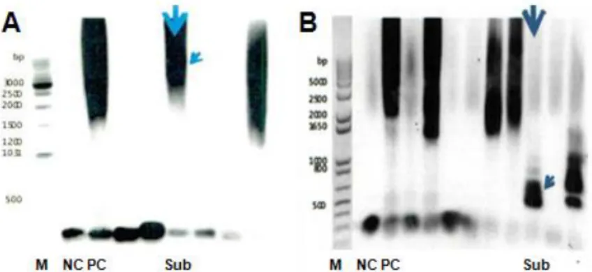
DMPK 유전자는 polymerase chain reaction (PCR)과 sou- thern blot analysis에 의해 검사하였다. 말초혈액에서 PCR 증 폭의 견본으로서 genomic DNA를 추출한 후 forward (5’-CA GTTCACAACCGCTCCGAGC-3’)와 reverse (5’-CGTGGA GGATGGAACACGGAC-3’)인 primer set을 포함한 LA-Taq polymerase (Takara, Shiga, Japan)을 이용하여 long-range PCR이 시행되었다. Long-range PCR product는 1% agarose gel electrophoresis로 용해시키고, nylon membrane으로 옮겨 져 80℃에서 1시간 동안 고정되었다. Biotin-labeled (CTG)10 probe로 45℃에서 overnight 동안 hybridization되었으며, 이
후 50분 동안 blocking solution으로 washing과 incu bation을 거쳐 50분 동안 alkaline phosphatase labeled streptoavidine 으로 membrane을 염색하였다. 그 다음 CDP-Star chemilumi- nescent substrate (KPL, Gaithersburg, MD, USA)를 투여하 고, ChemiDoc XRS+ (BioRad, Hercules, CA, USA)로 검사하 여 유전자 스캔 결과를 얻었다(Fig. 1).14
3. 임상 분석
환아와 어머니의 DMPK 유전자 CTG 삼핵산 반복수를 조사 하였고, 환아 어머니의 산과적 특성, 환아의 임상특성과 임상양 상, 질병 이환, 입원 경과, 사망률 및 장기 예후에 대하여 의무기 록을 후향적으로 분석하였다. 환아 어머니의 산과적 특징은 나 이, 임신방법, 분만력, 양수과다증, 비정상 태위, 태동 이상, 조 기 산통, 조기 양막파수, 제왕절개분만 여부를 조사하였다. 환 아의 임상특성으로 출생체중, 재태주령, 성별, Apgar 점수 및 분 만실에서의 소생술 여부를 조사하였고, 임상양상으로 전형적인
Table 1. The Number of CTG Copies in Patients and Their Mothers Case CTG copies of patient CTG copies of mother
1 >1,000 400
2 >1,000 200
3 >1,000 300
4 >1,000 150
5 >1,000 Not checked
6 >1,000 150
7 >1,000 Not checked
8 >1,000 400
9 >1,000 150
10 >1,000 Not checked
11 >1,000 150
12 >1,000 75
13 >1,000 100
14 >1,000 Not checked
15 400 Not checked
16 400 Not checked
17* 400 80
18 700 85
19 650 500
20* 300 80
21 550 Not checked
22 900 Not checked
23 400 90
Abbreviation: CTG, cytosine, thymine, guanine.
*Case 17 and 20 were sibling.
제시된 절단점에 근거하여 최저 점수 아래로 집계되어 심화평 가 권고에 해당한 경우로 정의하였다. 성장 부진은 한국 소아청 소년 표준 성장 도표에 따라 체중, 신장 또는 머리둘레가 각 연 령의 3백분위수 미만인 경우로 평가하였다.
4. 통계 분석
SPSS version 21.0 software (SPSS Inc., Chicago, IL, USA) 를 사용하여 데이터를 통계 처리하였으며, Mann-Whit ney test, Fisher’s exact test 및 Pearson's correlation 분석을 시행하였 다. 관찰 값은 평균±표준편차로 표시하였고, P값 0.05 미만인 경우를 통계적으로 유의하다고 하였다.
결과
1. CMD 빈도와 CTG 삼핵산 반복수
연구 기간 동안 신생아집중치료실 총 입원 환아 10,562명 중 23명이 선천 근긴장 퇴행위축 환아로 확진되어 0.2%를 차지하 였다(Table 1, Fig. 2).
확진 환아 23명을 CTG 삼핵산 반복수에 따라 1,000 이하군 (A군, 9명)과 1,000 초과군(B군, 14명) 두 군으로 분류하였다.
이 중 어머니의 유전자 검사는 14명(증례 17과 20은 남매)에서 실시하였는데, 모두에서 75-500반복수 사이로 나왔으며, CTG 삼핵산 반복수가 1,000을 넘는 환아의 반복수를 1,000으로 간 주하여 산모의 CTG 삼핵산 반복수와의 연관성을 확인하였을 때, 산모의 CTG 삼핵산 반복수와 환아의 반복수와는 관계가 없 었다(Table 1, Fig. 3; r=0.293, P=0.289).
얼굴 생김새, 근긴장도 저하, 내반첨족, 횡격막 상승 및 수유 곤 란 여부를 조사하였다. 출생체중이 재태주령에 따른 출생체중 의 10백분위수 미만인 경우 부당 경량아(small for gestational age, SGA)로 정의하였고, 90백분위수 이상인 경우 부당 중량아 (large for gestational age, LGA)로 정의하였다.
질병 이환은 호흡계, 심혈관계, 뇌신경계, 소화기계, 감염에 대해 조사하였다. 입원 경과는 재원 일수, 산소공급 기간, 기계 환기 사용 기간 및 장관영양 시기에 대해 조사하였다. 뇌실내 출 혈 grade III는 Papile grading system에 따라 두부초음파 검사 상 뇌실 확장을 동반한 뇌실내 출혈 소견시 진단하였다.15 괴사 성 장염 grade IIa는 modified Bell’s staging criteria에 따라 체 온 불안정, 무호흡, 서맥과 같은 전신 증상과 잔유량 증가, 복부 팽만, 잠혈 반응 양성, 장음 소실, 복부 압통과 같은 위장관 증상 이 있으면서 영상학적 검사상 장 폐색, 장벽내 공기 소견이 보 이는 경우 진단하였다.16 산소공급 기간은 한국 신생아 네트워 크(Korean Neonatal Network, KNN) 기준에 맞추어 지속적으 로 8시간 이상 투여한 경우만를 1일로 정의하였고, 비침습적 인 공 호흡기와 침습적 인공 호흡기 치료를 10분이라도 한 경우는 1일 한 것으로 인정하였고, 하루에 모드를 2회 이상 변경한 경우 하루 중에 더 많이 사용된 것을 기준으로 하였다.17
입원 동안 생존에 관계되는 인자에 대해 조사하였고, 퇴원 환 아들의 사망 여부와 장기 성장발달을 추적 관찰하였다. 발달평 가는 베일리 검사를 시행하지 못하였고, 모든 환아에서 한국형 영유아 발달선별검사(Korean Developmental Screening Test for Infants & Children, K-DST)로 연령별로 각 영역의 문항들 의 점수를 계산해서 평가하였다. 총 5개(대근육운동, 소근육운 동, 인지, 언어, 사회성) 영역별로 문항별 점수를 합하여 총점을 확인하였고, 발달지연의 경우 5개 영역의 총점이 각 영역별로
Fig. 1. Southern blot of long-range PCR products from the CTG copies in the DMPK gene. (A) More than 1,000 CTG copies expansion (patient of case 12), (B) 75 of CTG copies expansion (mother of case 12). The arrow indicate subject’s strand of PCR products. PCR, polymerase chain reaction; CTG, cytosine, thymine, guanine; M, gene ruler-size marker; NC, normal control; PC, posi tive control; Sub, subject.
2. 환아 어머니의 산과적 특징
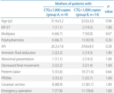
어머니 나이는 A군 31.9±5.2세, B군 32.0±3.0세로 두 군 간 차이는 없었다. 체외 수정 및 배아 이식 시술을 받아 임신한 경 우는 A군 1명(11.1%), B군 2명(14.3%)으로 차이가 없었고, 경 산부는 각각 6명(66.7%), 7명(50.0%)으로 차이가 없었다.
양수과다증은 A군 6명(66.7%)에 비해 B군은 13명(92.9%)으 로 많았으나 유의성은 없었고(P=0.26), 양수지수와 양수감소술 시행 유무 또한 차이를 보이지 않았다. 비정상 태위, 태동 이상, 조기 산통, 조기 양막파수, 제왕절개분만 모두 두 군 간 유의한 차이는 없었다(Table 2).
모든 어머니는 벌어진 입이나, 경직되고 부자연스러운 미소 등 myotonic dystrophy 환자의 전형적인 얼굴 모습이었고, 1명 의 어머니에서 근력 장애를 보이는 경우도 있었다.
3. 환아의 임상특성 및 임상양상
출생체중은 A군 2,071.1±519.2 g으로 B군의 2,435.7±
701.9 g에 비해 작았으나 유의성은 없었고, 재태주령도 각각 33.7±2.9주, 34.5±2.6주로 두 군 간의 차이는 없었다. 이외 성 별, Apgar 점수 및 분만실에서의 소생술 여부에서도 두 군 간 유 의한 차이는 없었다(Table 3).
임상양상으로 모든 환아에서 전형적인 얼굴 생김새와 근긴장 도 저하 및 수유 곤란이 있었다. 대부분 환아에서 내반첨족과 횡 격막 상승도 보였지만, 두 군 간 유의한 차이는 없었다(Table 3).
4. 질병 이환
신생아 호흡곤란증후군은 A군과 B군에서 각각 7명(77.8%) 과 9명(64.3%)으로 많은 빈도로 진단되었으나, 두 군 간의 유의 한 차이는 없었다. 두부초음파 검사상 뇌실 확장 소견이 A군과 B군에서 각각 7명(77.8%)과 10명(71.4%)으로 많았으나, 두 군 간 차이는 없었고, 3단계 이상 뇌실내 출혈 빈도에도 차이가 없 Fig. 2. Flow diagram of subjects. CMD, congenital myotonic dystrophy; CTG, cytosine, thymine, guanine.
Fig. 3. The relationship between CTG copies of patients and their mothers. CTG, cytosine, thymine, guanine.
었다. 이외에도 괴사성 장염, 패혈증 등의 질병 이환도 두 군 간 차이가 없었다(Table 4).
5. 입원 경과
재원일수는 A군 56.2±44.4일, B군 59.8±56.1일로, 두 군 간 유의한 차이는 없었다. 모든 환아가 산소치료를 받았고, 1명을 제외하고 나머지 모든 환아가 침습적 기계 환기치료를 받았다.
Table 2. Maternal Obstetric Characteristics between Group A and B Mothers of patients with
P- value CTG≤1,000 copies
(group A, n=9) CTG>1,000 copies (group B, n=14)
Age (yr) 31.9±5.2 32.0±3.0 0.98
IVFET 1 (11.1) 2 (14.3) 1.00
Multipara 6 (66.7) 7 (50.0) 0.67
Polyhydramnios 6 (66.7) 13 (92.9) 0.26
AFI 26.2±7.8 29.8±8.5 0.28
Amniotic fluid reduction 2 (22.2) 2 (14.3) 1.00
Abnormal presentation 1 (11.1) 2 (14.3) 1.00
Decreased fetal movement 2 (22.2) 3 (21.4) 1.00
Preterm labor 5 (55.6) 10 (71.4) 0.66
PROMs 3 (33.3) 5 (35.7) 1.00
Cesarean section 8 (88.9) 12 (85.7) 1.00
Emergency operation 7 (77.8) 11 (78.6) 1.00
Values are presented as mean±standard deviation or number (%).
Abbreviations: CTG, cytosine, thymine, guanine; IVFET, in vitro fertilization
embryo transfer; AFI, amniotic fluid index; PROMs, premature rupture of membranes.
Table 3. Neonatal Demographic and Clinical Findings between Group A and B
CTG≤1,000 copies
(group A, n=9) CTG>1,000 copies (group B, n=14) P-
value Demographic findings
Birth weight (g) 2,071.1±519.2 2,435.7±701.9 0.22
SGA* 1 (11.1) 0 0.39
LGA† 0 1 (9.1) 1.00
Gestational weeks 33.7±2.9 34.5±2.6 0.60
Female 4 (44.4) 9 (64.3) 0.42
Inborn 8 (88.8) 8 (72.7) 0.62
Apgar score
1 minute 3.3±2.4 3.0±2.4 0.78
5 minute 5.2±3.0 5.5±2.6 0.73
Resuscitation at delivery room
Intubation 6 (66.7) 11 (78.6) 1.00
Cardiac massage 2 (22.2) 2 (14.3) 1.00
Clinical findings
Typical facial appearance 9 (100.0) 14 (100.0) 1.00
Hypotonia 9 (100.0) 14 (100.0) 1.00
Club foot 5 (55.6) 5 (35.7) 0.42
Diaphragm elevation 5 (55.6) 10 (71.4) 0.66
Feeding problems 9 (100.0) 14 (100.0) 1.00
Values are presented as mean±standard deviation or number (%).
Abbreviations: CTG, cytosine, thymine, guanine; SGA, small for gestational age;
LGA, large for gestational age.
*Weight below the 10th percentile for the gestational age.
†Weight above the 90th percentile for the gestational age.
Table 4. Neonatal Morbidity between Group A and B CTG≤1,000 copies
(group A, n=9) CTG>1,000 copies (group B, n=14) P-
value Respiratory
RDS 7 (77.8) 9 (64.3) 0.66
BPD 5 (55.6) 8 (57.1) 1.00
PPHN 1 (11.1) 4 (28.6) 0.61
Atelectasis 6 (66.7) 5 (35.7) 0.21
Aspiration pneumonia 6 (66.7) 4 (28.6) 0.10
Pleural effusion 2 (22.2) 4 (28.6) 1.00
Pulmonary hemorrhage 0 3 (21.4) 0.25
Cardiovascular
PDA 5 (55.6) 9 (64.3) 1.00
Surgical ligation 0 1 (7.1) 1.00
Medically closed 5 (55.6) 5 (35.7) 0.42
Hypotension 3 (33.3) 7 (50.0) 0.67
Inotropics 3 (33.3) 7 (50.0) 0.67
Steroid 0 2 (14.3) 0.50
Cranial
IVH (≥grade II)* 2 (22.2) 0 0.14
PVL 0 0
Ventriculomegaly 7 (77.8) 10 (71.4) 1.00
Gastrointestinal
Ascites 0 4 (28.6) 0.13
NEC (≥stage IIa)† 0 1 (7.1) 1.00
TPN induced cholestasis 1 (11.1) 2 (14.3) 1.00
Infection
Sepsis (cultureproven) 2 (22.2) 4 (28.6) 1.00
Values are presented as number (%).
Abbreviations: CTG, cytosine, thymine, guanine; RDS, respiratory distress syn
drome; BPD, bronchopulmonary dysplasia; PPHN, persistent pulmonary hypertension of newborn; PDA, patent ductus arteriosus; IVH, intraventricular hemorrhage; PVL, periventricular leukomalacia; NEC, necrotizing enterocolitis;
TPN, total parenteral nutrition.
*Intraventricular hemorrhage with ventricular dilatation (based on Papile grading system).15
†Signs of temperature instability, apnea, bradycardia, elevated pregavage residuals, mild abdominal distension, occult blood in stool, absent bowel sounds, abdominal tenderness, ileus and pneumatosis intestinalis (based on Modified Bell’s staging criteria).16
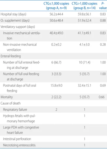
두 군 간 산소치료 일수와 기계 환기치료 일수에서 유의한 차이 는 없었다. 또한 체중 kg 당 하루 100 mL 이상을 섭취한 경우를 완전 장관영양으로 기준하였을 때, A군의 퇴원 전 사망 환아 2 명과 보호자 원하여 전원된 환아 1명을 제외한 6명(66.7%), B 군의 퇴원 전 사망 환아 4명을 제외한 10명(71.4%)에서 퇴원시 완전 장관영양이 가능하였다. 따라서 사망하거나 전원된 환아 를 제외하고는 모두 퇴원시 완전 장관영양을 달성하였다. 이 중 각각 3명(33.3%)과 5명(35.7%)은 경구영양만으로 하루 기준을 충족하였다. 완전 경구영양이 가능한 시기는 A군 15.8±9.0일 에 비해 B군이 32.4±15.1일로 오래 걸렸으나 유의하지는 않았 다(Table 5).
6. 사망률 및 생존 관련 인자
입원 동안 23명의 환아 중 7명이 사망하여 신생아집중치료 실에서의 사망률은 30.4%였다. A군과 B군에서 각각 2명(22.2
%)과 5명(35.7%)이었으며, 두 군 간 차이는 없었다. 7명의 사
망 환아 중 3명(42.9%)은 호흡부전으로 사망하여 사망 원인의 가장 많은 비율을 차지하였다. 나머지 4명은 각각 폐출혈이 동 반된 태아 수종, 동맥관 개존으로 인한 심부전, 장 천공 및 괴사 성 장염으로 사망하였다. 출생 당일 1명, 생후 2일-2주 1명, 생 후 2주-1개월 2명, 생후 1개월 이후 3명이 사망하였다(Table 5). 또한, 생존과 관계 있는 인자를 알아보기 위해 입원 중 생존
Table 5. Hospital Course between Group A and B CTG≤1,000 copies
(group A, n=9) CTG>1,000 copies (group B, n=14) P-
value
Hospital stay (days) 56.2±44.4 59.8±56.1 0.83
O2 supplement (days) 50.6±48.4 51.9±52.4 0.88
Ventilatory support (days) Invasive mechanical ventila
tion
40.4±49.0 41.1±49.1 0.83
Noninvasive mechanical ven tilation
0.2±0.2 4.1±3.0 0.28
Enteral feeding
Number of full enteral feed
ing at discharge
6 (66.7) 10 (71.4) 1.00
Number of full oral feeding at discharge
3 (33.3) 5 (35.7) 1.00
Postnatal days of full oral feeding
15.8±9.0 32.4±15.1 0.69
Mortality 2 (22.2) 5 (35.7) 0.66
Cause of death
Respiratory failure 2 1
Hydrops fetalis with pul
monary hemorrhage
1
Large PDA with congestive heart failure
1
Intestinal perforation 1
Necrotizing enterocolitis 1
Values are presented as mean±standard deviation or number (%).
Abbreviations: CTG, cytosine, thymine, guanine; PDA, patent ductus arteriosus.
Table 6. Differences in Demographics and Morbidities between Survivors and Mortality Cases
Alive at discharge
(n=16) Died at discharge
(n=7) P-
value Demographics
Gestational weeks 34.7±2.7 33.0±2.5 0.15
Birth weight (g) 2,347.5±626.6 2,168.6±735.4 0.67
Female 9 (56.3) 4 (57.1) 1.00
Inborn 14 (87.5) 4 (57.1) 0.14
Apgar score
1 minute 4.1±2.2 0.9±0.7 0.0001
5 minute 6.3±2.5 3.3±1.9 0.01
Resuscitation at delivery room
Intubation 10 (62.5) 7 (100.0) 0.12
Cardiac massage 0 2 (28.6) 0.08
Morbidity
RDS 9 (56.3) 7 (100.0) 0.06
BPD 9 (56.3) 4 (57.1) 1.00
PPHN 4 (25.0) 1 (14.3) 1.00
Atelectasis 8 (50.0) 3 (42.9) 1.00
Aspiration pneumonia 7 (43.8) 3 (42.9) 1.00
Pleural effusion 3 (18.8) 3 (42.9) 0.32
Pulmonary hemorrhage 1 (6.3) 2 (28.6) 0.21
PDA 9 (56.3) 5 (71.4) 0.66
Hypotension 5 (31.3) 5 (71.4) 0.17
Ventriculomegaly 12 (75.0) 5 (71.4) 1.00
IVH (≥grade III)* 1 (6.3) 1 (14.3) 0.53
Ascites 2 (12.5) 2 (28.6) 0.56
NEC (≥stage IIa)† 0 1 (14.3) 0.30
Sepsis (culture proven) 4 (25.0) 2 (28.6) 1.00
Values are presented as mean±standard deviation or number (%).
Abbreviations: RDS, respiratory distress syndrome; BPD, bronchopulmonary dysplasia; PPHN, persistent pulmonary hypertension of newborn; PDA, pa
tient ductus arteriosus; IVH, intraventricular hemorrhage; NEC, necrotizing enterocolitis.
*Intraventricular hemorrhage with ventricular dilatation (based on Papile grading system).15
†Signs of temperature instability, apnea, bradycardia, elevated pregavage residuals, mild abdominal distension, occult blood in stool, absent bowel sounds, abdominal tenderness, ileus and pneumatosis intestinalis (based on Modified Bell’s staging criteria).16
아와 사망아에서 주산기 특징과 질병 이환을 비교하였을 때, 생 존아에서 1분 Apgar 점수가 4.1±2.2로 사망아 0.9±0.7에 비 해 유의하게 높았고(P=0.0001), 5분 Apgar 점수도 6.3±2.5로 사망아 3.3±1.9에 비해 유의하게 높았다(P=0.01). 그 밖의 인 자는 두 군 간 유의한 차이가 없었다(Table 6).
7. 장기 예후
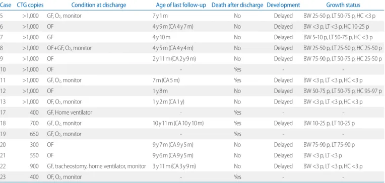
퇴원시 생존 환아 16명 중 추가 사망 6명이 발생하여 총 사망 률은 56.5%였으며, 이 중 4명은 퇴원 후 1주일 이내, 1명은 생후 7개월, 1명은 10세에 사망하였다. 6개월 이상 생존하였던 환아 12명 모두에서 발달지연을 보였고, 7명(58.3%)에서 성장 부진 을 보였다. 발달지연을 보인 환아들은 K-DST 5개 영역별 각 질 문 항목에 대부분 하지 못하는 편이다(1점) 또는 전혀 할 수 없 다(0점)로 표기되어 영역별 총점이 각 영역별로 제시된 절단점 최저 점수에도 모두 미치지 못하여 최종 총점에 대한 평가가 모 두 심화평가 권고로 확인되었다. 운동발달은 나이가 들수록 어 느 정도 호전되었으나, 모두 정상 범위에는 도달하지 못하였고, 모든 환아에서 언어 발달, 특히 발어(phona tion)에 지연이 있었 다(Table 7).
고찰
선천 근긴장 퇴행위축(CMD)은 상염색체 우성으로 유전되 는 근육의 퇴행성 병변으로 여러 기관을 침범하는 질환이다. 환 아의 임상 증상과 원인 유전자에 따라 근긴장성 이영양증 1형 (DM1)과 근긴장성 이영양증 2형(DM2)으로 분류되는데, DM1 은 19번 염색체의 DMPK 유전자의 CTG 삼핵산 반복의 이상 확 장에 의해 나타나고, DM2는 3q21 zinc finger protein 9 유전자 의 cytosine, cytosine, thymine, guanine (CCTG) 사염기 서열 반복의 이상 확장이라는 것이 밝혀졌다.9 따라서 과거 근전도상 특징적인 소견으로 진단되던 것이 근래는 분자 유전학적 검사 로 진단되고 있다.7,18,19
CMD 환자에서 CTG 삼핵산 반복수만으로 선천 근긴장 퇴행 위축으로 분류하는 것에 대해서는 현재까지는 불확실한 상태
이다.2,3,10 따라서, Campbell20은 선천 근긴장 퇴행위축의 정의
를 보다 명확하게 하기 위해, 유전학적으로 CTG 삼핵산 반복의 이상 확장이 있고, 임상적으로 신생아기에 근긴장 퇴행위축 증 상이 나타나 의학적 보조치료가 필요한 경우로 정의하자고 제 안하였다.
CMD는 모계를 통한 유전 양상이 특징이다.21,22 본 연구에서 어머니의 유전자 검사는 14명(증례 17과 20은 남매)에서 실시 하였는데 모두 검사 전까지 이환된 사실을 인지하지 못하고 있
Table 7. Long Term Outcomes of Survivors after Discharge
Case CTG copies Condition at discharge Age of last follow-up Death after discharge Development Growth status
5 >1,000 GF, O2, monitor 7 y 1 m No Delayed BW 2550 p, LT 5075 p, HC <3 p
6 >1,000 OF 4 y 9 m (CA 4 y 7 m) No Delayed BW <3 p, LT <3 p, HC 1025 p
7 >1,000 GF 4 y 10 m No Delayed BW 510 p, LT 5075 p, HC <3 p
8 >1,000 OF+GF, O2, monitor 4 y 5 m (CA 4 y 4 m) No Delayed BW 2550 p, LT 2550 p, HC 2550 p
9 >1,000 OF 2 y 11 m (CA 2 y 9 m) No Delayed BW 7590 p, LT 5075 p, HC 2550 p
10 >1,000 OF Yes
11 >1,000 GF, O2, monitor 7 m (CA 5 m) Yes Delayed BW <3 p, LT <3 p, HC <3 p
12 >1,000 OF 1 y 8 m No Delayed BW 5075 p, LT 5075 p, HC 9597 p
13 >1,000 OF, O2, monitor 1 y 2 m (CA 1 y) No Delayed BW <3 p, LT <3 p, HC <3 p
17 400 GF, Home ventilator Yes
18 700 GF, O2, monitor 10 y 11 m (CA 10 y 10 m) Yes Delayed BW 1025 p, LT 1025 p
19 650 GF, O2, monitor Yes
20 300 OF 9 y 7 m (CA 9 y 5 m) No Delayed BW 7590 p, LT 7590 p
21 550 OF 9 y 6 m (CA 9 y 5 m) No Delayed BW <3 p, LT <3 p
22 900 GF, tracheostomy, home ventilator, monitor 3 y 11 m (CA 3 y 9 m) No Delayed BW <3 p, LT <3 p, HC <3 p
23 400 OF, O2, monitor Yes
Abbreviations: CTG, cytosine, thymine, guanine; GF, gavage feeding; y, years; m, months; BW, body weight; LT, length; HC, head circumference; p, percentile; OF, oral feeding; CA, corrected age.
었으나, 벌어진 입이나, 경직되고 부자연스러운 미소 등 전형적 인 얼굴 모습이었고, 근력 장애를 보이는 경우도 있었다. 유전 자 검사가 실시된 14명 어머니(증례 17과 20은 남매) 모두 75- 500반복수를 보였으나, 환아의 반복수와는 상관관계가 없었다.
CMD는 세대로 내려갈수록 서열 반복수가 증가하고 임상양상 이 심하게 나타나는 ‘예견 효과(anticipation)’의 특징을 갖는다 고 한다.6,19,23 본 연구에서도 환아가 어머니에 비해 삼핵산 반복 수가 많았고, 근력 저하와 호흡곤란 증상이 심했던 점으로 미루 어 세대 간 ‘예견 효과’가 있음을 확인할 수 있었다.
CTG 삼핵산 반복수와 임상양상과의 관계에 대해서는 보고자 마다 결과가 다양하다. Marchini 등24은 24명의 DM1 환아에서 반복수가 증가할수록 더 이른 나이에 발병하고, 근육 장애가 심 하게 나타나며 지능과 단기 기억력이 더 떨어지지만, 근 위축이 나 호흡부전, 심장 이상, 녹내장 및 수면 장애 등과는 관련이 없 다고 보고하였다. 또한 Arsenault 등25은 102명의 DM1 환아들 에서 CTG 삼핵산 염기서열이 50-99반복수인 경우 대부분 무 증상이고, 100-200인 경우 근력 저하가 더 많음을 관찰하여, 200 미만의 비교적 적은 반복수에서도 반복수에 따라 임상양 상에 차이가 있음을 보고하였고, Logigian 등26은 15명의 DM1 환아에서 CTG 삼핵산 반복서열이 많을수록 수부 근육의 긴장 정도가 심해진다고 보고하였다. 그러나 Schara와 Schoser7과 Day와 Ranum27의 보고에 의하면 400반복수까지는 반복수와 환아의 임상양상이 상관관계를 갖지만, 그 이상에서는 관련성 이 없다고 보고하였다.
본 연구는 CTG 삼핵산 1,000반복수를 기준으로 신생아집중 치료실에서 신생아기에 진단된 선천 근긴장 퇴행위축 환아만을 대상으로 CTG 삼핵산 1,000반복수 초과군과 1,000반복수 이 하군 간에 임상양상, 산모의 특성, 질병의 이환 및 입원 경과에 서 유의할 만한 차이가 있는지 알아보고자 하였으며, 국내에서 단일 기관으로서는 가장 많은 수의 유전자 검사로 확진된 CMD 환아들이 포함되었다.
본 연구 결과, 환아 어머니의 산과적 특성, 환아의 임상적 특 징 및 질병 이환에서 차이 있는 항목은 없었다. 환아 어머니의 양수과다증 빈도는 두 군 간 통계학적 차이는 없었으나, 특히 1,000반복수 이상군에서 92.9%로 높아, Schild 등28의 보고와 유사하게 산모의 양수과다증의 원인으로 드물게 태아의 CMD 이환 가능성에 대해 고려해야 함을 알 수 있음을 알 수 있었다.
임상양상은 다른 연구보고29,30에서와 비슷하게 특징적인 얼굴 생김새와 근긴장도 저하 및 수유 곤란은 100%에서, 내반첨족은 43.5%, 횡격막 상승은 65.2%, 뇌실 확장소견 73.9%를 보였으 나, 동맥관 개존은 60.9%로 Fujii 등31이 보고한 것보다 많았다.
본 연구의 CMD 환아 중 18명(78.3%)이 미숙아였고, 이들의
평균 재태주수는 34.2±2.7주였다. 미숙아는 모두 응급 제왕절 개수술을 통해 출생하였으며, 그중 15명(83.3%)이 조기 산통이 원인이었다. Myotonic dystrophy와 조기 산통과의 연관성은 명 확히 밝혀지지 않았으나, myotonic dystrophy 산모에서 조기 산통의 흔하게 발생하는 것으로 보고되었다.32 또한 조기 산통 이 있는 산모 중 12명(80%)에서 양수과다증이 동반되어 있었으 며, 양수과다증이 조기 산통의 원인이 될 수 있기 때문에 미숙아 의 비율이 많은 것으로 생각된다.33
현재까지 침습적 기계환기 치료가 필요하였던 선천 근긴장 퇴행위축 환아 중 가장 적은 CTG 삼핵산 이상 확장은 400반복 수로 보고되었다.34 그러나 본 증례에서는 CTG 삼핵산 300반복 수인 환아(증례 20)에서 4일간 침습적 기계환기 치료가 필요하 여 더 낮은 반복수에서도 침습적 기계환기 치료가 필요할 수도 있음을 확인하였다.
CMD의 사망률은 16-41%로, 호흡부전이 사망의 주 요인으 로 보고되고 있다.3,30,35-38 115명의 환아에서 장기간 생존을 분 석한 Reardon 등37의 연구에 따르면 30대 중반까지의 사망률은 50%에 이르렀고, 후기 사망의 주 원인은 호흡 관련 질병과 부정 맥이었다. 본 연구에서는 총 환아 23명 중 13명(56.5%)이 사망 하였으며, 그중 6명(46.2%)이 호흡부전으로 사망하였다.
몇몇 연구에서 30일 이상의 장기 기계환기 치료를 받은 환아 에서 높은 사망률을 보였으나,3,30,39 본 연구에서는 기계환기 치 료와 산소치료 유지 일수는 사망률과 유의한 관계가 없었다. 본 연구에서 30일 이상 침습적 기계환기 치료를 유지하고 생존한 7명의 환아 중 3명(42.9%)에서 이탈 전 비침습적 호흡 보조기 를 사용하였고, 1명의 환아는 처음부터 비침습적 호흡보조기 치 료만으로 생존하였다. 이러한 비침습적 호흡보조기의 사용으로 장기간의 침습적 기계환기 치료 후 호흡기 이탈이 용이하게 됨 으로써 사망률도 감소한 것으로 생각된다.40 Apgar 점수는 환아 의 근육 긴장도와 호흡 노력을 반영한다고 하는데,20 본 연구에 서 1분과 5분 Apgar 점수만이 생존과 관련이 있는 것으로 관찰 되었다.
CMD 환아들에서 출생시 심했던 근력 저하는 연령이 증가하 면서 3-4세경 이후 점차 서서히 호전된다고 알려져 있다.7,38 본 연구에서도 3년 이상 추적관찰된 4명의 환아 모두 생후 초기에 심한 운동 발달지연을 보였으나 걸을 수 있을 정도로 서서히 호 전되었다. 한편, Roig 등38은 약 70%의 환아에서 정신지체가 동 반된다고 보고하였는데, 본 연구의 모든 생존 환아에서 언어발 달 특히 발어(phonation)에 지연을 보였다.
여러 보고 결과, CTG 삼핵산 반복 서열과 임상양상과의 관계 가 아직 명확하지 않은데, 이는 대부분의 연구가 통계적으로 의 미 있는 결과를 찾기에는 대상 환아 수가 적었고, 후향적 분석
으로 임상 정보가 불충분했기 때문일 수도 있겠다. 본 연구 또 한 단일 기관연구로 대상 환아 수가 적었고, 후향적 분석이라 는 제한점이 있지만, 신생아기에 진단된 환아로 대상을 한정하 여, CTG 삼핵산 반복수에 따른 임상적 특징과 예후와의 관계를 분석하였다는 점에서 의의가 있을 것으로 생각된다. 향후 더 많 은 환아를 대상으로 한 전향적 다기관 연구가 필요할 것으로 사 료된다.
결론적으로, 본 연구에서 신생아기에 선천 근긴장 퇴행위축 으로 확진된 환아들의 CTG 삼핵산 반복수는 어머니의 반복수 와는 관계가 없었다. CTG 삼핵산 1,000반복수 이하군과 CTG 1,000반복수 초과군으로 나누어 비교하였을 때, 두 군 간 환아 어머니의 산과적 특성, 환아의 임상특성과 임상양상, 질병 이환, 입원 경과, 사망률 및 장기 예후에 차이는 없었다. 선천 근긴장 퇴행위축 환아에서 입원 중 생존과 사망에 관계 있는 유의한 인 자는 1분 및 5분 Apgar 점수였다.
References
1) Vanier TM. Dystrophia myotonica in childhood. Br Med J 1960;2:1284-8.
2) Tsilfidis C, MacKenzie AE, Mettler G, Barceló J, Korneluk RG. Correlation between CTG trinucleotide repeat length and frequency of severe congenital myotonic dystrophy. Nat Genet 1992;1:192-5.
3) Campbell C, Sherlock R, Jacob P, Blayney M. Congenital myotonic dy- strophy: assisted ventilation duration and outcome. Pediatrics 2004;
113:811-6.
4) de Die-Smulders CE, Smeets HJ, Loots W, Anten HB, Mirandolle JF, Geraedts JP, et al. Paternal transmission of congenital myotonic dystro- phy. J Med Genet 1997;34:930-3.
5) Ranum LP, Cooper TA. RNA-mediated neuromuscular disorders. Annu Rev Neurosci 2006;29:259-77.
6) Pearson CE, Nichol Edamura K, Cleary JD. Repeat instability: mecha- nisms of dynamic mutations. Nat Rev Genet 2005;6:729-42.
7) Schara U, Schoser BG. Myotonic dystrophies type 1 and 2: a summary on current aspects. Semin Pediatr Neurol 2006;13:71-9.
8) Dyken PR, Harper PS. Congenital dystrophia myotonica. Neurology 1973;23:465-73.
9) Bird TD. Myotonic dystrophy type 1. [accessed on 22 Oct 2015]. Avail- able at http://www.ncbi.nlm.nih.gov/books/NBK1165.
10) Redman JB, Fenwick RG Jr, Fu YH, Pizzuti A, Caskey CT. Relationship between parental trinucleotide GCT repeat length and severity of myo- tonic dystrophy in offspring. JAMA 1993;269:1960-5.
11) Kim HK, Kim JH, Lee YA, Ko TS, Kim KS, Yoo HW, et al. A case of congeni tal myotonic dystrophy diagnosed by molecular genetics. J Korean Child Neurol Soc 1998;5:356-60.
12) Kim SH, Kim EY, Park SK, Choi SJ, Lim SC. A case of congenital myotonic dystrophy diagnosed by molecular genetics. J Korean Soc Neonatol
2006;13:194-8.
13) Jung SH, Bang MS. Belated diagnosis of congenital myotonic dystrophy in a boy with cerebral palsy. Am J Phys Med Rehabil 2007;86:161-5.
14) Yum MS, Lee BH, Kim GH, Lee JJ, Choi SH, Lee JY, et al. Southern analysis after long-range PCR: clinical application in Korean patients with myo- tonic dystrophy 1. J Genet Med 2013;10:33-7.
15) Papile LA, Burstein J, Burstein R, Koffler H. Incidence and evolution of sub ependymal and intraventricular hemorrhage: a study of infants with birth weights less than 1,500 gm. J Pediatr 1978;92:529-34.
16) Walsh MC, Kliegman RM. Necrotizing enterocolitis: treatment based on staging criteria. Pediatr Clin North Am 1986;33:179-201.
17) Chang YS, Ahn SY, Park WS. Committee on program and planning and advisory committee of Korean Neonatal Network. The establish ment of the Korean Neonatal Network (KNN). Neonatal Med 2013;20:169-78.
18) Mahadevan M, Tsilfidis C, Sabourin L, Shutler G, Amemiya C, Jansen G, et al. Myotonic dystrophy mutation: an unstable CTG repeat in the 3' untranslated region of the gene. Science 1992;255:1253-5.
19) Fu YH, Pizzuti A, Fenwick RG Jr, King J, Rajnarayan S, Dunne PW, et al. An unstable triplet repeat in a gene related to myotonic muscular dystro- phy. Science 1992;255:1256-8.
20) Campbell C. Congenital myotonic dystrophy. J Neurol Neurophysiol 2012;S7:001.
21) Harley HG, Rundle SA, MacMillan JC, Myring J, Brook JD, Crow S, et al. Size of the unstable CTG repeat sequence in relation to phenotype and parental transmission in myotonic dystrophy. Am J Hum Genet 1993;52:
1164-74.
22) Imbert G, Kretz C, Johnson K, Mandel JL. Origin of the expansion muta- tion in myotonic dystrophy. Nat Genet 1993;4:72-6.
23) Groenen P, Wieringa B. Expanding complexity in myotonic dystrophy.
Bioessays 1998;20:901-12.
24) Marchini C, Lonigro R, Verriello L, Pellizzari L, Bergonzi P, Damante G.
Correlations between individual clinical manifestations and CTG repeat amplification in myotonic dystrophy. Clin Genet 2000;57:74-82.
25) Arsenault ME, Prévost C, Lescault A, Laberge C, Puymirat J, Mathieu J.
Clinical characteristics of myotonic dystrophy type 1 patients with small CTG expansions. Neurology 2006;66:1248-50.
26) Logigian EL, Moxley RT 4th, Blood CL, Barbieri CA, Martens WB, Wiegner AW, et al. Leukocyte CTG repeat length correlates with severity of myo- tonia in myotonic dystrophy type 1. Neurology 2004;62:1081-9.
27) Day JW, Ranum LP. RNA pathogenesis of the myotonic dystrophies.
Neuromuscul Disord 2005;15:5-16.
28) Schild RL, Plath H, Hofstaetter C, Brenner R, Mann E, Mundegar RR, et al.
Polyhydramnios: an association with congenital myotonic dystrophy. J Obstet Gynaecol 1998;18:484-5.
29) Regev R, de Vries LS, Heckmatt JZ, Dubowitz V. Cerebral ventricular dila- tion in congenital myotonic dystrophy. J Pediatr 1987;111:372-6.
30) Rutherford MA, Heckmatt JZ, Dubowitz V. Congenital myotonic dys- trophy: respiratory function at birth determines survival. Arch Dis Child 1989;64:191-5.
31) Fujii T, Yorifuji T, Okuno T, Toyokuni S, Okada S, Mikawa H. Congenital myotonic dystrophy with progressive edema and hypoproteinemia.
Brain Dev 1991;13:58-60.
32) Awater C, Zerres K, Rudnik-Schöneborn S. Pregnancy course and out- come in women with hereditary neuromuscular disorders: comparison of obstetric risks in 178 patients. Eur J Obstet Gynecol Reprod Biol 2012;
162:153-9.
33) Hamza A, Herr D, Solomayer EF, Meyberg-Solomayer G. Polyhydra mnios:
causes, diagnosis and therapy. Geburtshilfe Frauenheilkd 2013;73:1241-6.
34) Echenne B, Rideau A, Roubertie A, Sébire G, Rivier F, Lemieux B. Myoto- nic dystrophy type I in childhood long-term evolution in patients sur- viving the neonatal period. Eur J Paediatr Neurol 2008;12:210-23.
35) Hageman AT, Gabreëls FJ, Liem KD, Renkawek K, Boon JM. Congenital myotonic dystrophy; a report on thirteen cases and a review of the lite- rature. J Neurol Sci 1993;115:95-101.
36) Wesström G, Bensch J, Schollin J. Congenital myotonic dystrophy. Inci- dence, clinical aspects and early prognosis. Acta Paediatr Scand 1986;
75:849-54.
37) Reardon W, Newcombe R, Fenton I, Sibert J, Harper PS. The natural his- tory of congenital myotonic dystrophy: mortality and long term clini cal aspects. Arch Dis Child 1993;68:177-81.
38) Roig M, Balliu PR, Navarro C, Brugera R, Losada M. Presentation, clinical course, and outcome of the congenital form of myotonic dystrophy.
Pediatr Neurol 1994;11:208-13.
39) Connolly MB, Roland EH, Hill A. Clinical features for prediction of sur vival in neonatal muscle disease. Pediatr Neurol 1992;8:285-8.
40) Keller C, Reynolds A, Lee B, Garcia-Prats J. Congenital myotonic dystrophy requiring prolonged endotracheal and noninvasive assisted ventilation:
not a uniformly fatal condition. Pediatrics 1998;101(4 Pt 1):704-6.