https://doi.org/10.14734/PN.2019.30.2.66 pISSN 2508-4887•eISSN 2508-4895
Kyung Suk Baek, MD1, Sol Han, MD1, Og Hyang Kim, MD1, Ju Sun Heo, MD2, Jihyun Jeon, MD1
1Department of Pediatrics, CHA Gangnam Medical Center, CHA University, Seoul; 2Department of Pediatrics, Korea University Anam Hospital, Seoul, Korea
Objective: In vitro fertilization (IVF) pregnancy is increasing. The recent reviews have reported the perinatal outcomes of IVF were preterm birth, low birth weight, small for gestational age, congenital malformations, neurologic disorders and epigenetic defects. We aimed to analyze the perinatal outcomes of IVF compared with natural pregnancy on very low birth weight infants.
Methods: Our study population was derived from Neonatal Intensive Care Unit of the Gangnam CHA Medical Center from 2010 to 2014, consisting of singleton live births in very low birth weight infants. We grouped IVF group (n=24) and control group (natural pregnancy, n=112). We analyzed two groups about maternal characteristics, neonatal characteristics, and outcomes (retinopathy of prematurity [ROP], bronchopulmonary dysplasia [BPD], periventricular leukomalacia [PVL], necro
tizing enterocolitis [NEC], death).
Results: Maternal age was significantly older in IVF group (34.9±0.9 vs. 33.0±0.4, P=0.03). The Apgar score 1 minute of the IVF group was significantly lower than control group (4.0±0.3 vs. 4.8±0.2, P=0.03).
But there were no other significant differences of neonatal morbidities. In univariate logistic regression analysis with IVF, the odds ratio of maternal age was 1.13 (95% confidence interval: 1.011.27, P=0.04).
The relative risk of RDS, BPD, PVL, and ROP was increased, but it was not significant in univariate and multivariate logistic regression.
Conclusion: As the maternal age increased, IVF pregnancy was significantly higher. The relative risk of RDS, BPD, PVL, and ROP was high in IVF group, but it was not significant in univariate and multivariate logistic regression.
Key Words: Infant, very low birth weight, Fertilization in vitro
서론
보조생식술(assisted reproductive technologies)은 정자와 난자를 인위적으로 실험 처 리하여 불임을 치료하는 것으로, 1978년 Edward 등1은 처음 체외 인공 수정을 통하여 출 산에 성공한 뒤 지난 40여 년간 지속적으로 시술 기관과 시행 수가 급격히 증가하였다.2,3 2015년 Centers for Disease Control and Prevention은 미국에서 시행되는 보조생식술 이 182,111회이며 59,334건의 출산이 이루어져 미국에서 출생한 신생아의 1.7%를 차지한 다고 하였다.4 2013년 보고된 한국의 보조생식술 현황에서는 27,947예의 보조생식술에서 5,196명의 신생아가 출생하였다고 보고하였고 정부의 지원도 확대되고 있다.5
체외 인공 수정(in vitro fertilization, IVF) 후 산모 및 신생아의 이환율 및 사망률에 대한 관심도 증가하였다. 체외 인공 수정을 통하여 출생한 신생아들이 재태 연령이 짧고 다태아 빈도가 높으며 자연 임신에 비하여 선천성 기형이 많다고 보고해 왔으나,6-8 최근 기술이 발 달함에 따라 이환율에 통계학적으로 유의한 차이가 없고, 성장 및 뇌신경 발달에도 자연 임 신과의 비교 시 유의하지 않다고 보고하였다.9,10 이러함에도 체외 인공 수정 출생 신생아에 Received: 17 September 2018
Revised: 13 October 2018 Accepted: 25 October 2018 Correspondence to Jihyun Jeon, MD
Department of Pediatrics, CHA Gangnam Medical Center, CHA University, 566 Nonhyeonro, Gangnamgu, Seoul 06135, Korea Tel: +82234682815
Fax: +82234682618 E-mail: [email protected] Copyright© 2019 by The Korean Society of Perinatology
This is an Open Access article distributed under the terms of the Creative Com
mons Attribution NonCommercial License (http://creativecommons.org/
license/bync/4.0/), which permits unrestricted noncommercial use, distribution, and reproduction in any
Perinatal Outcomes of In Vitro Fertilization
Versus Natural Pregnancy in Very Low Birth
Weight Infants
자들은 조기 분만된 극소 저체중 출생아 단태아만을 대상으로 분석하고자 하였다. 이에 본 저자들은 다태아를 제외한 극소 저 체중 출생 단태아를 대상으로 체외 인공 수정에 의한 임신과 자 연 임신에 의하여 출생한 신생아의 주산기 결과들을 비교하고 자 하였다.
대상 및 방법
1. 대상
2010년 1월부터 2014년 12월까지 강남 차병원에서 출생한 1,500 g 미만의 극소 저체중 출생아 중 신생아 집중치료실에 입 원하여 치료를 받은 253명을 대상으로 하였다. 이 중 쌍둥이와 삼둥이 117명을 제외하여 단태아로 출생한 136명을 최종 대상 으로 하였다. 이들 중 자연 임신으로 출생한 신생아를 control group (n=112)으로, 체외 인공 수정으로 출생한 신생아를 IVF group (n=24)으로 분류하여 분석하였다. 유전 질환과 동반된 심장 기형, 유전자 및 염색체 이상에 동반된 기형, 다발성 복합 기형을 가진 환아는 제외하였다.
2. 방법
대상 환자와 산모의 의무기록을 후향적으로 분석하여 병력을 조사하였다. 산모의 특성으로 분만 당시 나이, 분만력, 조기 양 막파수 시간, 전자간증 유무, 조직학적 융모양막염 유무, 산전 부신피질 호르몬 투여, 제왕절개술 유무에 대하여 조사하였다.
환아의 특성으로는 재태 연령, 출생 시 체중, 성별, 1분, 5분 아 프가 점수(Apgar score), 키, 두위를 포함하였다. 환아의 신생아 집중치료실 입원 치료 기간, 사망률, 기관 내 삽관 유지 기간, 산 소 치료 기간, 괴사성 장염(necrotizing enterocolitis, NEC), 신 생아 호흡곤란 증후군(respiratory distress syndrome, RDS), 동맥관개존증(patent ductus arteriosus, PDA), 미숙아 망막 병증(retinopathy of prematurity, ROP), 기관지 폐 이형성증 (bronchopulmonary dysplasia, BPD), 뇌실주위백질연화증 (periventricular leukomalasia, PVL)을 비교하였다. 신생아 호 흡곤란 증후군은 흉부 방사선 검사상 망상 과립상 음영을 보이 며 인공 폐 표면활성제의 치료가 필요한 경우로 정의하였고, 괴 사성 장염은 modified bell 분류에 의하여11 stage II 이상의 환자 를 대상으로 하였다. ICROP (The international classification of retinopathy of prematurity 진단기준을 이용하여 미숙아 망 막병증을 진단하였다.12 기관지 폐 이형성증은 월경 후 주령 36 주에 산소 또는 양압 환기가 필요한 경우로 정의 하였으며 Jobe 과 Bancalari13의 정의에 따라 중등도 이상의 경우를 대상으로
하였다. 뇌실주위백질연화증은 뇌 초음파 검사 및 퇴원 전 시행 한 뇌 자기공명영상검사를 통하여 이루어졌다.
3. 통계적 분석
Stata 13.0 (StataCorp LP, College Station, TX, U.S.A.) 통 계 프로그램을 사용하여 IVF group과 control group을 t-test, χ2 test, Fisher’s exact test를 이용하여 두 군을 분석하였다.
Univariate logistic regression을 이용하여 IVF와 단변수와의 odd’s ratio를 구하였으며, IVF와 perinatal outcomes와의 연관 성을 분석하기 위하여 perinatal outcomes에 영향을 줄 수 있는 다른 변수들을 보정한 multivariate logistic regression을 사용 하였다.
결과
1. 산모의 임상적 특징
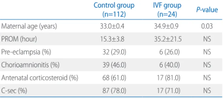
분만 당시 산모의 평균 나이는 IVF군이 34.9세(±0.9), control 군이 33.0세(±0.4)로 IVF군이 유의하게 높았다(P=0.03). 조기 양막파수 시간이 35.2시간(±21.1)으로 IVF군이 control군에 비 하여 길었으나, 통계적 유의성은 없었다. 산전 부신피질 호르몬 투여가 IVF군에서 81%, control군에서 61%로 차이가 있었으나 통계적 차이는 없었다. 전자간증 유무, 융모양막염, 제왕절개술 은 두 군 간에 통계학적 차이를 보이지 않았다(Table 1).
2. 신생아의 임상적 특징과 이환율
IVF군에서 재태 연령은 29.6주(±0.6), 출생 체중 1,382.5 g (±104.5)이고 control군의 재태 연령은 30.4주(±0.3), 출생 체 중은 1,477.4 g (±46.6g)으로 두 군 간에 유의한 차이는 없었 다. 그러나 1분 Apgar 점수가 IVF군에서 4.0점(±0.3), control 군에서 4.8점(±0.2)으로 유의하게 IVF군에서 낮았다(P=0.04).
Table 1. Maternal Characteristics and Morbidities between Two Groups Control group
(n=112) IVF group
(n=24) P-value
Maternal age (years) 33.0±0.4 34.9±0.9 0.03
PROM (hour) 15.3±3.8 35.2±21.5 NS
Preeclampsia (%) 32 (29.0) 6 (26.0) NS
Chorioamnionitis (%) 39 (46.0) 6 (40.0) NS
Antenatal corticosteroid (%) 68 (61.0) 17 (81.0) NS
Csec (%) 87 (78.0) 17 (71.0) NS
Abbreviations: IVF, in vitro fertilization; PROM, premature rupture of membrane;
NS, nonspecific; Csec, Cesarean section.
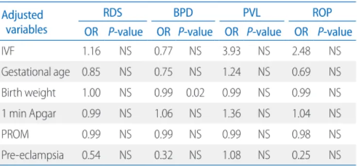
하게 증가하였다(Table 3). 재태 연령, 출생 체중, 1분 Apgar 점 수, 양막파수, 전자간증 변수를 보정하였을 때 IVF군에서 RDS, PVL, ROP 위험도가 증가하였으나 통계적으로 유의하지 않았 다(Table 4).
고찰
올해로 체외 인공 수정을 시작한지 40년이 되었다. 그동안 체 외 인공 수정을 통하여 출생한 신생아가 전 세계적으로 많아짐 에도 불구하고, 끊임없는 신생아 주산기 결과에 대한 우려는 아 직도 완전히 해소되지 않았다.
Källén 등14은 체외 인공 수정 시 미숙아, 자궁 내 성장 지연, 기형아 출생 위험이 높은 다태아 임신이 증가함에 따라 신생아 이환율이 자연 임신군에 비하여 나쁘고 재원 기간, 사망률 또한 높다고 하였다. 본 연구에서는 전체 환아 253명 중 심한 기형 을 가진 환아는 없었다. 최종 대상 환아 136명 중에서도 PDA는 IVF군에서 유의하게 많지 않았다. PDA 외에 다른 심장 기형은 심실 중격 결손(ventricular septal defect, VSD)이 control군에 서만 1명 있었다. 조기 진통에 의한 미숙아 출산은 산모의 고령 화와도 연관되어 있는데,15-17 Hastie 등18은 임신 시 산모의 연령 이 높아질수록 혈관 노화와 혈관 내피 기능장애가 생기고 이로 인하여 조기 진통 및 부당 경량아의 위험도가 높아지기 때문이 라고 하였다. 우리나라의 경우, 최근 들어 산모의 평균 출산 연 령이 2014년 32.0세에서 2017년 32.6세로 점점 높아지고 있으 며,19 35세 이상의 고위험 산모도 증가하는 추세이다.20 본 연구 에서도 IVF군의 평균 산모의 나이가 34.9±0.9세로 2017년 평 균 출산 연령보다 많았고, 산모의 나이가 1.13배 증가할수록 체 외 인공 수정이 유의하게 증가되었다. 산모의 고령화는 체외 인 그 외 출생 시 키, 머리둘레, 5분 Apgar 점수는 통계학적으로 유
의한 차이는 없었다. 신생아의 이환율 비교 시 기관 삽관 기간, 산소 치료 기간, 수혈 횟수, NEC, RDS, PDA, ROP, BPD, PVL에 서 두 군 간에 통계학적 차이는 없었다(Table 2).
IVF에 의한 극소 저체중 출생아의 주산기 결과 상관성을 확인 하기 위하여 로지스틱 회귀분석을 시행하였고, 단변수 분석에 서 산모의 나이가 1세 많아질수록 체외 인공 수정이 1.13배 유의 Table 2. Neonatal Characteristics and Outcomes between Two Groups
Control group
(n=112) IVF group
(n=24) P-value
Gestational age (weeks) 30.4±0.3 29.6±0.6 NS
Birth weight (g) 1,477.4±46.6 1,382.5±1,04.5 NS
Male 63 (56.0) 15 (63.0) NS
1 min Apgar 4.8±0.2 4.0±0.3 0.04
5 min Apgar 6.3±0.1 6.0±0.3 0.19
Length (cm) 39.8±0.4 38.9±0.1 NS
Head circumference (cm) 28.1±0.3 27.1±0.7 0.13
Hospital duration (day) 47.3±2.0 53.7±5.3 NS
Duration of intubation (day) 8.1±1.2 10.4±2.4 NS Duration of O2 treatment (day) 1.6±0.2 1.3±0.4 NS
NEC 11 (10.0) 0 (0) NS
ROP 8 (7.0) 4 (16.0) NS
RDS 87 (78.0) 21 (88.0) NS
PDA 25 (22.0) 5 (21.0) NS
BPD 21 (19.0) 7 (29.0) NS
PVL 2 (2.0) 1 (4.0) NS
Death 2 (2.0) 0 (0) NS
Values are presented as mean±standard deviation or number (%).
Abbreviations: IVF, in vitro fertilization; NS, nonspecific; NEC, necrotizing entero
colitis; ROP, retinopathy of prematurity; RDS, respiratory distress syndrome; PDA, patent ductus arteriosus; BPD, bronchopulmonary dysplasia; PVL, periventri cular leukomalacia.
Table 3. Univariate Logistic Regression Analysis with In Vitro Fertiliza- tion
Unadjusted OR 95% CI P-value
Maternal age* 1.13 1.011.27 0.04
RDS† 1.22 0.383.94 NS
BPD† 1.78 0.664.85 NS
PVL† 2.39 20.2127.49 NS
ROP† 2.60 0.719.47 NS
*Independent variables.
†Dependent varibles.
Abbreviations: OR, odds ratio; CI, confidence interval; RDS, respiratory distress syndrome; NS, nonspecific; BPD, bronchopulmonary dysplasia; PVL, periventri
cular leukomalacia; ROP, retinopathy of prematurity.
Table 4. Multivariate Logistic Regression Analysis for Perinatal Morbi- dities
Adjusted variables
RDS BPD PVL ROP
OR P-value OR P-value OR P-value OR P-value
IVF 1.16 NS 0.77 NS 3.93 NS 2.48 NS
Gestational age 0.85 NS 0.75 NS 1.24 NS 0.69 NS
Birth weight 1.00 NS 0.99 0.02 0.99 NS 0.99 NS
1 min Apgar 0.99 NS 1.06 NS 1.36 NS 1.04 NS
PROM 0.99 NS 0.99 NS 0.99 NS 0.98 NS
Preeclampsia 0.54 NS 0.32 NS 1.08 NS 0.25 NS
Abbreviations: RDS, respiratory distress syndrome; BPD, bronchopulmonary dysplasia; PVL, periventricular leukomalacia; ROP, retinopathy of prematurity; OR, odds ratio; IVF, in vitro fertilization; NS, nonspecific; PROM, premature rupture of membranes.
공 수정을 증가 시키고, 고령 산모는 조기 진통으로 인한 부당 경량아 및 저체중 출산이 많았다.21
위와 반대되는 보고들도 많았는데, Fan 등22은 375명의 28주 이상의 쌍둥이를 대상으로 한 연구에서 체외 인공 수정이 산 모와 환아의 이환율에 영향을 끼치지 않는다고 보고하였고, Messerschmidt 등23도 1,423명의 1,500 g 미만의 극소 저체중 출생아를 대상으로 다태아를 포함한 연구에서 체외 인공 수정이 주산기 합병증 위험성 및 이환율과의 연관성이 없다고 하였다.
국내 보고에서는 Choi와 Kim24이 재태 연령 32주 미만 126명의 쌍생아를 대상으로 하여 성장과 뇌신경 발달이 자연 임신군과 체외 인공 수정군 간의 차이가 없다고 하였다. 하지만 위의 보고 들은 대부분 체외 인공 수정으로 인한 다태아를 대상으로 진행 된 연구로, 체외 인공 수정 기술에 의한 임신과 자연 임신간을 분 석하는데 오류가 발생할 가능성이 있었다. 본 연구는 단태아만 을 대상으로 하여 분석하여 체외 인공 수정으로 인한 다태아 요 인을 처음부터 보정한 경우로, 재태 연령, 출생 체중, 키, 머리둘 레 차이가 없었고, 신생아 집중 치료실에서 입원 시 산소 사용 기 간, 기관 삽관 기간, 총 입원 기간, RDS, PDA, BPD, NEC, ROP, PVL, 사망 등 자연 임신으로 출생한 극소 저체중 출생 미숙아와 차이가 없었다.
체외 인공 수정의 경우 산모 질환 양막 파수, 고혈압, 전자간 증 등의 이환율이 증가한다고 하였는데,8,25 본 연구에서는 산모 나이 외에 양막 파수 시간, 전자간증 유무, 융모양막염 등 두 군 의 차이가 없었다. Romundstad 등9도 본 연구 결과와 동일하게 산모 질환 이환율이 자연 임신군과의 차이가 없다고 하였다. 본 연구에서처럼 산모 질환이 체외 인공 수정군에서 유의하게 증 가하지 않은 것은 위험요인인 다태아를 제외하였고, 산모 질환 은 임신 방법뿐만 아니라 생활습관, 산전 관리 등 여러 요인이 작용하기 때문에 두 군의 차이가 없었던 것으로 사료된다.26 또 한 45세 이상 산모들에게서는 고혈압 및 임신성 당뇨가 2-3배 이상 증가한다는 보고가 있어 연령에 따른 두 군간의 비교가 앞 으로 필요할 것으로 생각된다.27,28
본 논문에서는 두 군 간에 제왕절개 빈도에도 차이는 없었다.
체외 인공 수정 출생 시 제왕절개 빈도가 높다고 보고해 왔으나 이번 연구에서는 차이가 없었다. 이는 단태아에 비하여 다태아 의 제왕절개를 통한 분만이 많고,29,30 체외 인공 수정이 성공률 을 높이기 위하여 여러 배아를 이식하여 다태아 임신이 많다고 알려져 있어31 이를 배제한 본 연구에서는 두 군 간의 차이가 없 는 것으로 생각된다.
본 연구 결과에서는 1,500 g 미만 극소 저체중 출생아에서 체 외 인공 수정과 자연 임신에 의한 출산의 신생아 이환율이 큰 차 이가 없었다. 이는 본 연구에서 위험요인이 될 수 있는 다태아를
배제하였을 뿐만 아니라 체외 인공 수정 시 난소 자극을 최소화 하며, 착상 기술 발달 등의 요인이 영향을 끼쳐 이전 보고와 다 르게 차이가 없었던 것으로 생각된다.32 그러나 체외 인공 수정 이 40년 이상 됨에 따라 신생아의 단기 이환율뿐만 아니라 장기 간의 신경학적 발달, 학업 성취 차이, 성인 대사 질환 발생 차이 에 대한 연구가 필요할 것으로 생각된다.
이번 연구는 몇 가지 제한점을 가지고 있다. 첫째, 체외 인공 수정 환아수가 적어 결과에 영향을 끼쳤을 가능성이 있고 후향 적 연구이다. 둘째, 이번 연구에서는 1,500 g 미만의 극소 저체 중 출생아만을 대상으로 이루어져 전체 신생아를 대상으로 한 연구가 함께 이루어져야 할 것으로 생각된다. 셋째, 이번 연구는 1,500 g 미만의 극소 저체중 출생아를 대상으로 하였고 이들을 세분화하여 1,000-1,499 g, 1,000 g 미만의 초극소 저체중 출 생아군의 차이점의 비교가 필요할 것으로 생각된다.
결론적으로 극소 저체중 출생아에서 산모의 나이가 증가할수 록 체외 인공 수정에 의한 임신이 유의하게 높으나, 체외 인공 수정에 의한 임신이 극소 저체중 출생아의 호흡곤란 증후군, 기 관지 폐 이형성증, 뇌실주위백질연화증, 미숙아 망막병증의 위 험도를 유의하게 증가시키지 않았다.
References
1) Edwards RG, Steptoe PC, Purdy JM. Establishing full-term human preg- nancies using cleaving embryos grown in vitro. Br J Obstet Gynaecol 1980;87:737-56.
2) Kushnir VA, Barad DH, Albertini DF, Darmon SK, Gleicher N. Systematic review of worldwide trends in assisted reproductive technology 2004- 2013. Reprod Biol Endocrinol 2017;15:6.
3) Inhorn MC, Patrizio P. Infertility around the globe: new thinking on gender, reproductive technologies and global movements in the 21st century. Hum Reprod Update 2015;21:411-26.
4) Sunderam S, Kissin DM, Crawford SB, Folger SG, Boulet SL, Warner L, et al. Assisted reproductive technology surveillance United States, 2015.
MMWR Surveill Summ 2018;67:1-28.
5) Committee for Assisted Reproductive Technology, Korean Society of Obstetrics and Gynecology, Choi Y, Chun SS, Han HD, Hwang JH, et al.
Current status of assisted reproductive technology in Korea, 2009.
Obstet Gynecol Sci 2013;56:353-61.
6) Ceelen M, van Weissenbruch MM, Vermeiden JP, van Leeuwen FE, Delemarre-van de Waal HA. Growth and development of children born after in vitro fertilization. Fertil Steril 2008;90:1662-73.
7) Sutcliffe AG, Ludwig M. Outcome of assisted reproduction. Lancet 2007;370:351-9.
8) Schieve LA, Cohen B, Nannini A, Ferre C, Reynolds MA, Zhang Z, et al. A population-based study of maternal and perinatal outcomes associat-
ed with assisted reproductive technology in Massachusetts. Matern Child Health J 2007;11:517-25.
9) Romundstad LB, Romundstad PR, Sunde A, von Düring V, Skjaerven R, Gunnell D, et al. Effects of technology or maternal factors on perinatal outcome after assisted fertilisation: a population-based cohort study.
Lancet 2008;372:737-43.
10) Klemetti R, Sevón T, Gissler M, Hemminki E. Health of children born as a result of in vitro fertilization. Pediatr 2006;118:1819-27.
11) Kliegman RM, Walsh MC. Neonatal necrotizing enterocolitis: pathoge- nesis, classification, and spectrum of illness. Curr Probl Pediatr 1987;
17:219-88.
12) International Committee for the Classification of Retinopathy of Pre- maturity. The international classification of retinopathy of prematurity revisited. Arch Ophthalmol 2005;123:991-9.
13) Jobe AH, Bancalari E. Bronchopulmonary dysplasia. Am J Respir Crit Care Med 2001;163:1723-9.
14) Källén B, Finnström O, Lindam A, Nilsson E, Nygren KG, Olausson PO.
Selected neonatal outcomes in dizygotic twins after IVF versus non-IVF pregnancies. BJOG 2010;117:676-82.
15) McDONALD SD, Han Z, Mulla S, Murphy KE, Beyene J, Ohlsson A.
Preterm birth and low birth weight among in vitro fertilization single- tons: a systematic review and meta-analyses. Eur J Obstet Gynecol Reprod Biol 2009;146:138-48.
16) Zollner U, Dietl J. Perinatal risks after IVF and ICSI. J Perinatal Med 2013;
41:17-22.
17) Goldenberg RL, Culhane JF, Iams JD, Romero R. Epidemiology and causes of preterm birth. Lancet 2008;371:75-84.
18) Hastie CE, Smith GC, MacKay DF, Pell JP. Maternal risk of ischaemic heart disease following elective and spontaneous pre-term delivery: retro- spective cohort study of 750 350 singleton pregnancies. Int J Epidemiol 2011;40:914-9.
19) Statistics Korea. Birth statistics in 2017. Daejeon: Statistics Korea, 2017:7.
20) Sohn K. Parents are rapidly getting older in South Korea. Hum Fertil (Camb) 2017;20:212-6.
21) Chung IH, Kim S, Jo HS, Lee KH. Perinatal outcomes of in vitro fertilized twins in women of advanced age. J Korean Soc Neonatol 2011;18:197-
203.
22) Fan C, Sun Y, Yang J, Ye J, Wang S. Maternal and neonatal outcomes in dichorionic twin pregnancies following IVF treatment: a hospital-based comparative study. Int J Clin Exp Pathol 2013;6:2199-207.
23) Messerschmidt A, Olischar M, Birnbacher R, Weber M, Pollak A, Leitich H. Perinatal outcome of preterm infants <1500 g after IVF pregnancies compared with natural conception. Arch Dis Child Fetal Neonatal Ed 2010;95:F225-9.
24) Choi KY, Kim EK. Growth and Neurodevelopmental outcomes of pre- term twins conceived by in vitro fertilization. Neonatal Med 2013;20:
137-45.
25) Pandey S, Shetty A, Hamilton M, Bhattacharya S, Maheshwari A. Obste- tric and perinatal outcomes in singleton pregnancies resulting from IVF/ICSI: a systematic review and meta-analysis. Hum Reprod Update 2012;18:485-503.
26) Sazonova A, Källen K, Thurin-Kjellberg A, Wennerholm U-B, Bergh C.
Factors affecting obstetric outcome of singletons born after IVF. Hum Reprod 2011;26:2878-86.
27) Jacobsson B, Ladfors L, Milsom I. Advanced maternal age and adverse perinatal outcome. Obstet Gynecol 2004;104:727-33.
28) Carolan M. Maternal age ≥45 years and maternal and perinatal out- comes: a review of the evidence. Midwifery 2013;29:479-89.
29) Dera A, Breborowicz GH, Keith L. Twin pregnancy - physiology, compli- cations and the mode of delivery. Arch Perinat Med 2007;13:7-16.
30) Ombelet W, De Sutter P, Van der Elst J, Martens G. Multiple gestation and infertility treatment: registration, reflection and reaction--the Bel- gian project. Hum reprod update 2005;11:3-14.
31) European IVF-Monitoring Consortium (EIM); European Society of Human Reproduction and Embryology (ESHRE), Kupka MS, D'Hooghe T, Ferraretti AP, de Mouzon J, et al. Assisted reproductive technology in Europe, 2011: results generated from European registers by ESHRE.
Hum reprod 2016;31:233-48.
32) Pinborg A, Wennerholm UB, Romundstad LB, Loft A, Aittomaki K, Söderström-Anttila V, et al. Why do singletons conceived after assisted reproduction technology have adverse perinatal outcome? Systematic review and meta-analysis. Hum reprod update 2012;19:87-104.