https://doi.org/10.14734/PN.2021.32.1.18 pISSN 2508-4887•eISSN 2508-4895
Mi Hye Bae, MD1, Su Jung Park, MD1, Na Rae Lee, MD2, Seong Hee Jeong, MD2, Mun Hui Jeong, MD2, Young Mi Han, MD2, Shin Yun Byun, MD2, Kyung Hee Park, MD1
1Department of Pediatrics, Pusan National University Hospital, Pusan National University School of Medicine, Busan; 2Department of Pediatrics, Pusan National University Children’s Hospital, Pusan National University School of Medicine, Yangsan, Korea
Objective: Meconium related ileus (MRI) was defined as functional ileus in neonates characterized by impaired meconium excretion with preferential occurrence in very low birth weight (VLBW) infants.
The aim of this study was to investigate the clinical characteristics and outcome of VLBW infants who underwent surgical treatement, especially exploratory laparotomy and ileostomy, due to MRI.
Methods: This retrospective study included VLBW infants born in our hospital between January 2010 and December 2018. Infants were included if they were diagnosed with MRI and underwent exploratory laparotomy. In order to compare the clinical characteristics, matched controls for each subject were chosen.
Results: Among the 178 VLBW infants delivered during the study period, total 19 cases and 19 controls were identified. The mean GA and birth weight in the MRI group were 27.6±1.9 weeks and 966±233 g, respectively, and 28.7±2.2 weeks and 1,005±198 g, respectively, in the control group. There was no significant difference between the both groups in perinatal, maternal and neonatal characteristics such as respiratory distress syndrome (RDS). Day of full enteral feeding and hospitalization were significantly longer in MRI group compared to control group). The proportion of less than 3 percentile for postmenstrual age at discharge were significantly higher in MRI group (73%
vs. 21%, P=0.001).
Conclusion: The results of our study showed that delayed treatment of MRI lead to longer hospital stay and lower weight at discharge. Early treatment including exploratory laparotomy should be considered to improve outcome when the baby was suspected MRI with risk factors.
Key Words: Ileostomy, Meconium related ileus, Very low birth weight infant
Introduction
The concept of each type of meconium obstruction is still not definitive, although several types of meconium obstruction have been described.1 The term of meconium ileus is a disease of bowel obstruction due to inspissated meconium in cystic fibrosis patients.1 The other terms of meconium plug syndrome (MPS) or meconium disease (MD) refer to meconium obstruction in patients without cystic fibrosis. Especially, MD is frequently associated to severe prematurity and low birth weight and results from combination of highly viscous meconium and poor intestinal mortility.2,3 Since Kubota et al.4 first used the them meconium related ileus (MRI) in 1999, the term MRI has described both MPS and MD because there is no rationale to differentiate MPS and MD.
MRI was defined as functional ileus in neonates characterized by impaired meconium excretion with preferential occurrence in premature infants.5 The etiology is thought to be a combination of the highly viscous meconium of prematurity and the poor motility of the Received: 13 April 2020
Revised: 12 June 2020 Accepted: 29 September 2020 Correspondence to
Kyung Hee Park, MD
Department of Pediatrics, Pusan National University Hospital, 179 Gudeok-ro, Seo-gu, Busan 49241, Korea
Tel: +82-51-240-7293 Fax: +82-51-248-6205 E-mail: [email protected] Copyright© 2021 by The Korean Society of Perinatology
This is an Open Access article distributed under the terms of the Creative Com- mons Attribution Non-Commercial License (http://creativecommons.org/
license/by-nc/4.0/), which permits unrestricted non-commercial use, distribution, and reproduction in any
Clinical Characteristics and Outcome of
Very Low Birth Weight Infants with Surgical
Meconium Related Ileus
The aim of this study was to investigate the clinical charac
teristics of very low birth weight (VLBW) infants who underwent surgical treatement, especially exploratory laparotomy and ileostomy, due to MRI, which did not respond to conservative treatment.
Methods
1. Patients
A retrospective study was conducted on VLBW infants born between January 2010 and December 2018 in Pusan National University Hospital. In this study, MRI was defined if the preterm infants showed 1) micro or small sized colon extending to the distal ileum on contrast enema with or without filling defects, 2) a distended abdomen, bilous or nonbilous vomiting and/or gastric residual volume of more than 50% of the previous feeding volume clinically and 3) fixed distended bowel loops without air fluid level or pneumatosis interstinalis.
In our neonatal intensive care unit, trophic feeding was started within 48 hours after birth, if the vital signs were stabilized in VLBW infants. A glycerin (2 mL/kg) enemas was given once or twice daily as a routine procedure for 1 week after birth. If the patient showed presumed MRI symptoms, fourfold diluted gastrografin enema (5 mL/kg) was given once or twice daily until the symptoms began to resolve. After 2 weeks of life, gastrografin enema of 10 mL/kg was given once or twice daily.
Exploratory laparotomy was considered if the symptoms were not improved and failed with conservative treatment.
Infants were included if they were presumed MRI and needed finally surgical treatment including explarotory laparotomy.
Infants were excluded if they had any of the following: 1) infants who had major congenital anomalies and/or chromosomal ab
normality, 2) infants whose final diagnosis were not MRI post
operatively and 3) infants died during hospitalization. For the purpose of this study, this patient was named as surgical MRI group. In order to compare the clinical characteristics, infants of surgical MRI group were strictly matched to 1 to 1 control infants based on same sex, gestational age (GA) (±3 days) and birth weight (BW) (±50 g) among the infants without undergoing any other abdominal surgery.
2. Data collection
The data including clinical characteristics and morbidities were reviewed retrospectively. The perinatal data were collec
ted from the medical chart of infants and included the following parameters: GA , BW, intrauterine growth retardation (defined as a weight below the 10th percentile for the gestational age), gender, 1 minute and 5 minutes Apgar, and mode of delivery.
The maternal data were collected from the medical chart of mothers and included the following parameters: premature rupture of membranes (PROM, defined as duration >18 hours after membrane rupture), preeclampsia, oligohydramnios, ante
natal fever, antenatal steroid medication, antenatal magnesium medication, gestational diabetes mellitus.
The neonatal characteristics were reviewed including res
piratory distress syndrome (RDS); patent ductus arteriosus (PDA, defined as the need for ibuprofen medication or surgical ligation); chronic lung disease (CLD, defined as moderate and severe bronchopulmonary dysplasia by Jobe and Bancalari);
postnatal steroid medication; intraventricular hemorrhage (efined as grade 3 and above by Papile criteria); retinopathy of prematurity (ROP, defined as stage 2 and above). Immediate postoperative clinical course was assessed by postoperative diagnosis, ileostomy site, clinical outcome and complication.
3. Standard protocol approval
Ethical approval for this study was granted by the institutional review board of Pusan National University Hospital (2006006
091).
4. Statistical analysis
Data were stored in a dedicated access database and verified for accuracy. Statistical analyses were performed with SPSS 22.0 (IBM Corp., Chicago, IL, USA) software using raw scores.
Analyses presented here focus on the comparisons between the two groups (surgical MRI group and control groups). Statistical comparison of the continuous and categorical variables was tested by means of ttest and chisquare test as appropriate for the data. In addition to univariate nonparametric statistical tests, the results of the ttest and chisquare tests were confirmed with the MannWhitney test and the Fisher’s exact test. Pearson correlation analysis was used to relationship between the days of exploratory laparotomy and the outcomes
group (r=0.41, P=002, data was not shown). Although body weight per se at discharge were not statistically different, the proportion of less than 3 percentile for postmenstrual age at discharge were significantly higher in surgical MRI group (73%
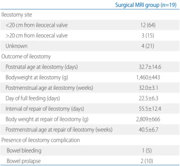
vs. 21%, P=0.001). The mean age of exploratory laparotomy was 32.7±14.6 days and mean body weight was 1,460±443 g. The mean postmenstrual age at surgery was 32.0±3.1 week (Table 4).
of surgery. A Pvalue <0.05 was regarded as statistically signi
ficant.
Results
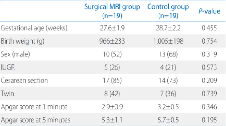
During the study period, 178 VLBW infants delivered at our hospital. Total 19 cases and 19 controls were identified. The demographic and clinical characteristics of both groups are shown in Table 1. The mean gestational age and birth weight were 27.6±1.9 weeks and 966±233 g, respectively, in the surgical MRI group. In the control group, the mean gestational age was 28.7±2.2 weeks and the mean birth weight was 1,005±198 g. There was no significant difference between the surgical MRI group and the control group in perinatal charac
teristics including gestational age, birth weight and Apgar score.
Of the 19 infants in surgical MRI group, 26% had a history of preeclampsia and 42% had premature rupture of the membranes (Table 2). There was no difference in maternal characteristics between the two groups.
Table 3 reports comparison of neonatal morbidities between both groups. The prevalences of RDS, PDA and CLD in preterm infants of surgical MRI were 94%, 52% and 74%, respec tively. Day of full enteral feeding and hospitalization were significantly longer in surgical MRI group compared to control group (57.8±18.3 vs. 32.2±11.7 days, P<0.001; 124.9±7.6 vs. 78.5±18.5 days, P<0.001; respectively). Pearson correlation demonstrated a positive correlation between the mean days (age) of exploratory laparotomy and mean duration of hospitalization in surgical MRI
Table 1. Comparison of Perinatal Characteristics between Surgical MRI Group and Control Group
Surgical MRI group
(n=19) Control group (n=19) P-value
Gestational age (weeks) 27.6±1.9 28.7±2.2 0.455
Birth weight (g) 966±233 1,005±198 0.754
Sex (male) 10 (52) 13 (68) 0.319
IUGR 5 (26) 4 (21) 0.573
Cesarean section 17 (85) 14 (73) 0.209
Twin 8 (42) 7 (36) 0.739
Apgar score at 1 minute 2.9±0.9 3.2±0.5 0.346
Apgar score at 5 minutes 5.3±1.1 5.7±0.5 0.195
Values are presented as mean±standard deviation or number (%).
Abbreviations: MRI, Meconium related ileus; IUGR, intrauterine growth restriction.
Table 2. Comparison of Maternal Characteristics between Surgical MRI Group and Control Group
Surgical MRI group
(n=19) Control group
(n=19) P-value
Maternal age (years) 33.2±3.2 33.8±5.4 0.415
IVF 6 (31) 5 (28) 0.720
Preeclampsia 5 (26) 4 (21) 0.702
PROM 8 (42) 7 (36) 0.739
Oligohydramnios 7 (36) 5 (26) 0.485
Antenatal steroid 14 (73) 18 (94) 0.075
Antenatal magnesium 12 (63) 11 (57) 0.739
Fever 2 (10) 1 (5) 0.547
Gestational DM 3 (15) 4 (21) 0.675
Values are presented as mean±standard deviation or number (%).
Abbreviations: MRI, Meconium related ileus; IVF, in vitro fertilization; PROM, premature rupture of membrane; DM, diabetes mellitus.
Table 3. Comparison of Neonatal Morbidities between Surgical MRI Group and Control Group
Surgical MRI group
(n=19) Control group (n=19) P-value
RDS 18 (94) 17 (89) 0.547
PDA 10 (52) 9 (47) 0.745
CLD 14 (74) 13 (68) 0.721
Postnatal steroid 10 (52) 8 (42) 0.515
IVH 3 (15) 1 (5) 0.290
ROP 9 (47) 6 (31) 0.319
Day of reaching full enteral feeding (>120 mL/kg)
57.8±18.3 32.2±11.7 <0.001
Day of hospitalization 124.9±7.6 78.5±18.5 <0.001 Postmenstrual age to discharge 45.6±4.4 38.5±3.5 <0.001
Body weight to discharge 3.3±0.7 3.1±0.5 0.437
<3 percentile of body weight for GA at discharge
14 (73) 4 (21) 0.001
Values are presented as mean±standard deviation or number (%).
Abbreviations: MRI, Meconium related ileus; RDS, respiratory distress syndrome;
PDA, patent ductus arteriosus; CLD, chronic lung disease; IVH, intraventricular hemorrhage; ROP, retinopathy of prematurity.
In the neonatal morbidities of our study, majority of diseases including RDS, PDA and ROP were not different in both groups.
However, day of full enteral feeding and hospitalization were significantly longer in surgical MRI group. In our study, the mean age of exploratory laparotomy was 32.7 days. And mean durations of hospitalization in both groups were 114.9 and 78.5 days respectively and the difference between both groups was 36.4 days. As a result, it could be seen that the length of hospital days was increased by the period until the surgery was decided.
Pearson correlation demonstrated a positive correlation between the mean days (age) of exploratory laparotomy and mean duration of hospitalization. Therefore, delay in descion for surgery might lead to prolonged hospital stay. And the rate of less than 3 percentile of weight for postmenstrual age was much higher at discharge, which might be due to poor enteral nutrition before surgery.
Many previous reports have mentioned that a delay in the initiation of therapy leads to decreased chance of avoiding surgery.1,9 Emil et al.9 indicated that a 10day duration of ob
struction is the limit for which an enema is effective.8 Hatanaka et al.1 concluded that for extremely low birth weight (ELBW) neonates with bowel obstruction of unclear etiology, mainly MRI, the early and frequent administration of a gastrografin enema is reasonable and surgery should be considered if the obstruction lasts beyond approximately 14 days after birth. Those were quite shorter than our report, in which surgery was done median 32 days after birth.
On the contrary, we should consider the risks of surgery.
In our study, there were no severe complications of surgical treatment except minor complications such as bowel prolapse and bowel bleeding that were resolved spontaneously without any other interventions. Emil et al.9 suggested that surgical therapy in uncomplicated MRI cases that have failed medical management should be minimally aggressive and surgical treat
ment including enterotomy and evacuation of the plug suffice was quite safe and simple to perform.
Although the MRI has been increasingly described in the last 20 years, there has been no agreement on medical manage
ment.11 We could know that the previous studies reported that they used the enema at an earlier age and in a more frequent manner. Emil et al.9 used 10 mL/kg of saline rectal irrigations every 6 hours daily in patients who were obstructed for 10 days
Discussion
We tried to investigate clinical characteristics and outcome of preterm infants clinically diagnosed MRI and failed with conservative treatment such as contrast enemas of gastrografin or glycerin, that required eventually explaratory laparotomy.
The results of this study showed that the clinical characteristics, including perinatal and maternal history of VLBW infants in surgical MRI group were not different from those in the control group.
Regarding risk factors of MRI, several factors including cesarean section, maternal hypertension, maternal magnesium therapy and maternal diabetes have been identified, based on small cohort studies.79 Okuyama et al.7 reported that twin pregnancy and premature rupture of membrane is significantly associated with the development of MRI in VLBW infants, which required surgical treatment.6 In that study, maternal steroid treatment was found to be protective to development of MRI. Recently, Byun et al.10 reported that male gender and low birth weight were associated with surgical MRI compared with medically treated MRI. However, we did not find any differences of maternal history in the surgical MRI groups compared with control group.
Table 4. Postoperative Clinical Course of Surgical MRI Group Surgical MRI group (n=19) Ileostomy site
<20 cm from ileocecal valve 12 (64)
>20 cm from ileocecal valve 3 (15)
Unknown 4 (21)
Outcome of ileostomy
Postnatal age at ileostomy (days) 32.7±14.6
Bodyweight at ileostomy (g) 1,460±443
Postmenstrual age at ileostomy (weeks) 32.0±3.1
Day of full feeding (days) 22.5±6.3
Interval of repair of ileostomy (days) 55.5±12.4 Body weight at repair of ileostomy (g) 2,809±666 Postmenstrual age at repair of ileostomy (weeks) 40.5±6.7 Presence of ileostomy complication
Bowel bleeding 1 (5)
Bowel prolapse 2 (10)
Values are presented as mean±standard deviation or number (%).
Abbreviations: MRI, meconium related ileus; NEC, necrotizing enterocolitis; FIP, focal intestinal perforation.
after birth.8 And Hatanaka et al.1 started gastrografin enema of 10 mL/kg initially and increased amount up to 15 mL/kg.
However, Amodio et al.12 reported colonic and ileal perfora
tions secondary to a contrast enema in a premature baby.
Caniano and Beaver13 reported a 13% complication rate after therapeutic enemas for meconium ileus. Therefore, management of MRI in premature infants, especially ELBW infants, should be need more cautions. We had followed a somewhat passive enema method because of the fear of intestinal perforation by the enema. Promptly recognition associated to an early and aggressive conservative management is essential to prevent complications and spare surgical interventions.2 However, if not improved despite aggressive conservative treatment, surgical treatment should be considered after 10 to 14 days after birth.2,14,15
Our study has several limitations. First, the small number of case patients were included. And also, we were not able to inroll the control group as 1 to more than 2 match manner. Second, there are the limitations inherent to retrospective review study.
Further studies with adequate sample size and prospective studies are needed.
Despites of these limitations, our study has been speculated that delayed decsion of surgery in treatment of MRI might prolong the hospitalization and adversely affect growth in VLBW infants. Therefore, early treatment including exploratory laparotomy should be considered to improve outcome when especially the baby has more than one of risk factors including male gender and low birth weight.
Acknowledgement
This study was supported by a clinical research grant from Pusan National University Hospital 2020.
Conflict of interest
No potential conflict of interest relevant to this article was reported.
References
1) Hatanaka A, Nakahara S, Takeyama E, Iwanaka T, Ishida K. Management of extremely low birth weight neonates with bowel obstruction within 2 weeks after birth. Surg Today 2014;44:2269-74.
2) Paradiso VF, Briganti V, Oriolo L, Coletta R, Calisti A. Meconium obstruction in absence of cystic fibrosis in low birth weight infants: an emerging challenge from increasing survival. Ital J Pediatr 2011;37:55.
3) Dimmitt RA, Moss RL. Meconium diseases in infants with very low birth weight. Semin Pediatr Surg 2000;9:79-83.
4) Kubota A, Imura K, Yagi M, Kawahara H, Mushiake S, Nakayama M, et al.
Functional ileum in neonates: Hirschsprung’s disease-allied disorders versus meconium related ileus. Eur J Pediatr Surg 1999;9:392-5.
5) Kubota A, Shiraishi J, Kawahara H, Okuyama H, Yoneda A, Nakai K, et al.
Meconium-related ileus in extremely low-birthweight neonates:
etiological considerations from histology and radiology. Pediatr Int 2011;53:887-91.
6) Kim HY, Kim SH, Cho YH, Byun SY, Han YM, Kim ALY. Meconium-related ileus in very low birth weight and extremely low birth weight infants:
immediate and one-year postoperative outcomes. Ann Sur Treat Res 2015;89:151-7.
7) Okuyama H, Ohfuji S, Hayakawa M, Urushihara N, Yokoi A, Take H, et al.
Risk factors for surgical intestinal disorders in VLBW infants: case-control study. Pediatr Int 2016;58:34-9.
8) Ziegler MM. Meconium ileus. Curr Probl Surg 1994;31:731-77.
9) Emil S, Nguyen T, Sills J, Padilla G. Meconium obstruction in extremely low-birth-weight neonates: guidelines for diagnosis and management.
J Pediatr Surg 2004;39:731-7.
10) Byun J, Han JW, Youn JK, Yang HB, Shin SH, Kim EK, et al. Risk factors of meconium-related ileus in very low birth weight infants: patients- control study. Sci Rep 2020;10:4674.
11) Shinohara T, Tsuda M, Koyama N. Management of meconium-related ileus in very low-birthweight infants. Pediatr Int 2007;49:641-4.
12) Amodio J, Berdon W, Abramson S, Stolar C. Microcolon of prematurity:
a form of functional obstruction. Am J Roentgenol 1986;146:239-44.
13) Caniano DA, Beaver BL. Meconium ileus: a fifteen-year experience with forty-two neonates. Surgery 1987;102:699-703.
14) Garza-Cox S, Keeney SE, Angel CA, Thompson LL, Swischuk LE.
Meconium obstruction in the very low birth weight premature infant.
Pediatrics 2004;114:285-90.
15) Chan KL, Ng SP, Chan KW, Wo YH, Tam PK. Pathogenesis of neonatal necrotizing enterocolitis: a study of the role of intraluminal pressure, age and bacterial concentration. Pediatr Surg Int 2003;19:573-7.