https://doi.org/10.4174/astr.2017.93.1.50 Annals of Surgical Treatment and Research
Evaluation of botulinum toxin type A effectiveness in preventing postoperative intraperitoneal adhesions
Mehmet Dokur, Erdal Uysal1
Department of Emergency Medicine, Necip Fazil City Hospital, Kahramanmaras, 1Department of General Surgery, Sanko University School of Medicine, Gaziantep, Turkey
INTRODUCTION
Postoperative intraperitoneal adhesions (PIAs) are one of the most important problems surgeons have to face after laparotomies. PIAs may form after many abdominal surgeries.
Pre valence is around 67%–93%. However, the percentage of total formations necessitating surgical operation caused by PIAs is around 15%–18% [1]. These percentages are lowered on account of development and common usage of minimally invasive techniques such as laparoscopy [2]. PIAs can cause hospitalization and surgical operations due to mechanic
ileus or pelvic pain. Additionally, PIAs may cause secondary infertility in women in the reproductive age group. Iatrogenic injuries are very common in operations needed on the grounds of adherence because exploration is extremely hard. These complications on the other hand may cause an increase in mortality and morbidity. Additionally, one of the most important consequences of ileus caused by PIAs is the increase in workforce loss and patient costs. For example, a study conducted in the United States shows that the cost of operations due to PIAs is around 1,3 billion United States dollars [3].
Purpose: Postoperative intraperitoneal adhesions (PIAs) are one of the most important problems surgeons have to face after laparotomies. In this study, we aimed to evaluate the effectiveness of local application of botulinum toxin type A (BoNT-A) in various dosages on the prevention of intra-abdominal adhesions in rats with experimental intra-abdominal adhesions.
Methods: Forty Wistar Albino female rats were randomly separated into 4 groups. The 4 groups were determined as follows: Control (group 1, n = 10); Sham (group 2, n = 10); 10-mg/kg low-dose BoNT-A (group 3, n = 10) and 30-mg/kg high- dose BoNT-A (group 4, n = 10). Subserosal injuries were created on the caecum of all rats. Laparotomy was performed on the fifth day. Adhesion scores, histopathological examination, and E-cadherin expression levels were evaluated.
Results: General adhesion scores for groups 1 and 2 were determined to be significantly high when compared to group 4 (P < 0.001). A significant difference was also determined between groups 3 and 4 in terms of general adhesion scores (P <
0.05). In pair comparisons, a significant decrease in high-dose BoNT-A group (group 4) when compared to groups 1 and 2 in terms of neovascularization, fibroblast density, collagen deposition and inflammatory cell count was determined (P < 0.05).
Conclusion: A significant decrease was observed only in postoperative PIAs in the high-dose BoNT-A group between all 4 rat-groups with experimentally created postoperative PIAs. In this study, high-dose BoNT-A is determined to be an effective agent in preventing postoperative PIAs.
[Ann Surg Treat Res 2017;93(1):50-56]
Key Words: Type A botulinum toxin, Rats, Tissue adhesions, Cadherins
Reviewed January February March April May June JulyAugust September October November December
Received December 19, 2016, Revised January 20, 2017, Accepted January 24, 2017
Corresponding Author: Mehmet Dokur
Department of Emergency Medicine, Necip Fazil City Hospital, Gaziantep Yolu 14. Km Karacasu Mevkii, Dulkadiroğlu, 46050-Kahramanmaras, Turkey Tel: +90-344-2282800, Fax: +90-344-2515103
E-mail: drdokur@gmail.com
Copyright ⓒ 2017, the Korean Surgical Society
cc Annals of Surgical Treatment and Research is an Open Access Journal. All articles are distributed under the terms of the Creative Commons Attribution Non- Commercial License (http://creativecommons.org/licenses/by-nc/4.0/) which permits unrestricted non-commercial use, distribution, and reproduction in any medium, provided the original work is properly cited.
Steroids and nonsteroidal antiinflammatory drugs, immuno
suppressive drugs, ClinOleic, high molecular weight hyaluronic acid and low molecular weight hydroxypropyl methylcellulose, fibrinogen degradation products, recombinant tissue plasmi no
gen activator, ankaferd blood stopper, VitaminE and carboxy
methylcellulose was used as adhesion barrier in order to de crease and prevent adherence [4,5]. Different results were obtained in each of these agents. The agents, except adhesion barrier, could not be applied clinically. This is the main reason the prevention of PIAs and fighting off adhesions are always an important subject for surgeons, especially those working in abdominal surgery.
It was determined that postoperational inflammatory re
sponse plays a great role in the formation of PIAs [6]. A study examining the formation of peritoneal adhesion formation at the molecular level indicates that TGFβ1 and interleukin6 (IL6), which are pleiotropic molecules, shows an increase in peritoneal fluid during and after abdominal operations.
It was asserted that TGFβ1 and IL6 interacted with each other, promoted mesothelial to mesenchymal transition and facilitated peritoneal adhesion formation. This is why the inflammatory response is thought to be efficient in decreasing PIAs, especially in repressing TGFβ1 and IL6 [7]. Thus, it is important that the agent used for preventing PIAs contain antiinflammatory features. It was also reported that adhesion molecules such as Ecadherin play a role in intraabdominal adhesion formation. Thus, antiinflammatory agents effecting adhesion molecules can be seen as ideal agents in preventing PIAs.
It was also determined that botulinum toxin type A (BoNTA) disturbs the bond between cells on the epithelial barrier by binding epithelial molecules, and has antiinflammatory effectiveness [8]. In this study, the hypothesis that BoNTA may decrease and even prevent PIAs was emphasized due to its anti
inflammatory effectiveness. There are no studies examining the effect of BoNTA on intraabdominal adhesions.
The aim of this study was to examine the effectiveness of
local application of BoNTA in various dosages on the prevention of intraabdominal adhesions on rats with experimental intra
abdominal adhesions.
METHODS
Rat groups
Forty Wistar Albino female rats weighing 260–280 g were randomly separated into 4 groups, 10 rats in each group; Control (group 1), sham (group 2), 10mg/kg lowdose BoNTA (group 3), and 30mg/kg highdose BoNTa (group 4). Subserosal injuries were created on the group 1 rats by rubbing sterile gauze pads on the caecum serosa without applying any drugs. Group 2 rats were also wounded with caecum subserosal injuries with an application of 1 mL 0.09% NaCl into the peritoneal cavity. Ten
and 30mg/kg BoNTA (BOTOX, Allergan, Dublin, Ireland) with 1 mL 0.09% NaCl into the peritoneal cavity were respectively applied on groups 3 and 4 rats.
Anesthesia and surgical technique
Rats were kept in a room with a fixed temperature (18°C–24°C) and light (12hour lightdark cycle), 4–6 rats in a single cage, with free water and feed supplies. Rats were given standard rat feed and normal water. Rats were left hungry for 6 hours before surgery. As anesthetics, 75mg/kg ketamine (Ketalar, Pfizer, Istanbul, Turkey) and 10mg/kg xylazine (Rompun, Bayer AG, Leverkusen, Germany) were applied intraperitoneally, respectively. Polyvinylpyrolidone iodine (polividoniodine) 10%
was applied for asepsis. 100mg/kg ceftriaxone was applied for intramuscular prophylaxis purposes. All rats were given 0.25% bupivacaine on the incision for postoperative analgesia control purposes. Postoperative analgesia was performed with 3mg/kg ketoprofen (Profenid, Sanofi Aventis, Paris, France) subcutaneously, daily for 3 days. Each rat was given 10 mL 0.09%
NaCl subcutaneously for dehydration. The rats were placed in different cages postsurgery and followed up. The rats were given free feed and water 5 hours after surgery.
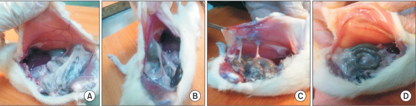
Fig. 1. Macroscopic appearance of tissues. (A) Control group: intermediate increase adhesion is observed. (B) Sham group:
inter mediate increase adhesion is observed. (C) Low-dose botulinum toxin type A (BoNT-A) group: small amount adhesion is observed. (D) High-dose BoNT-A group: the lowest amount adhesion is observed.
A B C D
A 6cm abdominal midline incision was applied on all rats after anesthesia. Additionally, subserosal injuries were created on the all rats by rubbing sterile gauze pads on the caecum serosa. After the experimental processes were applied on each group, rat derma and subcutaneous tissue were closed by 4/0 polyglactin continuous suture.
Evaluation of adhesions and tissue sampling
The rats were operated on once more on the 15th day under ketamine and 10mg/kg xylazine intraperitoneal anesthesia. In order to prevent injuries due to adhesions and to evaluate the adhesions due to incision, laparotomy with left paramedian incision was applied on all rats. A blinded researcher was brought in and asked to evaluate the intraabdominal adhesion levels between 0 and 9 according to the Linsky Scale [9].
Adhesion severity, degree, and adhesion rates in the defected area were evaluated (Fig. 1A–D). A general score was also obtained from the total of these three parameters. After the evaluation, tissue samples were taken from caecum and sur
rounding area for histopathology. Samples were determined in formaldehyde for histopathologic evaluation. After the opera
tion, all rats under anesthesia were euthanized through decor
tication.
Histopathologic examination
The same pathologist performed all histopathologic exami
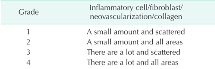
nations. Samples were prepared on paraffinembedded blocks and thin sections painted with hematoxylin and eosin stains were examined under light microscope. Images were saved to a computer. Histopathologic staging was done in accordance with EhrlichHunt Model [10] (Table 1).
Evaluation criteria in this model are determined as following:
inflammatory cell, fibroblast, neovascularization, and collagen amount. Cellular and histopathologic scoring is evaluated in 4 stages semiquantitatively. Different calculations were made for inflammatory cell, fibroblast proliferation, neovascularization, and collagen deposition.
Immunohistochemical examination
Sample tissues were taken from all rats in study groups and control group for immunohistochemical examination. The fol lowing processes were applied on samples respectively:
for malin fixation, paraffin application, blocking and immuno
histo chemical dyeing. Ecadherin protein amounts were semi
quan titatively evaluated on the samples belonging to different groups. (absent: 0, slight [1]: up to 20% positive, moderate 21%–
50% positive, potent 51%–100%).
Statistical analysis
The SPSS ver. 15.0 (SPSS Inc., Chicago, IL, USA) was used for statistical analysis. KruskalWallis test was used for group comparisons. KruskalWallis post hoc tests and SidakDunn test were used for pair comparisons. Results were expressed with a confidence interval of 95%. Results were given as mean ± standard error. Values of P < 0.05 were considered significant.
25th and 75th percentiles were calculated from definitive statis
tics. Group size calculation was performed by the resource equa
tion method.
Ethics statement
The protocol was approved by the Animal Ethics Review Com
mittee (permit number: 2015/0308, KSU Faculty of Medicine Ethics Committee). All experiments were conducted in com
pliance with the relevant laws and institutional guidelines.
RESULTS
Adhesion scores
General adhesion scores for each group was determined to be as follows; 6,2 ± 1.17 for group 1, 5.8 ± 0.49 for group 2, 4.2 ± 0,7 for group 3 and 2,4 ± 0,6 for group 4. General adhesion scores for groups 1 and 2 were determined to be significantly high Table 1. Histological grading scalea)
Grade Inflammatory cell/fibroblast/
neovascularization/collagen
1 A small amount and scattered
2 A small amount and all areas
3 There are a lot and scattered
4 There are a lot and all areas
a)Ehrlich-Hunt model.
Table 2. Comparison of adhesion scores of the Groups
Parameter Group 1 Group 2 Group 3 Group 4 P-valuea)
Participation percentage 1.8 ± 0.12 1.7 ± 0.14 1.2 ± 0.2 0.9 ± 0.3 <0.001***, <0.05*
Adhesion severity 2.1 ± 0.15 1.9 ± 0.11 1.4 ± 0.3 0.8 ± 0.2 <0.001***, <0.05*
Degree of adhesion 2.3 ± 0.9 2.2 ± 0.24 1.6 ± 0.2 0.7 ± 0.1 <0.001***, <0.05*
General adhesion score 6.2 ± 1.17 5.8 ± 0.49 4.2 ± 0.7 2.4 ± 0.6 <0.001***, <0.05*
Values are presented as mean ± standard error.
Group 1, control; group 2, sham; group 3, 10-mg/kg low-dose botulinum toxin type A (BoNT-A); group 4, 30-mg/kg high-dose BoNT-A.
a)Kruskal-Wallis test. *P < 0.05, group 4 vs. group 3. ***P < 0.001, group 4 vs. groups 1 and 2.
when compared to group 4 (P < 0.001). There was no significant difference in general adhesion scores between groups 1 and 2 (P > 0.05). There was also no significant difference in general adhesion scores of group 3 and groups 1 and 2. A significant difference was also determined between groups 3 and 4 in terms of general adhesion scores (P < 0.05) (Table 2).
Histopathological examination results
A significant difference was determined in the comparisons
between groups in terms of neovascularization, fibroblast density, collagen deposition, and inflammatory cell (P < 0.001).
In pair comparisons, a significant decrease in highdose BoNTA group (group 4) when compared to groups 1 and 2 in terms of neovascularization, fibroblast density, collagen deposition, and inflammatory cell was determined (P < 0.05). (Table 3; Fig. 2A–D).
Immunohistochemical examination results
No significant difference in terms of Ecadherin positive
Table 3. Comparison of histopathological findings of the groups
Parameter Group 1 Group 2 Group 3 Group 4 P-valuea)
Inflammation 3.1 ± 0.5 2.9 ± 0.3 2.2 ± 0 1,4 ± 0.3 <0.05*
Fibroblast activation 2.8 ± 0.4 2.8 ± 0.5 2,4 ± 0.7 1.6 ± 0.7 <0.05*
Neovascularization 3.2 ± 0.4 3.1 ± 0.2 2.6 ± 0.2 1.3 ± 0.4 <0.05*
Collagen deposition 2.7 ± 0.8 2.6 ± 0.5 2.3 ± 0.2 1.5 ± 0.5 <0.05*
E-cadherin 2.2 ± 0.3 2.4 ± 0.5 2.2 ± 0.2 2.0 ± 0 >0.05
Values are presented as mean ± standard error.
Group 1, control; group 2, sham; group 3, 10-mg/kg low-dose botulinum toxin type A (BoNT-A); group 4, 30-mg/kg high-dose BoNT-A.
a)Kruskal-Wallis test. *P < 0.05, group 4 vs. groups 1 and 2.
No statistically significant difference was found between groups 1 and 2 in all of the comparasions (P > 0.05).
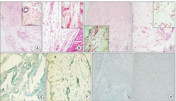
Fig. 2. Histopathologic changes in rat groups. (A) Control group: normal fibroblast intensity, neovascularization and collagen fiber formation are observed (H&E, ×100). (B) Sham group: normal fibroblast intensity, neovascularization and collagen fiber formation are observed (H&E, ×100). (C) Low-dose botulinum toxin type A (BoNT-A) group: decrease in fibroblast inten sity, neovascularization and collagen fiber formation are observed (H&E, ×100). (D) High-dose BoNT-A group: significant de crease in fibroblast intensity, neovascularization and collagen fiber formation are observed (H&E, ×100). (E) The pattern of grade 1 staining with E-cadherin in control group (immunohistochemistry, ×200). (F) The pattern of grade 0 staining with E-cadherin in sham group (immunohistochemistry, ×200). (G) The pattern of grade 0 staining with E-cadherin in low-dose BoNT-A group (immunohistochemistry, ×200). (H) The pattern of grade 0 staining with E-cadherin in high-dose BoNT-A group (immuno- histochemistry, ×200).
A B C D
E F G H
dyeing pattern was determined in the comparison between groups (P = 0.79) (Table 3; Fig. 2E–H).
DISCUSSION
PIAs are one of the most important problems surgeons have to face with [3]. Clinical problems caused by adhesions cause an increase in mortality and morbidity. Thus, finding a solution against PIAs is an important subject for surgeons.
Many agents were used in clinical and experimental studies in order to prevent PIA formation [4,5]. However, the search for an effective agent still continues. Additionally, there is no common approach for PIA treatment.
Even though the physiopathological mechanism of PIA formation is not fully explained, it has been indicated that the inflammatory reaction in the defected tissue plays an impor
tant role [6]. Macrophages and fibroblasts are active for the treat
ment of wounded tissue. Collagens and matrix proteins also play a role in tissue repair. As a result of the physio pathological events started by inflammatory reaction, fibrous adhesions are formed. This is the main idea behind using antiinflammatory agents to prevent PIAs.
BoNTA is a neurotoxin with a neurotoxic protein structure produced by Clostridium botulinum bacteria [11]. It is used for many diseases such as BoNTA upper motor neuron syndrome, focal hyperhydrosis, blepharospasm, strabismus, chronic migraine, and anal fissure. In most of the clinical cases in which BoNTA is used, BoNTA’s antiinflammatory effectiveness is beneficial [12]. BoNTA demonstrates its antiinflammatory effectiveness through macrophages, which are the main cells of the immune system [8]. Macrophages are also responsible for secretion of many proinflammatory mediators and small molecules playing part in the inflammation such as nitric oxide. BoNTA mainly affects proteins such as nuclear factorκB in the immune system signal pathway, transcription factors and mitogenactivated protein kinases including cJun Nterminal kinase, p38, and extracellular signalregulated kinase [13]. As a result, these proteins affect the genes responsible for the secretion of pro and antiinflammatory cytokine and chemicals.
Ultimately, BoNTA receives antiinflammatory features. Our study presents the antiinflammatory effectiveness of BoNTA and focuses on the hypothesis that BoNTA may decrease and even prevent intraabdominal adhesions.
During the comparisons in our study, it was determined that the inflammatory cell count significantly decreased in highdose BoNTA group when compared to control and sham groups. Recent studies have suggested that BoNTA repressed inflammation and presented an antiinflammatory effect [14]. Antiinflammatory effects of BoNTA in Sjögren Syndrome and chronic inflammatory pain treatments were also reported [15]. A study indicates that dural neurogenic
inflammation plays a role in migraine pathophysiology and also that BoNTA may have a positive effect on eliminating migraine and other headaches by decreasing dural neurogenic inflammation with its antiinflammatory effects [16]. BoNTA causes a decrease in inflammatory cell count as a result of its antiinflammatory effect [17]. In a study conducted by Kim et al. [18] on flap necrosis, it was shown that the inflammatory cell count decreased in the group with BoNTA applied. In another study conducted on the effect of BoNTA on a rat surgical wound model, it was indicated that BoNTA causes a decrease in inflammatory cell count. The same study also shows that TGFβ1 expression, which is known to be active in inflammatory processes and peritoneal adhesion formation, also decreases as a result of BoNTA application [19]. Our study also showed that the inflammatory cell count in the highdose BoNTA group decreased and created a correspondence with the existing literature. Even though the inflammatory cell counts in the lowdose BoNTA group decreased, no significant difference was determined when compared to control and sham groups.
This result proves that the antiinflammatory effect of BoNTA in the peritoneum is highly dependent on the dosage and increases accordingly.
Our study showed a significant decrease in neovasculariza
tion in highdose BoNTA group when compared to control and sham groups. Neovascularization plays an important role in adhesion formation development. An increasing neo vas cula
ri zation also causes an increase in adhesion formation [20].
The significantly decreasing adhesion score in the highdose BoNTA group that was determined in our study may be related to decreasing neovascularization. The significant decrease in neovascularization was determined to be dependent on BoNTA application dosage. The effect was more significant in higher dosages.
Fibroblasts are responsible of the secretion of extra matrix proteins and collagen. Increasing fibroblast density also causes an increase in collagen secretion, granulation tissue and adhesion [21]. In our study, a significant decrease in fibroblast density in highdose BoNTA group was determined when com
pared to control and sham groups. In the study conducted by Xiao et al. [22] on hypertrophic scars, a decrease in fibroblast count in the groups applied with BoNTA was reported. In the study conducted by Oh et al. [23] on human dermal fibroblasts, BoNTA was determined to cause a decrease in fibroblast count and collagen amount. Decreasing fibroblast count in the high
dose BoNTA group detected in our study was determined to be in correspondence with existing literature data.
In this study, it was determined that the collagen count in highdose BoNTA group significantly decreased when compared with sham and control groups. Collagen is the main protein of extracellular matrix and secreted by fibroblasts. Collagen takes an active part in wound healing processes. Our study
determined that highdose intraperitoneal BoNTA application causes a significant decrease in fibroblast count. Decreasing fibroblast count may be responsible for the decrease in collagen density. The in vitro study suggested that BoNTA application caused a decrease in collagen expression levels [24]. In the study conducted by Kim et al. [25], a decrease in collagen levels in tissues was determined in the BoNTA group. Additionally, in the study conducted by Sahinkanat et al. [26] on the effects of BoNTA on urethral wound healing, a significant decrease in collagen count was determined in BoNTA group. On the other hand, in a study on BoNTA’s effect on muscle contractile and structural properties, a significant increase in the collagen content of tissues in the BoNTA group was reported [27].
The increase in collagen deposition after BoNTA induced muscle fiber atrophy was responsible for collagen increase.
Another study failed to show an effect of BoNTA on collagen synthesis [28]. Data obtained from the current literature can be summarized to have different effects on different tissues in terms of collagen synthesis and count.
Ecadherin is one of the molecules that are responsible for intracellular adhesion and communication. The increase in Ecadherin expression leads to peritoneal adhesion formation [29]. No significant difference between groups in terms of Ecadherin expression was determined in our study. Even though molecular studies regarding the relationship between Ecadherin and BoNTA were performed, no significant result could be obtained [30].
Our study included several limitations. This study is the first study to work on the effects of BoNTA on postoperative intraperitoneal adhesion formation. Thus, this study can be considered a pilot study. Therefore, a detailed ultrastructural
examination of the histopathological changes BoNTA causes on the peritoneum is necessary. Gene studies and electron microscopy studies can be developed on the subject. The relationship between Desmocollin 3, Cadherin 3, ARHGDIA, CYFIP2, and LAMA4 gene expressions and α5β1 proteins and BoNTA can be examined in detail. This study examined peritoneal adhesion formation in a single timeframe. New examinations on different timeframes and different doses can be performed.
In conclusion, it can be said that a significant decrease was observed only in postoperative intraperitoneal adhesions in the highdose BoNTA group between all 4 ratgroups with experimentally created postoperative intraperitoneal adhesions.
Neovascularization, fibroblast count, collagen deposition, and inflammatory cell count significantly decreased in highdose BoNTA group. BoNTA is determined to be an effective agent in preventing postoperative intraperitoneal adhesions. A further ultrastructural examination on the histopathologic changes BoNTA causes on the peritoneum is necessary.
CONFLICTS OF INTEREST
No potential conflict of interest relevant to this article was reported.
ACKNOWLEDGEMENTS
We thank to Assistant Professor Dr. Ibrahim IBILOGLU M.D.
(Dicle University Medical FacultyDepartment of Pathology) for his endeavors and also we thank to the Staff of Experimental Animal Center of Kahramanmaras Sutcu Imam University.
REFERENCES
1. Ouaissi M, Gaujoux S, Veyrie N, Deneve E, Brigand C, Castel B, et al. Postoperative adhesions after digestive surgery: their incidence and prevention: review of the literature. J Visc Surg 2012;149:e10414.
2. Takagi K, Araki M, Fukuoka H, Takeshita H, Hidaka S, Nanashima A, et al. Novel powdered antiadhesion material: pre
venting postoperative intraabdominal adhe sions in a rat model. Int J Med Sci 2013;10:46774.
3. R ay N F, D enton WG, Th a mer M, Henderson SC, Perry S. Abdominal adhe
siolysis: inpatient care and expenditures
in the United States in 1994. J Am Coll Surg 1998;186:19.
4. Karaca G, Aydin O, Pehlivanli F, Kocael A, Pekcici R, Duymus E, et al. Effect of ankaferd blood stopper in experimental peritoneal adhesion model. Ann Surg Treat Res 2016;90:2137.
5. Kirdak T, Uysal E, Korun N. Assessment of effectiveness of different doses of me thylprednisolone on intraabdominal adhesion prevention. Ulus Travma Acil Cerrahi Derg 2008;14:18891.
6. Saed GM, Diamond MP. Molecular charac
teri zation of postoperative adhesions: the
adhesion phenotype. J Am Assoc Gynecol Laparosc 2004;11:30714.
7. Jin X, Ren S, Macarak E, Rosenbloom J.
Pathobiological mechanisms of peritoneal adhesions: The mesenchymal transition of rat peritoneal mesothelial cells induced by TGFβ1 and IL6 requires activation of Erk1/2 and Smad2 linker region phos
phorylation. Matrix Biol 2016;51:5564.
8. Kim YJ, Kim JH, Lee KJ, Choi MM, Kim YH, Rhie GE, et al. Botulinum neurotoxin type A induces TLR2mediated inflam ma
tory responses in macrophages. PLoS One 2015;10:e0120840.
9. Linsky CB, Diamond MP, Cunningham T, Constantine B, DeCherney AH, diZerega GS. Adhesion reduction in the rabbit uterine horn model using an absorbable barrier, TC7. J Reprod Med 1987;32:1720.
10. Ehrlich HP, Tarver H, Hunt TK. Effects of vitamin A and glucocorticoids upon in
flam mation and collagen synthesis. Ann Surg 1973;177:2227.
11. Hsu YC, Wang HJ, Chuang YC. Intra pro
static Botulinum Neurotoxin Type A Injec
tion for Benign Prostatic Hyper pla siaA Spotlight in Reality. Toxins (Basel) 2016;8.
pii: E126.
12. Nelson RL. Efficacy of Fissurectomy and Botox for Chronic Anal Fissure. Dis Colon Rectum 2016;59:e41.
13. Houston FE, Hain BA, Adams TJ, Houston KL, O’Keeffe R, Dodd SL. Heat shock protein 70 overexpression does not atte
nuate atrophy in botulinum neuro toxin type Atreated skeletal muscle. J Appl Physiol (1985) 2015;119:8392.
14. Liu HT, Kuo HC. Intravesical botulinum toxin A injections plus hydrodistension can reduce nerve growth factor produc
tion and control bladder pain in inter
stitial cystitis. Urology 2007;70:4638.
15. O’Neil LM, Palme CE, Riffat F, Mahant N.
Botulinum Toxin for the Management of Sjogren SyndromeAssociated Recur rent Parotitis. J Oral Maxillofac Surg 2016;74:
242830.
16. Lackovic Z, Filipovic B, Matak I, Helyes Z. Activity of botulinum toxin type A in cranial dura: implications for treatment
of migraine and other headaches. Br J Pharmacol 2016;173:27991.
17. Aoishi K, Takahashi H, Hato N, Gyo K, Yokota M, Ozaki S, et al. Treatment of allergic rhinitis with intranasal infusion of botulinum toxin type A in mice. Life Sci 2016;147:1326.
18. Kim SY, Lee SH, Lee B, Park YJ, Park JH, Lee YS, et al. The protective effects of botulinum Toxin A against flap necrosis after perforator twisting and its underlying molecular mechanism in a rat model. Ann Plast Surg 2016;77:2428.
19. Lee BJ, Jeong JH, Wang SG, Lee JC, Goh EK, Kim HW. Effect of botulinum toxin type a on a rat surgical wound model.
Clin Exp Otorhinolaryngol 2009;2:207.
20. Molinas CR, Binda MM, Manavella GD, Koninckx PR. Adhesion formation after laparoscopic surgery: what do we know about the role of the peritoneal environ
ment? Facts Views Vis Obgyn 2010;2:149
60.
21. Boys F. The prophylaxis of peritoneal adhesion. Review of literature. Surgery 1942;11:11868.
22. Xiao Z, Zhang F, Lin W, Zhang M, Liu Y. Effect of botulinum toxin type A on trans forming growth factor beta1 in fibro
blasts derived from hypertrophic scar: a preli minary report. Aesthetic Plast Surg 2010;34:4247.
23. Oh SH, Lee Y, Seo YJ, Lee JH, Yang JD, Chung HY, et al. The potential effect of botulinum toxin type A on human der mal fibroblasts: an in vitro study.
Dermatol Surg 2012;38:168994.
24. Kim S, Ahn M, Piao Y, Ha Y, Choi DK, Yi MH, et al. Effect of botulinum toxin type A on TGFβ/Smad pathway signaling:
implications for siliconeinduced capsule
formation. Plast Reconstr Surg 2016;138:
821e829e.
25. Kim YS, Hong JW, Yoon JH, Hwang YS, Roh TS, Rah DK. Botulinum toxin A affects early capsule formation around sili cone implants in a rat model. Ann Plast Surg 2015;74:48895.
26. Sahinkanat T, Ozkan KU, Ciralik H, Ozturk S, Resim S. Botulinum toxinA to improve urethral wound healing: an experimental study in a rat model. Urology 2009;73:405
9.
27. Minamoto VB, Suzuki KP, Bremner SN, Lieber RL, Ward SR. Dramatic changes in muscle contractile and structural pro per
ties after 2 botulinum toxin injections.
Muscle Nerve 2015;52:64957.
28. Gauglitz GG, Bureik D, Dombrowski Y, Pavicic T, Ruzicka T, Schauber J. Botuli
num toxin A for the treatment of keloids.
Skin Pharmacol Physiol 2012;25:3138.
29. Cheong YC, Laird SM, Li TC, Shelton JB, Ledger WL, Cooke ID. Peritoneal healing and adhesion formation/reformation.
Hum Reprod Update 2001;7:55666.
30. Lee K, Zhong X, Gu S, Kruel AM, Dorner MB, Perry K, et al. Molecular basis for disruption of Ecadherin adhesion by botulinum neurotoxin A complex. Science 2014;344:140510.