• 검색 결과가 없습니다.

The Relationship Between Serum Pentraxin 3 and Central Obesity in ST-Segment Elevation Myocardial Infarction Patients

N/A
N/A
Protected

Academic year: 2022

Share "The Relationship Between Serum Pentraxin 3 and Central Obesity in ST-Segment Elevation Myocardial Infarction Patients"

Copied!
6
0
0

로드 중.... (전체 텍스트 보기)

전체 글

(1)

308

Open Access

The Relationship Between Serum Pentraxin 3 and Central Obesity in ST-Segment Elevation Myocardial Infarction Patients

Byung-Ju Shim, MD, Hui-Kyung Jeon, MD, Seung-Jae Lee, MD, Sung-Sik Kim, MD, Mi-Youn Park, MD, Dong-Hyeun Lee, MD, Woo-Seung Shin, MD, Jong-Min Lee, MD, Ho-Joong Youn, MD, Wook-Sung Chung, MD and Ki-Bae Seung, MD

Division of Cardiology, Department of Internal Medicine, College of Medicine, The Catholic University of Korea, Seoul, Korea ABSTRACT

Background and Objectives: As shown in previous studies, pentraxin 3 (PTX3) can be a useful inflammatory marker for metabolic syndrome and central obesity. Serum PTX3 levels are also an independent factor associated with visceral fat area.

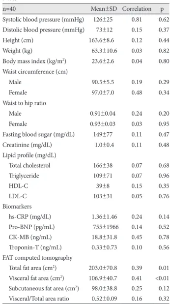
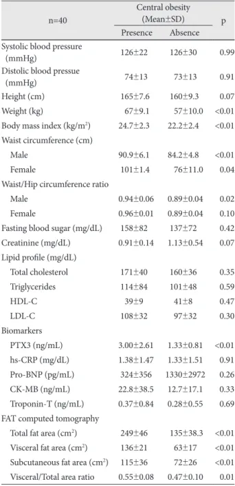
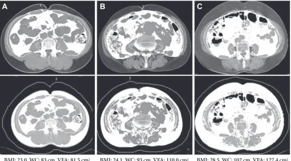
The aim of this study was to assess the role of PTX3 as an inflammatory maker in patients with central obesity undergoing pri- mary percutaneous coronary intervention (PCI) following an ST-segment elevation myocardial infarction (STEMI). Sub- jects and Methods: From December 2007 to June 2008, 40 subjects (mean age: 61±11 years, M : F=34 : 6) with STEMI who underwent primary PCI were enrolled. We determined waist circumference, waist/hip ratio, body mass index (BMI), and visceral and total fat area via fat computed tomography (FAT-CT), and compared them with serum PTX3 concentrations.

Results: The serum PTX3 concentration was closely related to FAT-CT-estimated visceral fat area (r=0.41, p<0.01) and to- tal fat area (r=0.38, p=0.01), respectively. The serum PTX3 concentration was not related to waist circumference (r=0.27, p=

0.20), waist circumference/hip ratio (r=0.25, p=0.16), BMI (r=0.04, p=0.80) and lipid profiles, respectively. Among the parame- ters determining metabolic syndrome, an increasing visceral fat area had the strongest association with PTX3 concentrations.

Conclusion: In patients with STEMI, PTX3 is associated with central obesity and it is significantly and independently corre- lated with visceral fat area. FAT-CT-estimated visceral fat area is the most reliable factor associated with serum PTX3 levels in patients with STEMI and central obesity. (Korean Circ J 2010;40:308-313)

KEY WORDS: Pentraxin 3; Myocardial infarction; Central obesity.

Received: October 10, 2009 Revision Received: January 4, 2010 Accepted: January 12, 2010

Correspondence: Hui-Kyung Jeon, MD, Division of Cardiology, Depart- ment of Internal Medicine, College of Medicine, The Catholic University of Korea, 65-1 Geumo-dong, Uijeongbu 480-821, Korea

Tel: 82-31-820-3593, Fax: 82-31-847-0461 E-mail: [email protected]

cc

This is an Open Access article distributed under the terms of the Cre- ative Commons Attribution Non-Commercial License (http://creativecom- mons.org/licenses/by-nc/3.0) which permits unrestricted non-commer- cial use, distribution, and reproduction in any medium, provided the ori- ginal work is properly cited.

Introduction

Arteriosclerosis has been shown to progress because of a persistent inflammatory reaction. Numerous studies have been conducted on the relationship between coronary artery dis- eases and the inflammatory reaction,

1)

with recent studies re- porting that the inflammatory reaction is mutually associat- ed with lipid metabolism and the metabolic syndrome.

2-4)

Mar-

kers of inflammatory reaction are also markers in the diag- nosis and prognosis of cardiovascular diseases, and in parti- cular have had practical applications in the diagnosis and prog- nosis of myocardial infarction, unstable angina, and heart fail- ure. C-reactive protein (CRP), serum amyloid P, and pentraxin 3 (PTX3) are among well known inflammatory markers.

5)6)

CRP and PTX3 belong to the same PTX superfamily. Where- as CRP is a short PTX and is synthesized in the liver upon sti- mulation with primarily interleukin (IL)-6, PTX3 is a long PTX and is synthesized upon stimulation with diverse cytokines, such as IL-1 and tumor necrosis factor (TNF)-α, and it is spe- cifically present in abundance in cardiac muscle.

7-9)

Because of the latter characteristic, PTX3 is reported to be a more specific marker than CRP in cardiovascular diseases, and studies have been conducted on its usefulness as a marker for early diagno- sis and prognosis in acute myocardial infarction patients and as an inflammatory marker in chronic heart failure patients.

10)11)

Previous studies have reported that PTX3 is useful as an in-

flammatory marker in metabolic syndrome patients, and that

(2)

there is a correlation between PTX3 levels and abdominal obesity.

12)

A recent study found that in terms of type of ab- dominal obesity, PTX3 is closely associated with visceral fat tissues and it may also be associated with arteriosclerosis.

13)

However, studies on the correlation of PTX3 levels with meta- bolic syndrome or abdominal obesity in cardiovascular dis- ease patients remain limited. Therefore, our study assessed whether there are associations among the concentration of se- rum PTX3, the metabolic syndrome, and abdominal obesity in myocardial infarction patients undergoing primary per- cutaneous coronary intervention (PCI) for ST-segment ele- vation myocardial infarction.

Subjects and Methods

Study participants

The study was conducted on 40 patients who had acute myo- cardial infarctions and were hospitalized via the emergency room at Uijeongbu Saint Mary’s Hospital from December 2007 to June 2008 and treated with primary PCI. In all pa- tients, the serum PTX3 level was measured during the pro- cedure and after admission; blood tests, echocardiography, physical measurements, and fat computed tomography (FAT- CT) were also performed. Acute myocardial infarction was defined as ST segment changes corresponding to ischemia ac- cording to American College of Cardiology/American Heart Association (ACC/AHA) guidelines, and acute symptoms of myocardial ischemia or the elevation of myocardial enzyme levels. Cases with liver cirrhosis, chronic renal failure (plasma creatinine >2.0 mg/dL), autoimmune diseases, or acute inflam- matory diseases were excluded. Metabolic syndrome was defin- ed according to the standard suggested by the National Cho- lesterol Education Program, and the waist circumference was considered according to the recommended standard for the Asia-Pacific region; individuals with more than three of the fol- lowing criteria were considered as having the metabolic syn- drome: 1) waist circumference (male ≥90 cm, female ≥85 cm), 2) triglyceride level (≥150 mg/dL), 3) high density lipoprotein- cholesterol (HDL-C) level (male <40 mg/dL, female <50 mg/dL), 4) blood pressure (≥130/85 mmHg), and 5) fasting blood glu- cose level (≥100 mg/dL). Abdominal obesity was defined as visceral fat >103.8 cm

2

.

14)

Hemotological analysis

For patients diagnosed with acute myocardial infarction, cor- onary artery intervention was performed within 2 hours af- ter arriving at the hospital and 10 mL of blood was collected from the femoral artery into ethylenediaminetetraacetic acid (EDTA) tubes, separated by centrifugation, and plasma was st- ored at -70℃ . For the measurement of PTX3 levels, enzyme linked immuno sorbent assay (ELISA) plates were coated with PTX3 capture antibody (Alexis Biochemicals, ALX-804-

464) at 4℃ overnight, blocked with 5% skim milk, and in- cubated with EDTA-plasma samples and the standard PTX3 protein at 37℃ for 2 hours. The samples and the standard pro- tein were removed, washed, incubated with PTX3 detection antibody (Alexis Biochemicals, ALX-210-365B) at 37℃ for 1 hour and incubated with streptavidin-HRP (Zymed, 43- 4323) at room temperature for 1 hour. The samples were incu- bated with TMB (tetramethyl benzidine; chromogenic method) (R&D, DY999) at room temperature for 20 minutes; after staining, 2N H

2

SO

4

was added to stop the staining reaction and the plates were read. In addition, basic biochemical tests were performed.

Physical measurements

Height and weight were measured by standardized devices and body mass index (BMI=kg/m

2

) was calculated. The waist circumference was measured in the middle of the bottom of lower rib and the pelvic iliac crest. The hip circumference was measured from the widest circumference of the greater tro- chanter area, and from this, the waist to hip ratio was calcu- lated. Abdominal obesity was measured by FAT-CT (Soma- tom Plus, Siemens, Germany), and the visceral fat area, the subcutaneous fat area, and the total fat area were measured.

The visceral-subcutaneous fat ratio was calculated by divid- ing the visceral fat area by the subcutaneous fat area.

FAT-CT scanned the 4th and 5th radius area within a 10 mm range. Dyes were not used. Areas with Hounsfield Units from -150 to -50 on CT were considered as fat, and the total fat, visceral fat and subcutaneous fat areas were automatically calculated.

Statistical analysis

Statistical data analysis was performed using the Statistical Package for the Social Sciences (SPSS) version 16.0 (SPSS Inc., Chicago, IL, USA), and statistical values are presented as a mean±standard error. Serum PTX3 concentration and con- tinuous variables between the non-obese group and the obese group were compared using independent sample t-test, and the correlation of the continuous variables of serum PTX3 concentration and abdominal obesity was analyzed with the Pearson coefficient. Cases with a p less than 0.05 were consid- ered to be statistically significantly different.

Results

Clinical characteristics of patients

The study included 40 patients with a mean age of 61±11

years, 34 (85%) of whom were men. In terms of associated dis-

eases, 15 (37.5%) of patients had hypertension, 11 (27.5%) had

diabetes, 9 (22.5%) had hyperlipidemia, 17 (42.5%) met crite-

ria for metabolic syndrome, 24 (60%) had abdominal obesity,

and 28 (70 %) were smokers (Table 1).

(3)

Among these patients, 4 (10%) were on statins; nonethe- less, the use of statins did not appear to affect levels of PTX3 (2.35±1.89 vs. 2.32±2.45, p=0.98), CRP (1.85±1.77 vs. 1.09±

1.22, p<0.13) or serum lipids and it was not associated with abdominal obesity.

The relationship between serum pentraxin 3 level and cardiovascular risk factors

Among the 40 patients, serum PTX3 concentration corre- lated with the presence or absence of hypertension, diabetes, smoking, metabolic syndrome, or hyperlipidemia. When PTX3 levels were compared between those with or without each factor associated with the metabolic syndrome, there was no correlation with the waist circumference in male or female patients (male: r=0.19, p=0.29, female: r=0.48, p=

0.34), BMI (r=0.04, p=0.80), triglycerides (r=0.07, p=0.96) HDL-C (r=0.15, p=0.35), blood pressure (SBP: r=0.81, p=

0.62, DBP: r=0.15, p=0.37), or fasting blood glucose level (r=

0.11, p=0.47). In addition, there was no correlation between PTX3 and CRP levels (r=0.24, p=0.14) (Table 2).

The relationship between serum pentraxin 3 con- centration and abdominal obesity measured by fat computed tomography

Abdominal obesity was defined by the visceral fat area measured by FAT-CT (>103.8 cm

2

), and serum PTX3 level and cardiovascular risk factors were compared according to the presence or absence of abdominal obesity.

Serum PTX3 concentrations tended to be increased in pa-

tients with a large visceral fat area (r=0.41, p<0.01) and total fat area (r=0.39, p=0.01); however, a correlation to subcutaneous fat area (r=0.25, p=0.12) was not detected (Fig. 1).

According to the presence or absence of abdominal obesity, in abdominal obesity patients, serum PTX3 concentration was significantly increased (3.00±2.61 vs. 1.33±0.81, p<0.01) (Fig. 2). Nevertheless, a difference of serum PTX3 concentra- tion according to the presence or absence of metabolic syn- drome was not seen (3.01±2.88 vs. 1.83±1.47, p=0.10).

When the CRP level (1.38±1.47 vs. 1.33±1.51, p=0.91), which is known to be associated with abdominal obesity and cholesterol values, was compared according to the presence or absence of abdominal obesity, there was no significant differ- ence (Table 3).

Therefore, PTX3 levels measured in acute myocardial in- Table 1. Baseline characteristics

n=40 (%)

Age (year) 61±11

Male 34 (85.0)

Hypertension 15 (37.5)

Diabetes mellitus 11 (27.5)

Smoking 28 (70.0)

CVA history 1 (2.5)

Metabloic syndrome 17 (42.5)

Dyslipidemia 09 (22.5)

Statin medication 4 (10)

Central obesity 24 (60)0

Culprit lesion

LAD 23 (57.5)

LCx 4 (10)

RCA 13 (32.5)

LVEF 53.2±11.7

PTX3 (ng/mL) 2.33±2.23

CVA: cerebrovascular accident, LAD: left anterior descending coro- nary artery, LCx: left circumflex coronary artery, RCA: right coronary artery, LVEF: left ventricular ejection fraction, PTX3: pentraxin 3

Table 2. The relationship between cardiovascular risk factors and PTX3

n=40 Mean±SD Correlation p

Systolic blood pressure (mmHg) .0126±25 0.81 0.62 Distolic blood pressure (mmHg) .0073±12 0.15 0.37

Height (cm) 163.6±8.6 0.12 0.44

Weight (kg) 063.3±10.6 0.03 0.82

Body mass index (kg/m

2

) 023.6±2.6 0.04 0.80 Waist circumference (cm)

Male 090.5±5.5 0.19 0.29

Female 097.0±7.0 0.48 0.34

Waist to hip ratio

Male .0.91±0.04 0.24 0.20

Female .0.93±0.03 0.03 0.95

Fasting blood sugar (mg/dL) .0149±77 0.11 0.47

Creatinine (mg/dL) 001.0±0.4 0.11 0.48

Lipid profile (mg/dL)

Total cholesterol .0166±38 0.07 0.68

Triglyceride .0109±71 0.07 0.96

HDL-C .0039±8 0.15 0.35

LDL-C .0103±31 0.05 0.76

Biomarkers

hs-CRP (mg/dL) 01.36±1.46 0.24 0.14

Pro-BNP (pg/mL) .0755±1966 0.14 0.52

CK-MB (ng/mL) 018.8±31.8 0.45 0.78

Troponin-T (ng/mL) 00.33±0.73 0.10 0.56 FAT computed tomography

Total fat area (cm

2

) 203.0±70.8 0.39 0.01

Visceral fat area (cm

2

) 106.9±40.7 0.41 <0.01

Subcutaneous fat area (cm

2

) 098.0±38.8 0.25 0.12

Visceral/Total area ratio 00.52±0.09 0.16 0.32

PTX3: pentraxin 3, SD: standard deviation, HDL-C: high density li-

poprotein-cholesterol, LDL-C: low density lipoprotein-cholesterol,

hs-CRP: high sensitivity C-reactive protein, BNP: B type natriuretic

peptide, CK-MB: creatine kinase myoglobulin

(4)

farction patients correlated significantly to abdominal obesity measured by FAT-CT.

Discussion

Our study suggests that among inflammatory markers wh- ose levels are elevated in acute myocardial infarction patients, serum PTX3 is associated with metabolic syndrome and ab- dominal obesity.

Numerous studies have been conducted on the significance Fig. 1. The relationship between serum pentraxine 3 concentration

and fat area of 3 groups. A: total. B: visceral. C: subcutaneous.

10.00 8.00 6.00 4.00 2.00 0.00

10.00 8.00 6.00 4.00 2.00 0.00

10.00 8.00 6.00 4.00 2.00 0.00

Total fat area (cm

2

)

Visceral fat area (cm

2

)

Subcutaneous fat area (cm

2

) Se ru m p en tra xi n 3 co nc en tra tio n (n g/ m L) Se ru m p en tra xi n 3 co nc en tra tio n (n g/ m L) Se ru m p en tra xi n 3 co nc en tra tio n (n g/ m L)

50 100 150 200 250 300 350 400

0 50 100 150 200

0 50 100 150 200 r=0.39

p=0.01

r=0.41 p<0.01

r=0.25 p=0.12

4.00 3.00 2.00 1.00

0.00

Central obesity Se ru m p en tra xi n 3 co nc en tra tio n (n g/ m L)

Presence 3.00±2.61

1.33±0.81 p<0.01

Absence

Fig. 2. The difference on serum pentraxine 3 concentration accord- ing to existence of central obesity.

Table 3. The difference between metabloic markers and serum PTX3 concentration according to existence of central obesity

n=40

Central obesity

(Mean±SD) p

Presence Absence Systolic blood pressure

(mmHg) .126±22 0.126±30 0.99

Distolic blood pressue

(mmHg) .074±13 00.73±13 0.91

Height (cm) .165±7.6 0.160±9.3 0.07

Weight (kg) .067±9.1 00.57±10.0 <0.01 Body mass index (kg/m

2

) 24.7±2.3 022.2±2.4 <0.01 Waist circumference (cm)

Male 90.9±6.1 084.2±4.8 <0.01

Female .101±1.4 00.76±11.0 0.04

Waist/Hip circumference ratio

Male 0.94±0.06 00.89±0.04 0.02

Female 0.96±0.01 00.89±0.04 0.10

Fasting blood sugar (mg/dL) .158±82 0.137±72 0.42 Creatinine (mg/dL) 0.91±0.14 01.13±0.54 0.07 Lipid profile (mg/dL)

Total cholesterol .171±40 0.160±36 0.35

Triglycerides .114±84 0.101±48 0.59

HDL-C .039±9 00.41±8 0.47

LDL-C .108±32 00.97±32 0.30

Biomarkers

PTX3 (ng/mL) 3.00±2.61 01.33±0.81 <0.01 hs-CRP (mg/dL) 1.38±1.47 01.33±1.51 0.91 Pro-BNP (pg/mL) .324±356 .1330±2972 0.26 CK-MB (ng/mL) 22.8±38.5 012.7±17.1 0.33 Troponin-T (ng/mL) 0.37±0.84 00.28±0.55 0.69 FAT computed tomography

Total fat area (cm

2

) .249±46 0.135±38.3 <0.01

Visceral fat area (cm

2

) .136±21 00.63±17 <0.01

Subcutaneous fat area (cm

2

) .115±36 00.72±26 <0.01

Visceral/Total area ratio 0.55±0.08 00.47±0.10 0.01

PTX3: pentraxin 3, SD: standard deviation, HDL-C: high density li-

poprotein-cholesterol, LDL-C: low density lipoprotein-cholesterol,

hs-CRP: high sensitivity C-reactive protein, BNP: B type natriuretic

peptide, CK-MB: creatine kinase myoglobulin

(5)

of serum PTX3 concentration, and in particular, on its role in the prognosis of acute myocardial infarction and chronic heart failure.

10)11)

Recently, a correlation between serum PTX3 con- centration and metabolic syndrome was investigated, and was shown to be related to arteriosclerosis or dyslipidemia.

15)

In our study, the PTX3 concentration and metabolic syn- drome were not correlated, likely because of the small num- ber of patients included and that they had had a myocardial infarction, which alone caused the elevation of the PTX3 level.

Peri et al.

10)

have reported the role of PTX3 as an early marker of ischemic damage such as that in acute myocardial infarction, and showed that in these patients, the PTX3 level usually peaks 7.5 hours after admission to the intensive care unit (ICU), while the CRP level is elevated 24 hours after admission to the ICU.

Our study revealed that within 2 hours after arrival to the emer- gency room, the serum PTX3 concentration was elevated to 2.33 ng/mL (normal value: <0.99 ng/mL). Latini et al.

16)

re- ported that in acute myocardial patients, serum PTX3 concen- trations are superior to levels of CRP or other biochemical mar- kers as a long-term prognosis marker.

CRP and serum PTX3 belong to the same PTX superfami- ly; nonetheless, CRP is a short PTX synthesized in the liver primarily in response to IL-6 stimulation, and serum PTX3 is a long PTX synthesized in response to stimulation by IL-1 and TNF-α, and it has been known to be specifically and abun- dantly present in cardiac muscle.

7-9)

It has been shown that, unlike CRP, serum PTX3 is rapidly induced by several inflam- matory cytokines and primarily secreted from vascular endo- thelial cells.

17)

PTX3 secreted from cardiac vascular endothe- lial cells in response to several immunological signals

18)

is ele- vated not only in coronary artery diseases, but also in heart

failure and has been reported to correlate with prognosis in heart failure.

11)

Recent studies have reported that adipocytes induce PTX3 synthesis by secreting various inflammatory cytokines

19)

Al- berti et al.

13)

have reported that serum PTX3 concentrations were higher in patients with visceral obesity than those with subcutaneous obesity. In our study, in acute myocardial in- farction patients, subcutaneous fat and visceral fat were mea- sured quantitatively by FAT-CT, and it was observed that in- creases in PTX3 concentration were significantly associated with increases in visceral fat and abdominal fat areas. Never- theless, there was no correlation between CRP or serum lipid levels and obesity; a statistical association with PTX3 concen- tration was not seen either, which might be explained by the fact that acute myocardial infarctions were associated with ele- vated levels of CRP and serum lipid values. Similarly, PTX3 le- vels did not correlate with CRP levels and it has been shown that in acute myocardial infarction, their temporal and site dis- tribution are different.

10)

In acute myocardial patients, metabolic syndrome has been known to be a major risk factor for major cardiac events be- cause the latter is associated with systemic inflammatory fac- tors.

20)21)

In addition, Yusuf et al.

22)

have reported a close rela- tionship between myocardial infarction and obesity; among obesity markers, the waist-hip circumference ratio was more im- portant than BMI, which in particular emphasized the impor- tance of visceral fat. In our study, similarly, 60% of patients had abdominal obesity; however, in cases with BMI ≥25 kg/m

2

, it was low, namely 25%, likely because even in patients with nor- mal BMI, abdominal obesity was shown in many cases (Fig. 3).

Kip et al.

23)

have reported that obesity itself is meaningful as

A B C

BMI: 23.0, WC: 83 cm, VFA: 81.5 cm

2

BMI: 24.1, WC: 93 cm, VFA: 110.0 cm

2

BMI: 28.5, WC: 102 cm, VFA: 127.4 cm

2

Fig. 3. Images demonstrate our method for determining abdominal fat distribution on a CT scan obtained at the umbilicus. The white areas

demonstrated on upper images were regarded as visceral fat tissue, and the white areas on lower images were regarded as total fat tissue

(visceral fat and subcutaneous fat). (A) is the CT scan in patient with normal BMI and normal VFA, (B) in patient with normal BMI and central

obesity, and (C) in patient with increased BMI and central obesity. BMI: body mass index, WC: waist circumference, VFA: visceral fat area.

(6)

a part of metabolic syndrome rather than as an independent risk factor for cardiovascular diseases. Therefore, abdominal obesity should be further emphasized as a risk factor for car- diovascular diseases, together with obesity.

The limitations of our study are that first, it was conducted on a small number of patients, and thus a correlation between serum PTX3 and metabolic syndrome could not be shown clearly. Second, it was conducted on myocardial infarction patients in a regional community hospital and thus it is dif- ficult to generalize sociodemographic factors. Third, it was conducted on acute myocardial patients and thus the effects of medications such as statins could not be distinguished, and the precise relationship between serum lipid levels which is usually elevated in obese patients or CRP and abdominal obesity associated with myocardial infarction could not be assessed.

In addition, long-term follow-up is ongoing to ascertain whether high serum PTX3 concentration is associated with abdominal obesity and thus correlates with long-term clini- cal cardiovascular complications.

Our study documents that in acute myocardial infarction patients, the elavation of serum PTX3 concentration is not only due to cardiovascular disease, but also to abdominal visceral obesity as measured by FAT-CT. Therefore, the ele- vation of serum PTX3 concentration could be used not only in the diagnosis and prognosis of cardiovascular diseases, but also as a marker predicting abdominal visceral obesity in acute myocardial infarction patients.

REFERENCES 1) Lusis AJ. Atherosclerosis. Nature 2000;407:233-41.

2) Noh HJ, Kwon NH, Joo SB. Severity of coronary atherosclerosis:

influence of metabolic syndrome risk factor clustering and hs-CRP.

Korean Circ J 2006;36:802-8.

3) Rattazi M, Faggin E, Bertipaglia B, Pauletto P. Innate immunity and atherogenesis. Lupus 2005;14:747-51.

4) Hansson GK, Robertson AL, Soderberg-Naucler C. Inflammation and atherosclerosis. Annu Rev Pathol 2006;1:297-329.

5) Koo BK, Choi DH, Ryu SK, et al. Role of inflammation on coronary artery disease in Koreans. Korean Circ J 2002;32:988-95.

6) Kim TI, Chae SC, Yang DH, Shin SC, et al. Short-term prognostic value of CRP in the patients with acute coronary syndrome. Korean Circ J 2000;30:1387-94.

7) Mantovani A, Garlanda C, Bottazzi B. Pentraxin 3, a non-redundant soluble pattern recognition receptor involved in innate immunity.

Vaccine 2003;21(Suppl 2):S43-7.

8) Mantovani A, Garlanda C, Bottazzi B, et al. The long pentraxin PTX3 in vascular pathology. Vascul Pharmacol 2006;45:326-30.

9) Napoleone E, Di Santo A, Bastone A, et al. Long pentraxin PTX3 upregulates tissue factor expression in human endothelial cells: a novel link between vascular inflammation and clotting activation.

Arterioscler Thromb Vasc Biol 2002;22:782-7.

10) Peri G, Introna M, Corradi D, et al. PTX3, A prototypical long pen- traxin, is an early indicator of acute myocardial infarction in hu- mans. Circulation 2000;102:636-41.

11) Suzuki S, Takeishi Y, Niizeki T, et al. Pentraxin 3, a new marker for vascular inflammation, predicts adverse clinical outcomes in patients with heart failure. Am Heart J 2008;155:75-81.

12) Moon DK, Lee DH, Youn HJ, et al. Pentraxin 3 as an inflammatory marker in subjects with metabolic syndrome. Korean J Med 2007;

451S. Abstract.

13) Alberti L, Gilardini L, Zulian A, et al. Expression of long pentraxin PTX3 in human adipose tissue and its relation with cardiovascular risk factors. Atherosclerosis 2009;202:455-60.

14) Kim JA, Choi CJ, Yum KS. Cut-off values of visceral fat area and waist circumference: diagnostic criteria for abdominal obesity in a Korean population. J Korean Med Sci 2006;21:1048-53.

15) Zenetti M, Bosutti A, Ferreira C, et al. Circulating pentraxin 3 levels are higher in metabolic syndrome with subclinical atherosclerosis:

evidence for association with atherogenic lipid profile. Clin Exp Med 2009;9:243-8.

16) Latini R, Maggioni AP, Peri G, et al. Prognostic significance of the long pentraxin PTX3 in acute myocardial infarction. Circulation 2004;110:2349-54.

17) Introna M, Alles VV, Castellano M, et al. Cloning of mouse PTX3, a new marker of the pentraxin gene family expressed at extrahepatic sites. Blood 1996;87:1862-72.

18) Presta M, Camozzi M, Salvatori G, Rusnati M. Role of the soluble pattern recognition receptor PTX3 in vascular biology. J Cell Mol Med 2007;11:723-38.

19) Tilg H, Moschen AR. Adipocytokines: mediators linking adipose tis- sue, inflammation and immunity. Nat Rev Immunol 2006;6:772-83.

20) Takeno M, Yasuda S, Otsuka Y, et al. Impact of metabolic syndrome on the long-term survival of patients with acute myocardial infarction- potential association with C-reactive protein. Circ J 2008;72:415-9.

21) Clavijo LC, Pinto TL, Kuchulakanti PK, et al. Metabolic syndrome in patients with acute myocardial infarction is associated with in- creased infarct size and in-hospital complications. Cardiovasc Rre- vasc Med 2006;7:7-11.

22) Yusuf S, Hawken S, Ounpuu S, et al. Obesity and the risk of myocar- dial infarction in 27000 participants from 52 countries: a case-con- trol study. Lancet 2005;366:1640-9.

23) Kip KE, Marroquin OC, Kelley DE, et al. Clinical importance of

obesity versus the metabolic syndrome in cardiovascular risk in

women. Circulation 2004;109:706-13.

수치

Table 2. The relationship between cardiovascular risk factors and  PTX3
Table 3. The difference between metabloic markers and serum  PTX3 concentration according to existence of central obesity
Fig. 3. Images demonstrate our method for determining abdominal fat distribution on a CT scan obtained at the umbilicus

참조

관련 문서

STEMI involves remodeling of the left ventricle (LV); and diseased LV is associated with future adverse outcomes. In this issue of the Journal, Na et al., 3) in their research using

Therefore, the phenotype of high platelet reactivity (HPR) in the presence of unpredictable and suboptimal antiplatelet agents might provoke excessive infiltration of

might be less valuable in STEMI, compared to acute coronary syn- drome whose ECG changes are more nonspecific; the absence and resolution of fragmented QRS and QRS narrowing on

Our study suggests an association among high plasma le- vels of TnI, NT-proBNP, and hsCRP at admission, incomplete Fig. Prognostic value of a multi-marker approach. The addition

Previous studies identified several major categories that might predict prehospital delay, such as socio-demographic factors, context at symptom onset, and clinical, cognitive, and

The goal of this study was to quantitatively evaluate microcirculatory impairment with IMR in patients with SCM and to compare it with the IMR in patients with

The aim of this study is to investigate the clinical outcomes of the elderly patients with Non ST-segment elevation myocardial infarction (NSTEMI) undergoing coronary ar- tery

This is related to progression of atherosclerosis and restenosis (Gerdes et al., 2002). Pathologic angiotensin II-induced signaling in vascular, endothelial and cardiac cells