CORRESPONDENCE
Research
Correspondence Quantitative Comparison of Microcirculatory Dysfunction in Patients With Stress Cardiomyopathy
and ST-Segment Elevation Myocardial Infarction
To the Editor:Stress cardiomyopathy (SCM) manifests as transient and reversible left ventricular dysfunction in the absence of obstructive coronary disease (1). Although abnormal microvascular function has been proposed as a cause of SCM, this factor has not been evaluated using the index of microcirculatory resistance (IMR), an invasive index of the status of the microvasculature. The goal of this study was to quantitatively evaluate microcirculatory impairment with IMR in patients with SCM and to compare it with the IMR in patients with ST-segment elevation myocardial infarction (STEMI).
From 2005 to 2010, a total of 11 of 36 patients with SCM underwent physiological assessment of the microcirculation at the time of coronary angiography. For comparison, we identified the 12 patients with anterior STEMI who underwent IMR assessment immediately after primary percutaneous coronary intervention in a previously published study (2). Coronary flow reserve (CFR) and IMR were measured in the left anterior descending artery. An IMR of 22 U was used as the upper limit of normal (3,4). Thrombolysis In Myocardial Infarction (TIMI) flow grade, TIMI myocardial perfu- sion grade (TMPG), and corrected TIMI frame count (cTFC) were assessed. Serum creatine kinase (CK) was measured every 8 h after presentation. Echocardiography was performed at admission and repeated within 6 months of presentation. Continuous variables are presented as median (interquartile range), and categorical variables are presented as numbers or percentages. Groups were compared using the Mann-Whitney U test or the Fisher exact test.
Patients with SCM were older than patients with STEMI (78 years [interquartile range (IQR): 57 to 81 years] vs. 61 years [IQR: 51 to 64 years]; p⫽ 0.033) and more were female (90.9% vs. 25.0%; p ⫽ 0.003). Seven of the 11 patients with SCM presented after a stressful situation, and 8 experienced chest pain. The most frequent electro- cardiogram changes were T-wave inversion in the precordial leads with or without ST-segment elevation. Coronary angiography re- vealed no significant epicardial disease and, in 9 of 11 patients, was performed urgently on the day of admission (15h [IQR: 3 to 25h]
from the time of admission). Left ventricular ejection fraction in the SCM group at admission and follow-up was 38% and 58%, respec- tively, which was not different from the STEMI group (40% and 50%, respectively). Also similar to the SCM and STEMI groups were TIMI flow grade 3 (100.0% vs. 83.3%), TMPG 0 or 1 (9.1% vs.
16.7%), cTFC (23 [IQR: 21 to 28] vs. 24 [IQR: 17 to 29]), and CFR (1.8 [IQR: 1.2 to 2.7] vs. 1.5 [IQR: 1.1 to 1.7]). Peak CK was lower in the SCM than in the STEMI group (117 U/l [IQR: 74 to 174 U/l]
vs. 3,634 U/l [IQR: 1,288 to 4,488 U/l]; p ⬍ 0.001). IMR was elevated⬎22 U in 9 SCM patients (81.8%). The mean IMR in SCM patients was significantly higher than that in the 15 normal patients in a previously published study (39⫾ 21 U vs. 19 ⫾ 5 U; p ⫽ 0.009) (4).
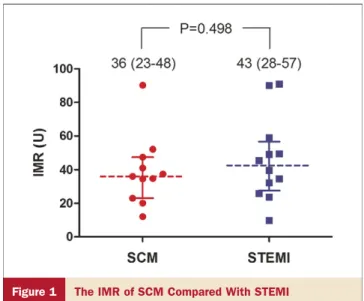
As shown inFigure 1, there was no difference in the IMR between
patients with SCM and STEMI (36 U [IQR: 23 to 48 U] vs. 43 U [IQR: 28 to 57 U]; p⫽ 0.498).
The main finding of this study is that microcirculatory function is impaired in SCM patients at the time of presentation. Micro- vascular dysfunction, as assessed by using IMR, was as high in patients with SCM as in those with STEMI, despite the signifi- cantly lower peak CK value in the SCM patients. Both groups may suffer a similar microvascular insult leading to a similar effect on IMR and left ventricular function, but the insult in the setting of SCM seems to be transient and does not result in significant cell death, whereas it is less reversible after STEMI. Microcirculatory dysfunction after reperfusion in STEMI is believed to be due to distal embolization of atherosclerotic and thrombotic debris and in situ thrombosis resulting in microvascular obstruction (5), which can lead to myonecrosis and CK elevation. A different pathophys- iological mechanism of microvascular dysfunction may be occur- ring in patients with SCM, accounting for their lower peak CK values and transient left ventricular dysfunction. Abnormal endo- thelial function has been noted in women with SCM, with excessive vasoconstriction and augmented sympathetic responses to acute stress (1). Future research aimed at differentiating endothelial-dependent versus endothelial-independent mecha- nisms of microvascular dysfunction in patients with SCM may provide further insight.
Figure 1 The IMR of SCM Compared With STEMI
Values are expressed as median (interquartile range). IMR⫽ index of microcir- culatory resistance; SCM⫽ stress cardiomyopathy; STEMI ⫽ ST-segment ele- vation myocardial infarction.
Journal of the American College of Cardiology Vol. 58, No. 23, 2011
© 2011 by the American College of Cardiology Foundation ISSN 0735-1097/$36.00
Published by Elsevier Inc.
Limitations of this study include that it is a retrospective single-center analysis with possible selection bias. Also, because of the small sample size, we may not have sufficient power to detect small differences in IMR between the 2 groups. IMR was calculated in approximately one-third of our patients with SCM, but there were no differences in clinical features between patients with and without IMR measurements. Because patients with SCM did not present to the hospital as quickly after symptom onset as those with STEMI, IMR was not measured at the same interval after the beginning of symptoms. Finally, sex differences may contribute to the findings because some data suggest that women may have more microvascular disease than men (6).
Although this study examined a small number of selected patients, it is the first, to our knowledge, to use IMR in evaluating the microcirculatory status in patients with SCM. Microcirculatory dys- function was frequently present during the acute phase of SCM, and microvascular damage was as marked as in patients with STEMI.
Hyun-SookKim, MD
Jennifer A. Tremmel, MD, MPH Chang-Wook Nam, MD
Jessica Zhou, MD Francois Haddad, MD Randall H. Vagelos, MD David P. Lee, MD Alan C. Yeung, MD
*William F. Fearon, MD
*Division of Cardiovascular Medicine Stanford University Medical Center 300 Pasteur Drive, Room H2103 Stanford, California 94305 E-mail:[email protected]
doi:10.1016/j.jacc.2011.08.046 Please note: Dr. Fearon has received research support from St. Jude Medical. All other authors have reported that they have no relationships relevant to the contents of this paper to disclose. The first 2 authors contributed equally to this paper.
REFERENCES
1. Nef HM, Młollmann H, Akashi YJ, Hamm CW. Mechanisms of stress (Takotsubo) cardiomyopathy. Nat Rev Cardiol 2010;7:187–93.
2. Fearon WF, Shah M, Ng M, et al. Predictive value of the index of microcirculatory resistance in patients with ST-segment elevation myo- cardial infarction. J Am Coll Cardiol 2008;51:560 –5.
3. Aarnoudse W, Fearon WF, Manoharan G, et al. Epicardial stenosis severity does not affect minimal microcirculatory resistance. Circulation 2004;110:2137– 42.
4. Melikian N, Vercauteren S, Fearon WF, et al. Quantitative assessment of coronary microvascular function in patients with and without epicardial atherosclerosis. EuroIntervention 2010;5:939 – 45.
5. Saber RS, Edwards WD, Bailey KR, McGovern TW, Schwartz RS, Holmes DR Jr. Coronary embolization after balloon angioplasty or thrombolytic therapy: an autopsy study of 32 cases. J Am Coll Cardiol 1993;22:1283– 8.
6. Han SH, Bae JH, Holmes DR, et al. Sex differences in atheroma burden and endothelial function in patients with early coronary athero- sclerosis. Eur Heart J 2008;29:1359 – 69.
JACC Vol. 58, No. 23, 2011 Correspondence 2431
November 29, 2011:2430 –1