저작자표시-비영리-변경금지 2.0 대한민국 이용자는 아래의 조건을 따르는 경우에 한하여 자유롭게 l 이 저작물을 복제, 배포, 전송, 전시, 공연 및 방송할 수 있습니다. 다음과 같은 조건을 따라야 합니다: l 귀하는, 이 저작물의 재이용이나 배포의 경우, 이 저작물에 적용된 이용허락조건 을 명확하게 나타내어야 합니다. l 저작권자로부터 별도의 허가를 받으면 이러한 조건들은 적용되지 않습니다. 저작권법에 따른 이용자의 권리는 위의 내용에 의하여 영향을 받지 않습니다. 이것은 이용허락규약(Legal Code)을 이해하기 쉽게 요약한 것입니다. Disclaimer 저작자표시. 귀하는 원저작자를 표시하여야 합니다. 비영리. 귀하는 이 저작물을 영리 목적으로 이용할 수 없습니다. 변경금지. 귀하는 이 저작물을 개작, 변형 또는 가공할 수 없습니다.
Doctoral Thesis in
Medical Sciences
Prognostic Impact of Left Ventricular
Mass Change in Patients with
ST-elevation Myocardial Infarction
Ajou University Graduate School
Medical Sciences Major
Prognostic Impact of Left Ventricular
Mass Change in Patients with
ST-elevation Myocardial Infarction
Seung-Jea Tahk, M.D., Ph.D., Advisor
I submit this thesis as the
Doctoral thesis in Medical Sciences.
February 2018
Ajou University Graduate School
Medical Sciences Major
This certifies that the dissertation of
Jin-Sun Park is approved.
SUPERVISORY COMMITTEE
Myeong-Ho Yoon
Seung-Jea Tahk
Gyo-Seung Hwang
So-Yeon Choi
Se-Joong Rim
Ajou Graduate School, Ajou University
i -ABSTRACT-
Prognostic Impact of Left Ventricular Mass Change
in Patients with ST-elevation Myocardial Infarction
Background: Prognostic significance between progression of left ventricular hypertrophy (LVH) and clinical outcomes in patients with ST-elevation myocardial infarction (STEMI) is uncertain. The objective of this study was to investigate prognostic impact of
progression of LV mass index (LVMI) in patients with STEMI.
Methods: We analyzed the data and clinical outcomes of patients with STEMI who received successful coronary intervention. A total of 200 patients who had
echocardiographic follow-up between 12 and 36 months were finally enrolled. According to change in LVMI compared to baseline LVMI, patients were classified into progression group and non-progression group. Progression of LVMI was defined when increment of LMVI was greater than 10% compared to baseline LVMI. End points were major adverse cardiac events within 5 years, including death, recurrent MI, target vessel
revascularization, and hospitalization due to heart failure.
Results: Progression of LVMI occurred in 55 patients. In the progression group, rate of recurrent MI was higher (13 vs. 2 %, P = 0.026) and the event-free survival of recurrent MI was significantly worse (log-rank P < 0.001) than that in the non-progression group. Adjusted hazard ratio of progression of LVMI for recurrent MI was 10.253 (95% confidence intervals 2.019-52.061, P = 0.005).
Conclusion: Increased LVMI was an independent predictor for adverse events, especially for recurrent MI, in patients with STEMI.
ii
List of Text
Abstract ··· ⅰ List of Text ··· ⅱ List of Figure ··· ⅲ List of Tables ··· ⅳ Ⅰ. Introduction ··· 1Ⅱ. Material and Method ··· 2
A. Subject Population ··· 2
B. Definition of Left Ventricular Mass and its Progression ··· 2
C. Study End-points ··· 3 D. Statistical analysis ··· 3 Ⅲ. Results ··· 5 Ⅳ. Discussion ··· 17 Ⅴ. Conclusion ··· 20 References ··· 21 국문요약 ··· 24
iii
List of Figures
Figure 1. Kaplan-Meier survival curves for free of adverse outcomes in non-progression group and progression group. ··· 12
iv
List of Tables
Table 1. Baseline clinical characteristics. ··· 6
Table 2. Baseline angiographic characteristics. ··· 7
Table 3. Baseline echocardiographic characteristics. ··· 8
Table 4. Follow-up echocardiographic characteristics. ··· 10
Table 5. Cox’s regression analysis for the adverse outcomes.··· 14
Table 6. Multivariate logistic regression analysis of the presence of progression of left ventricular mass index for clinical outcomes. ··· 16
1
I.
INTRODUCTION
Left ventricular hypertrophy (LVH) is closely related to adverse cardiovascular events in various etiologies (Levy et al., 1990; Bombelli et al., 2009; Cuspidi et al., 2014). We have previously reported that LVH is associated with increased rate of adverse clinical outcomes in 30-day survivors after ST-elevation myocardial infarction (STEMI) with successful percutaneous coronary intervention (PCI) (Park et al., 2015). In the previous study, we examined the prognostic significance of LVH only at baseline without evaluating the impact of their serial changes. The progression or regression of LVH might affect the clinical outcomes. However, prognostic significance between progression of LVH and clinical outcomes in patients with STEMI has not been established yet. Therefore, the objective of this study was to determine the prognostic impact of progression of LVH in patients with STEMI.
2
II. Material and Method
A. Subject Population
We consecutively enrolled 30-day survivors after STEMI who underwent successful revascularization. Successful revascularization was defined as thrombolysis in myocardial infarction trial (TIMI) grade 3 flow and < 30% residual stenosis in infarct related artery after primary PCI. Transthoracic echocardiography was performed within 48 hours of primary PCI. We finally enrolled 200 patients (133 males, 56 ± 11 year-old) with echocardiographic follow-up (F/U) between 12 and 36 months after index STEMI. Medical records of all patients were retrospectively reviewed. This study was approved by the Ajou University Hospital Institutional Review Board (approval number: AJIRB-MED-MDB-17-015). We excluded patients from this study if they had history of prior revascularization. We also excluded patients if the LV dysfunction was caused by any of the following: predisposing cardiomyopathy, severe valvular heart disease including symptomatic aortic stenosis, or more than moderate aortic and mitral regurgitation.
B
.Definition of Left Ventricular Mass and its Progression
Left ventricular mass (LVM) was calculated according to Devereux’s formula (Devereux
et al., 1986
) using linear measurements derived from two-dimensional echocardiography (Lang et al., 2005):LVM (g) =0.8 x (1.04 x [(LV internal diastolic diameter + posterior wall thickness in diastole + septal wall thickness in diastole)3-( LV internal diastolic diameter)3] + 0.6
3
to baseline LVMI, patients were classified into progression group and non-progression group. Progression of LVMI was defined when increment of LMVI was greater than 10% compared to baseline LVMI.
C. Study End-points
End points of the present study were major adverse cardiac events (MACEs) within five years, including death, recurrent myocardial infarction (MI), target vessel revascularization (TVR), and hospitalization due to heart failure (HF). Recurrent myocardial infarction was defined according to the universal definition of MI (
Thygesen
et al., 2007). Target vessel revascularization was defined as clinically indicated percutaneous or surgical revascularization of the index vessel during follow-up. At five years after index STEMI, follow-up data were obtained by reviewing medical records and/or telephone interview with patients. To demonstrate a correlation between progression of LVH and long-term clinical outcomes, we enrolled the patients from 2003 to 2009, who were followed up for more than 5 years.D. Statistical analysis
SPSS 13.0 statistical software package (SPSS Inc., Chicago, IL, USA) was used for all analyses. Data are shown as mean ± standard deviation for continuous variables or numbers and percentages for categorical variables. Comparisons were conducted by unpaired Student’s t test for continuous variables or Pearson chi-square test for categorical variables. Event free survival analysis for patients in these groups was performed using the Kaplan-Meier method. Differences between groups were assessed by log-rank test. To assess adjusted relative hazard ratio (HR) of progression of LVMI to the study end points, Cox’s proportional hazard model was used with potential variables associated with clinical outcomes. Adjusted covariates for the Cox’s proportional hazard model were well-known predictors of MACEs such as age, gender,
4
diabetes mellitus, hypertension, smoking, dyslipidemia, Killip classification, LV ejection fraction (EF), and progression of LVMI. Results of Cox’s regression analysis were expressed as adjusted HRs with 95% confidence intervals (CI) for clinical outcomes. Multivariate logistic regression analysis was performed to assess the effect of the presence of progression of LVMI on clinical outcomes. Null hypotheses of no difference were rejected if P values were less than 0.05.
5
III. Results
From 2003 to 2009, a total of 200 patients (164 males, 56 ±11 year-old) were enrolled. Mean value of baseline LVMI of these 200 patients was at 114 ± 30 g/m2. Baseline LVMI showed normal distribution. Progression of LVMI occurred in 55 patients (41 males, 58 ± 12 year-old). Fifty-five patients (27.5%) were included in the progression group while the remaining 145 patients (72.5%) were included in the non-progression group.
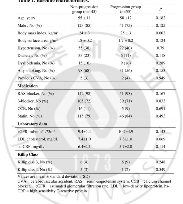
Baseline clinical characteristics according to the two groups are summarized in Table 1. There were no statistical differences in baseline characteristics such as medical history or medical treatments between the two groups.
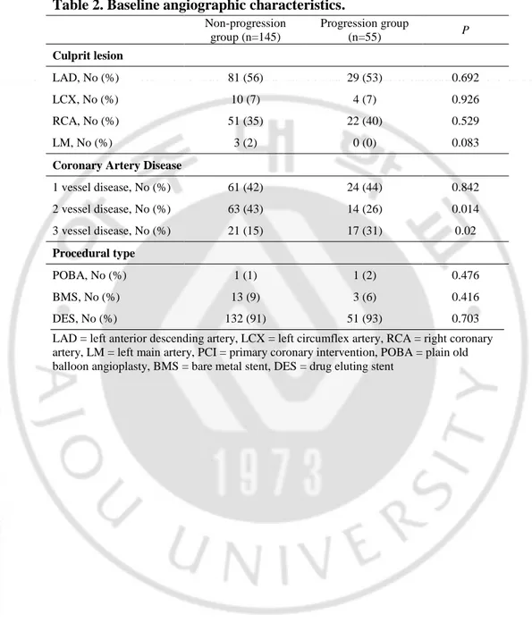
Patients with three-vessel disease were more common in the progression group (P = 0.02, Table 2) while patient with two-vessel disease were more common in the non-progression group (P = 0.014). Overall, patients with multi-vessel disease were not statistically different between the two groups. Distributions of culprit lesion and procedural type were also similar between the two groups.
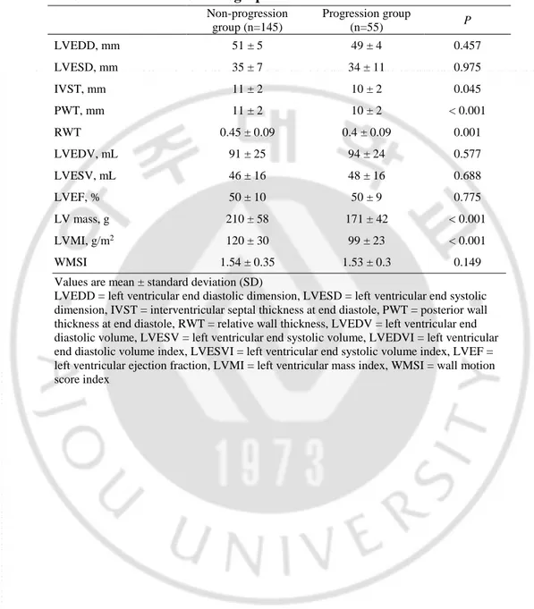
Baseline LVMI was significantly lower in the progression group compared to that in the non-progression group (99 ± 23 vs. 120 ± 30 g/m2, P < 0.001, Table 3). LV systolic function measured by EF and LV regional function measured by wall motion score index (WMSI) in the progression group were similar to those in the non-progression LVMI group (EF: 50 ± 9 vs. 50 ± 10 %, P = 0.775; WMSI: 1.53 ± 0.3 vs. 1.54 ± 0.35, P = 0.149). Parameters indicating LV chamber size such as LV end diastolic dimension (LVEDD) and LV end diastolic volume (LVEDV) were also similar between the two groups (49 ± 4 vs. 51 ± 5 mm, P = 0.457 and 94 ± 24 vs. 91 ± 25 mL, P = 0.708, respectively).
6
Table 1. Baseline characteristics.
Non-progression group (n=145) Progression group (n=55) P Age, years 55 ± 11 58 ±12 0.182 Male , No (%) 123 (85) 41 (75) 0.125 Body mass index, kg/m2 24 ± 3 25 ± 3 0.602 Body surface area, g/m2 1.8 ± 0.2 1.7 ± 0.2 0.124 Hypertension, No (%) 55 (38) 22 (40) 0.79 Diabetes, No (%) 33 (23) 6 (11) 0.118 Dyslipidemia, No (%) 15 (10) 9 (16) 0.289 Any smoking, No (%) 98 (68) 31 (56) 0.153 Previous CVA, No (%) 5 (3) 2 (4) 0.949 Medication RAS blocker, No (%) 142 (98) 51 (93) 0.167 β-blocker, No (%) 105 (72) 39 (71) 0.833 CCB, No (%) 16 (11) 5 (9) 0.691 Statin, No (%) 115 (79) 46 (84) 0.493 Laboratory data eGFR, ml/min/1.73m2 9.4±4.4 10.7±4.9 0.143 LDL cholesterol, mg/dL 7.4±1.0 7.8±1.0 0.069 hs-CRP, mg/dL 6.4±2.1 5.7±2.0 0.114 Killip Class Killip clss 3, No (%) 6 (4) 5 (9) 0.248 Killip clss 4, No (%) 5 (3) 1 (2) 0.549 Values are mean ± standard deviation (SD)
CVA = cerebrovascular accident, RAS = renin-angiotensin system, CCB = calcium channel blocker, eGFR = estimated glomerular filtration rate, LDL = low-density lipoprotein, hs-CRP = high sensitivity C-reactive protein
7
Table 2. Baseline angiographic characteristics.
Non-progression group (n=145) Progression group (n=55) P Culprit lesion LAD, No (%) 81 (56) 29 (53) 0.692 LCX, No (%) 10 (7) 4 (7) 0.926 RCA, No (%) 51 (35) 22 (40) 0.529 LM, No (%) 3 (2) 0 (0) 0.083
Coronary Artery Disease
1 vessel disease, No (%) 61 (42) 24 (44) 0.842 2 vessel disease, No (%) 63 (43) 14 (26) 0.014 3 vessel disease, No (%) 21 (15) 17 (31) 0.02 Procedural type POBA, No (%) 1 (1) 1 (2) 0.476 BMS, No (%) 13 (9) 3 (6) 0.416 DES, No (%) 132 (91) 51 (93) 0.703 LAD = left anterior descending artery, LCX = left circumflex artery, RCA = right coronary artery, LM = left main artery, PCI = primary coronary intervention, POBA = plain old balloon angioplasty, BMS = bare metal stent, DES = drug eluting stent
8
Table 3. Baseline echocardiographic characteristics.
Non-progression group (n=145) Progression group (n=55) P LVEDD, mm 51 ± 5 49 ± 4 0.457 LVESD, mm 35 ± 7 34 ± 11 0.975 IVST, mm 11 ± 2 10 ± 2 0.045 PWT, mm 11 ± 2 10 ± 2 < 0.001 RWT 0.45 ± 0.09 0.4 ± 0.09 0.001 LVEDV, mL 91 ± 25 94 ± 24 0.577 LVESV, mL 46 ± 16 48 ± 16 0.688 LVEF, % 50 ± 10 50 ± 9 0.775 LV mass, g 210 ± 58 171 ± 42 < 0.001 LVMI, g/m2 120 ± 30 99 ± 23 < 0.001 WMSI 1.54 ± 0.35 1.53 ± 0.3 0.149 Values are mean ± standard deviation (SD)
LVEDD = left ventricular end diastolic dimension, LVESD = left ventricular end systolic dimension, IVST = interventricular septal thickness at end diastole, PWT = posterior wall thickness at end diastole, RWT = relative wall thickness, LVEDV = left ventricular end diastolic volume, LVESV = left ventricular end systolic volume, LVEDVI = left ventricular end diastolic volume index, LVESVI = left ventricular end systolic volume index, LVEF = left ventricular ejection fraction, LVMI = left ventricular mass index, WMSI = wall motion score index
9
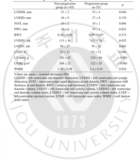
Mean changes of LVMI in the progression group and those in the non-progression group were 28 ± 14% and -12 ± 15 %, respectively. Follow-up echocardiography showed that LVMI was significantly higher in the progression group than that in the non-progression group (122 ± 25 vs. 104 ± 22 g/m2, P < 0.001, Table 4). LV systolic and regional functions were maintained in both groups (EF: 53 ± 12 vs. 53 ± 11 %, P = 0.966; WMSI: 1.4 ± 0.35 vs. 1.39 ± 0.38, P = 0.954). In the progression group, LVEDD and LVEDV, parameters indication LV chamber size, showed progressive LV remodeling compared to those in the non-progression group (53 ± 5 vs. 51 ± 2 mm, P = 0.046; 131 ± 28 vs. 113 ± 36 mL, P = 0.035, respectively).
10
Table 4. Follow-up echocardiographic characteristics
Non-progression group (n=145) Progression group (n=55) P LVEDD, mm 51 ± 2 53 ± 5 0.046 LVESD, mm 36 ± 9 37 ± 8 0.176 IVST, mm 10 ± 2 10 ± 1 0.004 PWT, mm 10 ± 2 10 ± 1 0.022 RWT 0.38 ± 0.09 0.39 ± 0.07 0.372 LVEDV, mL 113 ± 36 131 ± 28 0.035 LVESV, mL 58 ± 27 70 ± 21 0.065 LVEF, % 53 ± 11 53 ± 12 0.966 LV mass, g 181 ± 42 210 ± 46 < 0.001 LVMI, g/m2 104 ± 22 122 ± 25 < 0.001 WMSI 1.39 ± 0.38 1.4 ± 0.35 0.954 Values are mean ± standard deviation (SD)
LVEDD = left ventricular end diastolic dimension, LVESD = left ventricular end systolic dimension, IVST = interventricular septal thickness at end diastole, PWT = posterior wall thickness at end diastole, RWT = relative wall thickness, LVEDV = left ventricular end diastolic volume, LVESV = left ventricular end systolic volume, LVEDVI = left ventricular end diastolic volume index, LVESVI = left ventricular end systolic volume index, LVEF = left ventricular ejection fraction, LVMI = left ventricular mass index, WMSI = wall motion score index
11
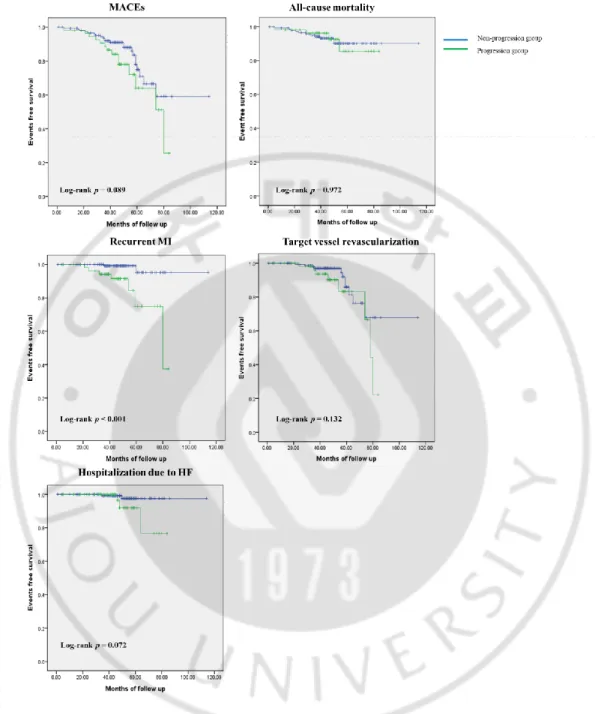
Patients were followed up for 47 ± 16 months after index STEMI. MACEs occurred in 37 patients (19%). Of 200 patients, 15 (8%) died, 10 (5%) experienced recurrent MI, 19 (10%) needed TVR and 5 (3%) were hospitalized due to HF. Occurrences of MACEs, death, TVR and hospitalization due to HF were similar between the two groups (26 vs. 16 %, 7 vs. 8 %, 15 vs. 8 % and 6 vs. 1 % respectively). Rate of recurrent MI was higher in the progression group than in the non-progression group (13 vs. 2 %, P = 0.026). Event-free survival of recurrent MI was significantly worse in the progression group than in the non-progression group (log-rank P < 0.001, Figure 1).
12
Fig 1. Kaplan-Meier survival curves for free of adverse outcomes in non-progression group and progression group.
13
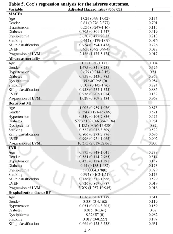
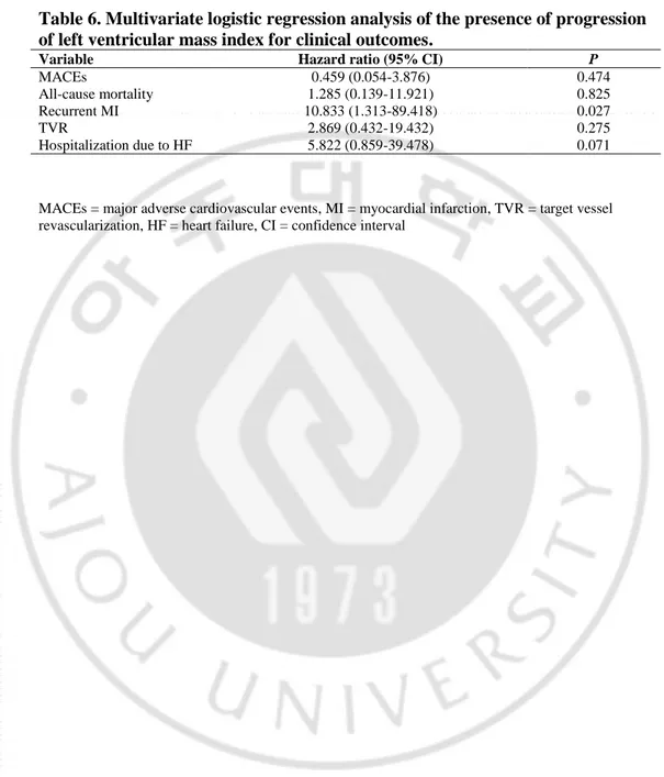
Results of multivariate survival analysis using Cox’s regression model are summarized in Table 5. In Cox’s proportional hazard model, LVEF (HR 0.956, 95% CI 0.92-0.994, P = 0.023) and progression of LVMI (HR 2.466, 95% CI 1.175-5.174, P = 0.017) were related to MACEs. Age was strongly related to all cause of death (HR 1.1, 95% CI 1.03-1.175, P = 0.004). Adjusted HR of progression of LVMI for recurrent MI was 10.253 (95% CI 2.019-52.061, P = 0.005). LVEF (HR 0.926, 95% CI 0.869-0.987, P = 0.019) and progression of LVMI (HR 3.709, 95% CI 1.257-10.945, P = 0.018) were related to TVR. In a multivariate regression model, progression of LVMI was independently associated with increased risk for recurrent MI (HR 10.833, 95% CI 1.313-89.418, P = 0.027, Table 6).
14
Table 5. Cox’s regression analysis for the adverse outcomes.
Variable Adjusted Hazard ratio (95% CI) P MACEs Age 1.026 (0.99-1.062) 0.154 Gender 0.81 (0.276-2.377) 0.701 Hypertension 0.536 (0.247-1.16) 0.113 Diabetes 0.705 (0.301-1.647) 0.419 Dyslipidemia 3.670 (0.475-28.37) 0.213 Smoking 0.442 (0.179-1.09) 0.076 Killip classification 0.924 (0.594-1.438) 0.726 LVEF 0.956 (0.92-0.994) 0.023 Progression of LVMI 2.466 (1.175-5.174) 0.017 All-cause mortality Age 1.1 (1.030-1.175) 0.004 Gender 1.675 (0.341-8.238) 0.526 Hypertension 0.679 (0.214-2.15) 0.51 Diabetes 0.959 (0.243-3.785) 0.953 Dyslipidemia 352307.965 (0) 0.984 Smoking 0.505 (0.145-1.76) 0.284 Killip classification 0.958 (0.532-1.725) 0.885 LVEF 0.956 (0.902-1.014) 0.132 Progression of LVMI 1.029 (0.309-3.434) 0.963 Recurrent MI Age 1.005 (0.939-1.076) 0.875 Gender 2.354 (0.121-45.689) 0.571 Hypertension 0.549 (0.106-2.836) 0.474 Diabetes 57789.182 (0-6.364E194) 0.961 Dyslipidemia 1.135 (0.096-13.438) 0.92 Smoking 0.522 (0.072-3.809) 0.522 Killip classification 0.806 (0.273-2.376) 0.696 LVEF 0.996 (0.931-1.065) 0.902 Progression of LVMI 10.253 (2.019-52.061) 0.005 TVR Age 0.993 (0.948-1.041) 0.778 Gender 0.581 (0.114-2.965) 0.514 Hypertension 0.423 (0.128-1.391) 0.157 Diabetes 0.44 (0.135-1.432) 0.173 Dyslipidemia 5900004.378(0) 0.979 Smoking 0.392 (0.102-1.51) 0.173 Killip classification 0.786 (0.371-1.666) 0.529 LVEF 0.926 (0.869-0.987) 0.019 Progression of LVMI 3.709 (1.257-10.945) 0.018 Hospitalization due to HF Age 1.036 (0.903-1.189) 0.611 Gender 0.004 (0-4.162) 0.119 Hypertension 0.051 (0.001-3.203) 0.159 Diabetes 0.015 (0-1.64) 0.08 Dyslipidemia 8.326E7 (0) 0.982 Smoking 0.017 (0-8.227) 0.197 Killip classification 0.664 (0.125-3.538) 0.631
15
LVEF 0.74 (0.545-1.005) 0.054 Progression of LVMI 267.439 (0.843-84801.068) 0.057
MACEs = major adverse cardiovascular events, LVEF = left ventricular ejection fraction, LVMI = left ventricular mass index, MI = myocardial infarction, TVR = target vessel revascularization, HF = heart failure, CI = confidence interval
16
Table 6. Multivariate logistic regression analysis of the presence of progression of left ventricular mass index for clinical outcomes.
Variable Hazard ratio (95% CI) P
MACEs 0.459 (0.054-3.876) 0.474 All-cause mortality 1.285 (0.139-11.921) 0.825 Recurrent MI 10.833 (1.313-89.418) 0.027 TVR 2.869 (0.432-19.432) 0.275 Hospitalization due to HF 5.822 (0.859-39.478) 0.071
MACEs = major adverse cardiovascular events, MI = myocardial infarction, TVR = target vessel revascularization, HF = heart failure, CI = confidence interval
17
IV. Discussion
The present study demonstrated that change of LVMI was associated with clinical outcomes in patients with STEMI who received successful PCI. After index STEMI, LV remodeling is divided into an early phase (within 72 hours) and a late phase (beyond 72 hours). Early remodeling is induced by acute loss of myocardium, resulting in abrupt increase in loading conditions (Sutton et al., 2000). In our previous study, LVH at index STEMI is associated with increased rate of adverse clinical outcomes, especially all-cause mortality. Myocardial structural and functional alterations beyond EF representing LV systolic function might affect adverse clinical outcomes in patients with early remodeling (Park et al., 2015). Late remodeling, including LVH and alterations in ventricular architecture, is an adaptive response that can offset chronically increased hemodynamic load, attenuate progressive dilatation, and stabilize contractile function (Pfeffer et al., 1980). The progression of LVMI was related to increased risk for recurrent MI in the present study. In Cox’s proportional hazard model, progression of LVMI was related to recurrent MI and TVR. Progression of LVMI might be more closely related to coronary vascular complication.
Myocyte hypertrophy is initiated by activation of neurohormonal system with local tissue renin-angiotensin (RAS) system and myocardial stretch. After MI, decreased cardiac output enhances cathecholamine production by adrenal medulla and sympathetic nerve terminals. It also activates RAS-aldosterone axis (Sutton et al., 2000). Stimulation of α1 adrenoreceptor by enhanced norepinephrine (NE) release, and angiotensin 1 receptor can induce myocyte hypertrophy via Gqα-dependent pathway which is upregulated in the viable border and scar tissue in post-MI hearts (Ju et al., 1998). Both NE and angiotensin II augment endothelin-1 release, which is another stimulus for myocyte hypertrophy (Levin et al., 1998). Mechanical myocardial stretch induced by elevated wall stresses sensed by infarcted and non-infarcted myocardium can result in secretion of angiotensin II from cytoplasmic granules and induce myocyte hypertrophy
18
mediated by angiotensin 1 receptor (Yamazaki et al., 1995).
In atherosclerotic plaque lesions, local RAS system is also activated. Angiotensin II can induce myocyte hypertrophy. It can also generates oxidative stress in vessel wall, resulting in stimulation of vascular thrombosis and inflammation (Mehta et al., 2007). In vascular smooth muscle cells, exposure to angiotensin II leads to increased levels of plasminogen-activator inhibitor type 1 which can inhibit tissue plasminogen activator and urokinase, and cell adhesion molecules such as vascular cell adhesion molecule-1 and intercellular adhesion molecule-1, resulting in prothrombotic status (Feener et al., 1995). In angiotensin II stimulated vascular smooth muscle cells, effects of inflammatory cytokine interleukin-18 are enhanced. This is related to progression of atherosclerosis and restenosis (Gerdes et al., 2002).
Pathologic angiotensin II-induced signaling in vascular, endothelial and cardiac cells can promote vascular thrombosis, neointima formation and LVH. In the present study, close correlation between progression of LVMI and adverse clinical outcomes, especially recurrent MI, might be affected by pathologic angiotensin-II signaling. Many studies have demonstrated that RAS blocker can prevent angiotensin II-induced LVH and vascular pathology (Kim et al., 1995; Keidar et al., 1997; 18. Yusuf et al., 2000), Therefore, RAS blocker might be able to reduce adverse long-term clinical outcomes in patients with progression of LVMI after index STEMI.
This study has several limitations. First, echocardiographic measuring LVM using linear measurements has potential limitations. It is based on the assumption that the LV is represented by a prolate ellipse (Devereux et al., 1986). Nevertheless, LVM obtained with this method has been well validated and widely used in clinical practice (Lang et al., 2005). Although there could be geometrical deformation, we measured LVMI at index STEMI and follow-up using same technical method and analyzed the presence of change in LVMI not the value of LMVI itself. Second, there is no validated definition of
19
progression of LVMI. In the present study, we defined progression of LVMI as an increment of LMVI greater than 10% compared with baseline LVMI. There have been no data demonstrating association between progression of LVMI and clinical outcomes. Further study is needed to define clinically significant progression of LVMI. Third, the present study could not demonstrate a possible benefit of regression of LVMI. A total of 82 patients had LVMI regression in the present study. There was no significant correlation between regression of LVMI and MACEs. It might be due to the relatively small number of our study population. Fourth, levels of angiotensin II and related cytokines were not checked in the present study. The present study logically implied that angiotensin II had a pathological role in the progression group. To prove this, further studies might be needed to evaluate levels of angiotensin II and related cytokines. Finally, we could not fully evaluate the impact of serial changes of LVMI. Since the present study was retrospective and annual echocardiographic follow-up was not recommended after index STEMI in current guidelines, we enrolled patients with echocardiographic follow-up (F/U) between 12 and 36 months after index STEMI. We could not know the effect of rate of progression in LVMI.
20
V. Conclusion
In summary, increased LVMI was found to be an independent predictor for adverse events, especially for recurrent MI in patients with STEMI who received successful coronary intervention. Therefore, maintenance and dose-adjustment of proper medical treatment including RAS blocker should be considered in patients with progression of LVMI after index STEMI.
21
REFERENCES
1. Bombelli M, Facchetti R, Carugo S, Madotto F, Arenare F, Quarti-Trevano F, Capra A, Giannattasio C, Dell'Oro R, Grassi G, Sega R, Mancia G: Left ventricular hypertrophy increases cardiovascular risk independently of in-office and out-of-office blood pressure values. J Hypertens 27(12): 2458-2464, 2009. 2. Cuspidi C, Facchetti R, Bombelli M,
Sala C, Grassi G, Mancia G:
DifferentialValue of Left Ventricular Mass Index and Wall Thickness in Predicting Cardiovascular Prognosis: Data From the PAMELA Population. Am J Hypertens 27(8): 1079-1086, 2014.
3. Devereux RB, Alonso DR, Lutas EM, Gottlieb GJ, Campo E, Sachs I, Reichek N: Echocardiographic assessment of left ventricular hypertrophy: comparison to necropsy findings. Am J Cardiol 57(6): 450-458, 1986.
4. Feener EP, Northrup JM, Aiello LP, King GL: Angiotensin II induces plasminogen activator inhibitor-1 and -2 expression in vascular endothelial and smooth muscle cells. J Clin Invest 95(3): 1353-1362, 1995.
5. Gerdes N, Sukhova GK, Libby P, Reynolds RS, Young JL, Schönbeck U: Expression of interleukin (IL)-18 and functional IL-18 receptor on human vascular endothelial cells, smooth muscle cells, and macrophages: implications for atherogenesis. J Exp Med 195(2): 245-257, 2002.
6. Ju H, Zhao S, Tappia PS, Panagia V, Dixon IM: Expression of Gq alpha and PLC-beta in scar and border tissue in heart failure due to myocardial infarction. Circulation 97(9): 892-899, 1998.
7. Keidar S, Attias J, Smith J, Breslow JL, Hayek T: The angiotensin-II receptor antagonist, losartan, inhibits LDL lipid peroxidation and atherosclerosis in apolipoprotein E-deficient mice. Biochem Biophys Res Commun 236(3): 622-625, 1997.
22
8. Kim S, Ohta K, Hamaguchi A, Yukimura T, Miura K, Iwao H: Angiotensin II induces cardiac phenotypic modulation and remodeling in vivo in rats. Hypertension 25(6): 1252-1259, 1995.
9. Lang RM, Bierig M, Devereux RB, Flachskampf FA, Foster E, Pellikka PA, Picard MH, Roman MJ, Seward J, Shanewise JS, Solomon SD, Spencer KT, Sutton MS, Stewart WJ; Chamber Quantification Writing Group; American Society of Echocardiography's Guidelines and Standards Committee; European Association of Echocardiography: Recommendations for chamber quantification: a report from the American Society of Echocardiography's Guidelines and Standards Committee and the Chamber Quantification Writing Group, developed in conjunction with the European Association of Echocardiography, a branch of the European Society of Cardiology. J Am Soc Echocardiogr 18(12): 1440-1463, 2005.
10. Levin ER, Gardner DG, Samson WK: Natriuretic peptides. N Engl J Med 339(5): 321-328, 1998.
11. Levy D, Garrison RJ, Savage DD, Kannel WB, Castelli WP: Prognostic implications of echocardiographically determined left ventricular mass in the Framingham Heart Study. N Engl J Med 322(22): 1561-1566, 1990.
12. Mehta PK, Griendling KK: Angiotensin II cell signaling: physiological and pathological effects in the cardiovascular system. Am J Physiol Cell Physiol 292(1): C82-C97, 2007.
13. Park JS, Shin JS, Lee YH, Seo KW, Choi BJ, Choi SY, Yoon MH, Hwang GS, Tahk SJ, Shin JH: Left ventricular hypertrophy on long-term cardiovascular outcomes in patients with ST-elevation myocardial infarction. Clin Exp Hypertens 37(8): 674-679, 2015.
14. Pfeffer MA, Braunwald E: Ventricular remodeling after myocardial infarction. Experimental observations and clinical implications. Circulation 81(4): 1161-1172, 1990.
23
pathophysiology and therapy. Circulation 101(25): 2981-2988, 2000.
16. Thygesen K, Alpert JS, White HD; Joint ESC/ACCF/AHA/WHF Task Force for the Redefinition of Myocardial Infarction, Jaffe AS, Apple FS, Galvani M, Katus HA, Newby LK, Ravkilde J, Chaitman B, Clemmensen PM, Dellborg M, Hod H, Porela P, Underwood R, Bax JJ, Beller GA, Bonow R, Van der Wall EE, Bassand JP, Wijns W, Ferguson TB, Steg PG, Uretsky BF, Williams DO, Armstrong PW, Antman EM, Fox KA, Hamm CW, Ohman EM, Simoons ML, Poole-Wilson PA, Gurfinkel EP, Lopez-Sendon JL, Pais P, Mendis S, Zhu JR, Wallentin LC, Fernández-Avilés F, Fox KM, Parkhomenko AN, Priori SG, Tendera M, Voipio-Pulkki LM, Vahanian A, Camm AJ, De Caterina R, Dean V, Dickstein K, Filippatos G, Funck-Brentano C, Hellemans I, Kristensen SD, McGregor K, Sechtem U, Silber S, Tendera M, Widimsky P, Zamorano JL, Morais J, Brener S, Harrington R, Morrow D, Lim M, Martinez-Rios MA, Steinhubl S, Levine GN, Gibler WB, Goff D, Tubaro M, Dudek D, Al-Attar N: Universal definition of myocardial infarction. Circulation 116 (22): 2634-2653, 2007.
17. Yamazaki T, Komuro I, Kudoh S, Zou Y, Shiojima I, Mizuno T, Takano H, Hiroi Y, Ueki K, Tobe K, Kadowaki T, Nagai R, Yazaki Y: Angiotensin II partly mediates mechanical stress-induced cardiac hypertrophy. Circ Res 77(2): 258-265, 1995.
18. Yusuf S, Sleight P, Pogue J, Bosch J, Davies R, Dagenais G: Effects of an angiotensin-converting-enzyme inhibitor, ramipril, on cardiovascular events in high-risk patients. N Engl J Med 342(3): 145-153, 2000.
24 -국문요약-
ST 분절 상승 심근경색증 환자에서
좌심실 질량 변화의 임상적 의의
목적: ST 분절 상승 심근경색증 환자에서 좌심실 질량 변화와 주요심장사건 발생에 대한 연구는 이루어지지 않았다. 본 연구는 ST 분절 상승 심근경색 증 환자에서 좌심실 질량 지수의 변화가 예후에 영향을 미치는 지에 대해 조사하였다. 방법: 성공적인 일차적 관동맥 중재시술을 받은 ST 분절 상승 심근경색증 환자의 임상 데이터를 분석하였다. ST 분절 상승 심근경색증 진단 후 12개 월에서 36개월 사이에 심초음파 추적관찰을 시행한 총 200명의 환자를 최 종적으로 연구에 등록하였다. 심초음파 추적 검사에서 좌심실 질량지수의 증 가 여부에 따라 환자의 군을 분류하였다. 좌심실 질량지수의 증가는 입원 당 시 측정한 좌심실 질량지수와 비교하여 10% 이상의 좌심실 질량지수 증가 가 있을 때로 정의하였다. 5년 이내의 환자의 사망, 재발성 심근경색, 표적 혈관의 재 개통, 심부전에 의한 입원으로 구성된 주요 심장사건을 연구하였 다. 결과: 55명의 환자에서 좌심실 질량지수의 증가가 있었다. 좌심실 질량지수 의 증가가 있었던 환자군에서, 재발성 심근경색의 발생율이 좌심실 질량지수 의 증가가 없었던 환자군보다 높았고 (13 vs. 2 %, P = 0.026) 재발성 심근 경색 무사건 생존률은 통계적으로 의미있게 낮았다(log-rank P < 0.001). 좌심실 질량지수의 증가와 재발성 심근경색에 대한 비교 위험도는 10.253 (95% 신뢰구간 2.019-52.061, P = 0.005) 이었다.25
결론: ST 분절 상승 심근경색증 환자에서 좌심실 질량 지수의 증가는 주요 심장 사건 중 특히 재발성 심근경색과 연관성이 있었다.