• 검색 결과가 없습니다.

Combined Assessments of Biochemical Markers and ST-Segment Resolution Provide Additional Prognostic Information for Patients With ST-Segment Elevation Myocardial Infarction

N/A
N/A
Protected

Academic year: 2022

Share "Combined Assessments of Biochemical Markers and ST-Segment Resolution Provide Additional Prognostic Information for Patients With ST-Segment Elevation Myocardial Infarction"

Copied!
7
0
0

로드 중.... (전체 텍스트 보기)

전체 글

(1)

372

Open Access

Combined Assessments of Biochemical Markers and

ST-Segment Resolution Provide Additional Prognostic Information for Patients With ST-Segment Elevation Myocardial Infarction

Jong Shin Woo, MD

1

, Jin Man Cho, MD

2

, Soo Joong Kim, MD

1

, Myeong Kon Kim, MD

1

, and Chong Jin Kim, MD

2

1

Division of Cardiology, Department of Internal Medicine, School of Medicine, Kyung Hee University, Seoul,

2

Cardiovascular Center of Kyung Hee University, Gang Dong Kyung Hee Medical Center, Seoul, Korea

ABSTRACT

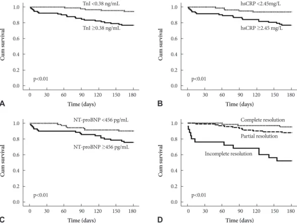
Background and Objectives: The prognostic value of biochemical markers and the resolution of ST-segment elevation on electrocardiogram are well established. However, how a combination of these two tools affects the evaluation of risk stratifi- cation has not yet been evaluated. Subjects and Methods: Between January 2006 and June 2008, 178 consecutive patients treated with primary percutaneous coronary interventions after ST-segment elevation myocardial infarctions (STEMI) were analyzed at two coronary care units. Patients were divided into the following three groups according to ST-segment resolu- tion: complete (≥70% depression of the elevated ST-segment, n=63), partial (30% to 70%, n=90), and incomplete (<30%, n=25). Demographic data, including history, electrocardiography, biochemical markers, initial ejection fraction, and angio- graphic findings were also evaluated. Results: There were 7 deaths, 3 repeated myocardial infarctions, and 17 readmissions for worsening heart failure during six months of follow-up. In a multivariate analysis to predict clinical outcomes, ejection frac- tion {hazard ratio (HR): 0.83 (0.76-0.91), p<0.01}, high-sensitivity C-reactive protein {HR: 1.15 (1.05-1.26), p<0.05}, and the degree of ST-segment resolution {HR: 0.96 (0.93-0.09), p<0.05} were independently associated with clinical outcomes. Accord- ing to the Cox-proportional hazards model, the addition of ST-segment resolution markedly improved the prognostic utility of the model containing biochemical markers and ejection fraction. Conclusion: Assessment of biomarkers upon admis- sion and ST-segment resolution are strong predictors of clinical outcomes. The combination of these data provides additive information about prognosis at an early point in the disease progression and further improves risk stratification for STEMI.

(Korean Circ J 2011;41:372-378)

KEY WORDS: Myocardial infarction; Prognosis; C-reactive protein; N-terminal pro-B-type natriuretic peptide;

Electrocardiogram.

Received: August 20, 2010 Accepted: October 26, 2010

Correspondence: Jin Man Cho, MD, Division of Cardiology, Depart- ment of Internal Medicine, School of Medicine, Kyung Hee University, 149 Sangil-dong, Gangdong-gu, Seoul 134-727, Korea

Tel: 82-2-440-6107, Fax: 82-2-440-6295 E-mail: [email protected]

• The authors have no financial conflicts of interest.

cc

This is an Open Access article distributed under the terms of the Cre- ative Commons Attribution Non-Commercial License (http://creativecom- mons.org/licenses/by-nc/3.0) which permits unrestricted non-commer- cial use, distribution, and reproduction in any medium, provided the origi- nal work is properly cited.

Introduction

Early risk stratification is essential in the management of patients with suspected or confirmed acute coronary synd-

rome. Traditionally, risk stratification involves an evaluation

of medical history, a physical examination, and electrocardio-

graphy (ECG) upon admission. In recent years, additional

variables for early risk stratification in patients with ST elev-

ation myocardial infarction (STEMI) have been identified,

and include the elevation of cardiac biochemical markers. For

example, the myocardial release of troponin I (TnI) and N-ter-

minal pro-B-type natriuretic peptide (NT-proBNP) represent

sensitive serum markers for minimal myocardial injury. When

present, these markers are associated with an increased likeli-

hood of a cardiac event.

1)

The elevation of another marker,

high-sensitivity C-reactive protein (hsCRP), upon admission

with STEMI is associated with an increased risk of compli-

cations both during hospitalization and after discharge.

2)

These

biochemical markers are noninvasive indicators of the risk of

(2)

first or recurrent cardiovascular events and also predict the success of therapeutic and preventive interventions.

1)

Thus, several studies have demonstrated the independent progno- stic value of elevated TnI, NT-proBNP, or hsCRP at admission in patients with STEMI.

3-9)

Additionally, in patients treated for STEMI, early ST-segment resolution has been associated with a better outcome based on improved early infarct-related ar- tery patency, smaller infarct size, and less impairment of left ventricular function.

10-12)

However, the combination of these two early available tools for predicting risk soon after a patient’s presentation has not been evaluated. To address these issues, we retrospectively assessed the clinical characteristics and follow-up outcomes of consecutive patients who received primary percutaneous coronary intervention (PCI) after STEMI. The aim of this stu- dy was to evaluate the combined effect of using multiple bio- chemical markers and ST-segment resolution assessments for early risk stratification in patients with STEMI and treated by PCI.

Subjects and Methods

Study participants

Between January 2006 and June 2008, 178 consecutive pa- tients who presented with their first STEMI and were subse- quently treated with primary PCI were enrolled in the study, which took place at two coronary care units at the Kyung Hee University Hospital, Seoul, Korea. All patients were treated according to the standard of care for STEMI and underwent primary PCI. The diagnostic work-up in all patients consist- ed of history, 12-lead ECG, blood chemistries, and a two-di- mensional (2D) echocardiography. The inclusion criteria were as follows: 1) presentation within 12 hours of symptom onset; 2) chest pain lasting ≥30 minutes and resistant to ni- trates; and 3) ≥0.2 mV ST-segment elevation in at least two contiguous leads on 12-lead ECG. The exclusion criteria were as follows: 1) prior myocardial infarction, defined by pre-exist- ing pathologic Q-wave; 2) non-ST elevation myocardial in- farction; and 3) thrombolytic therapy taken instead of PCI. Pa- tents with infectious disease, collagen disease, malignant dise- ase, or severe hepatic or renal failure were also excluded. Ini- tial clinical demographic data, including Killip classification, symptom-to-balloon times, door-to-balloon times, and pre- and post-thrombolysis in myocardial infarction (TIMI) flow grades were evaluated. All patients included in the study sam- ple provided written informed consent for all procedures and our institutional review board approved this research.

Electrocardiographic and echocardiographic assessment

For all 178 patients, a standard 12-lead ECG was obtained at initial presentation and at 60 minutes after PCI, and was

obtained by investigators blinded to the clinical characteris- tics and coronary angiography findings of the patients. Using calipers, the sum of the ST-segment elevation 20 ms after the J point was calculated and compared with the baseline ECG.

The percent resolution of the ST-segment on ECG was cate- gorized as complete (≥70%), partial (30% to 70%), or incom- plete (≤30%), as originally described by Schroder et al.

10)

In all patients, standard echocardiography was performed within one hour after coronary intervention. Left ventricular (LV) ej- ection fraction (EF) was also assessed.

Biochemical markers

Blood samples used to obtain serum levels of TnI, NT-pro- BNP, and hsCRP were taken on presentation at the emerg- ency department of each center. Creatine phosphokinase (CK), CK-MB isoenzyme, and TnI levels were measured quantita- tively at admission, six hours later, and then twice daily over the first 48 hours. Levels were detected routinely in the labo- ratory using commercially available assays (Abbott Immu- noassay, Abbott Laboratories, Abbott Park, IL, USA). The nor- mal value of TnI in our institution is 0.5 ng/mL. TnI levels

≥0.5 ng/mL on admission was used as a serum marker for myocardial ischemia. NT-proBNP and hsCRP levels were also measured quantitatively at admission. NT-proBNP was quantified using a one-step enzyme immunoassay based on electrochemiluminescence technology (Elecsys 2010, Roche Diagnostics). Finally, hsCRP was determined with the Hita- chi 717 system (Roche Diagnostics).

Follow-up and end points

All study participants underwent the implantation of drug- eluting stents (DES). The choice of the specific type of DES {i.e., sirolimus-eluting (Cypher, Cordis, Johnson & Johnson, Miami Lakes, FL, USA) or paclitaxel-eluting (Taxus, Boston Scientific, Natick, MA, USA) stents} was left to the surgeon’s discretion. Patients were prescribed aspirin and clopidogrel for at least six months, regardless of DES type. All partici- pants were followed for at least six months (13.6±3.1 mon- ths). The outcome events in this study were all cause mortali- ty, repeated myocardial infarction, and readmission for wor- sening of heart failure up to six months after PCI.

Statistical analysis

Statistical analysis was performed using the SPSS package

for Windows, Version 17.0. Continuous variables are present-

ed as a mean±a standard deviation or median, as appropriate,

and categorical variables as a percentage. Continuous data

were compared using one-way analysis of variance among ST-

segment resolution groups. Categorical data were compared

using the chi-square test. Correlations between two continu-

ous variables were performed using the Pearson correlation

coefficient. A multivariate analysis using forward stepwise

(3)

logistic regression was performed to check for independent variables related to major adverse cardiac events (MACE) re- ported up to six months of follow-up. The overall MACE- free survival rates were calculated using the Kaplan-Meier me- thod, and the differences were determined using the log-rank test. The independent effects of variables on prognosis were calculated using a Cox proportional hazards regression mo- del. Additionally, we performed a univariate and multivari- ate analysis using a Cox proportional hazards regression mo- del, in which we explored the effects of different combinations of biomarkers on patient prognosis. A p<0.05 was consider- ed statistically significant.

Results

Initial clinical characteristics in each ST-segment resolution group

The 178 patients treated with primary PCI were divided into the following three groups according to ST-segment resolu- tion: complete (n=63, 59.9±12.3 years, male 81%), partial (n=90, 63.9±10.9 years, male 68%), and incomplete (n=25, 67.6±13.3 years, male 76%). Baseline characteristics, risk factors, LV EF, and biochemical markers at admission are li- sted in Table 1. Patients in the incomplete resolution group were older, had worse Killip classifications, and had signifi-

cantly prolonged symptom-to-door times and door-to-bal- loon times. Risk factors such as diabetes, hypertension, dys- lipidemia, smoking history, and ischemic heart disease his- tory were not different among the three groups. There were no significant differences among the groups regarding use of medications, such as aspirin, clopidogrel, beta-blockers, an- giotensin converting enzyme inhibitors, angiotensin recep- tor blockers, and statins either during admission or after dis- charge (data not shown).

Laboratory and echocardiographic findings in each ST-segment resolution group

The LV EF was significantly lower in patients with incom- plete resolution (Table 1). Biochemical markers, such as TnI, NT-proBNP, and hsCRP at admission were significantly hi- gher among patients in the incomplete resolution group, and were well correlated with the percent of ST-segment resolu- tion (r=-0.38, p<0.01; r=-0.41, p<0.01; r=-0.43, p<0.01, res- pectively). However, the maximal values of CK-MB and TnI after PCI were not significantly different among the three gr- oups (data not shown).

Angiographic findings and clinical outcomes

As shown in Table 2, there were no differences among the 3 groups in the number of diseased vessels, location of the cul- Table 1. Baseline characteristics classified by degree of ST-segment resolution after PCI in patients with ST-segment elevation myocardial infarction

Complete (n=63) Partial (n=90) Incomplete (n=25) p

Age (year) 59.9±12.3 63.9±10.9 67.6±13.3 <0.05

Male gender (%) 51 (81) 61 (68) 19 (76) 0.18

sBP (mmHg) 129.3±28.80 120.3±35.10 115.0±28.00 0.10

dBP (mmHg) 76.0±14.2 73.5±18.7 68.5±19.7 0.19

Heart rate (beat/min) 73.4±14.8 79.2±23.7 79.6±21.1 0.19

Killip class (more than II) (%) 10 (16) 41 (45) 16 (64) <0.01

Symptom-to-door times (minute) 209.8±130.8 263.5±171.0 422.3±206.7 <0.01

Door-to-balloon time (minute) 80.6±24.7 96.2±41.3 185.4±74.90 <0.01

Anterior wall MI (%) 34 (54) 48 (53) 15 (60) 0.83

Risk factors (%)

Diabetes 21 (33) 26 (29) 08 (32) 0.84

Dyslipidemia 21 (33) 24 (27) 06 (24) 0.57

Hypertension 36 (57) 23 (65) 15 (60) 0.56

Smoking 41 (65) 54 (60) 15 (60) 0.75

History of IHD 21 (33) 25 (28) 13 (52) 0.07

Ejection fraction 53.5±9.60 47.3±10.3 40.3±9.90 <0.01

Initial TnI (ng/mL) 02.6±12.8 05.9±16.9 16.3±26.2 <0.01

NT-proBNP (pg/mL) 353.9±723.9 1082.5±2205.1 2035.3±1903.0 <0.01

hsCRP (mg/L) 3.5±7.9 4.9±7.6 9.9±9.8 <0.01

Continuous data are presented as the mean value±standard deviation and categorical variables are presented as a percentage. P were calcu-

lated from comparisons across the three groups. PCI: percutaneous coronary intervention, sBP: systolic blood pressure, dBP: diastolic blood

pressure, MI: myocardial infarction, IHD: ischemic heart disease, TnI: troponin I, NT-proBNP: N-terminal pro-B-type natriuretic peptide,

hsCRP: high-sensitivity C-reactive protein

(4)

prit artery, type of lesion, or target lesion. Pre- and post-TI- MI flow grade 0 or 1 was more commonly observed in the in- complete group, but this finding was not statistically signifi- cant. The duration of coronary care unit admission in days was significantly prolonged in the incomplete group (2.3±

1.4, 3.2±2.3 and 4.7±2.9, respectively; p<0.05). Three (1.7%) patients died before discharge, and another four (2.2%) pa- tients died during the follow-up period. The in-hospital mor- tality was only in the incomplete group.

The all-cause mortality rate increased stepwise according to ST-segment resolution status (0%, 1%, and 24%, respecti- vely; p<0.01). Three patients were re-treated due to repeated infarction, as shown in Table 2. During follow-up, 17 patients were readmitted for worsening congestive heart failure. The readmission rate was 3% (2/63), 10% (9/90), and 24% (6/25) in complete, partial, and incomplete ST resolution groups, res- pectively (p<0.01).

Prognostic values of biochemical markers and ST-segment resolution

Using median values, the study sample was divided into two groups according to the initial TnI (median=0.38 ng/mL), hsCRP (median=2.45 mg/L), and NT-proBNP (median=456 pg/mL) levels. Kaplan-Meier curves showing MACE-free-sur- vival based on the biochemical levels and ST-segment resolu- tions are presented in Fig. 1. There was a significant increas- ed risk of six-month MACE with increasing baseline circul- ating levels of either TnI (6%, and 23% for lower and higher levels, respectively; p<0.01), hsCRP (6%, and 23% for lower and higher levels, respectively; p<0.01), or NT-proBNP (10%, and 24% for lower and higher levels, respectively; p<0.05).

There was also a significant, gradually increased risk of six- month MACE according to the ST resolution status (5%, 12%, and 48% for complete, partial and incomplete ST resolution, respectively; p<0.01).

To evaluate the independent contribution of ST-segment Table 2. Angiographic characteristics and outcomes according to degree of ST-segment resolution after PCI in patients with STEMI

Complete (n=63) Partial (n=90) Incomplete (n=25) p

Coronary flow before PCI (%) 0.15

TIMI 0 or 1 46 (73) 68 (75) 23 (92)

TIMI 2 or 3 17 (27) 22 (25) 2 (8)

Coronary flow after PCI (%) 0.11

TIMI 0 or 1 5 (8) 6 (7) 05 (20)

TIMI 2 or 3 58 (92) 84 (93) 20 (80)

No. of diseased vessels (%) 0.48

1 22 (35) 39 (43) 08 (32)

2 20 (32) 29 (36) 10 (40)

3 21 (33) 19 (21) 07 (28)

Culprit artery (%) 0.75

Left main 0 (0) 3 (3) 0 (0)

LAD 31 (49) 50 (55) 13 (52)

LCX 6 (9) 5 (5) 2 (8)

RCA 26 (41) 32 (35) 10 (40)

Lesion type (%) 0.24

A or B1 14 (22) 14 (15) 2 (8)

B2 or C 49 (78) 76 (85) 23 (92)

Target lesion

Stent diameter 03.2±0.5 03.1±0.3 03.3±0.4 0.15

Stent length 25.5±5.1 26.3±5.2 26.0±5.7 0.67

CCU adm (days) 02.3±1.4 03.3±2.3 03.7±2.9 <0.01

In-hospital mortality (%) 0 (0) 0 (0) 3 <0.01

Overall mortality (%) 0 (0) 1 (1) 06 (24) <0.01

Re-infarction (%) 1 (1) 1 (1) 1 (4) 0.61

Re-hospitalization due to CHF (%) 2 (3) 09 (10) 06 (24) <0.01

Continuous data are presented as the mean value±standard deviation and categorical variables are presented as a percentage. P were calculated from comparisons across the three groups. PCI: percutaneous coronary intervention, STEMI: ST-segment elevation myocardial infarction, TIMI: Thrombolysis in Myocardial Infarction, LAD: left descending artery, LCX: left circumflex artery, RCA: right coronary artery, CCU:

coronary care unit, CHF: congestive heart failure

(5)

resolution to the prediction of clinical outcomes after STEMI, multiple regression analysis was performed (Table 3). Variables used for this analysis were age, Killip classification, symp- tom-to-balloon time, door-to-balloon time, LV EF, TnI, hs- CRP, NT-proBNP, and the degree of ST-segment resolution.

For multiple regression analysis, univariate analysis was se- lected for factors showing a p<0.05. The most important pre- dictors of clinical outcomes after STEMI were the LV EF {ha- zards ratio (HR): 0.83 (0.76-0.91), p<0.01}, hsCRP {HR: 1.15

(1.05-1.26), p<0.05}, and the degree of ST-segment resolution {HR: 0.96 (0.93-0.09), p<0.05}. The additional benefit of us- ing ST-segment resolution to predict cardiac events is shown in Fig. 2. With regard to the incremental value, ST-segment resolution is a good parameter to predict MACE after STEMI.

However, the addition of biochemical markers markedly im- proved the prognostic utility of the model containing ST-seg- ment resolution.

Table 3. Univariate and multivariate predictors of major adverse cardiac events after ST-segment elevation myocardial infarction

Univariate analysis Multivariate analysis

p Hazard ratio (95% Cl) p Hazard ratio (95% Cl)

Age <0.08 1.03 (0.99-1.07) - -

Killip class <0.07 2.18 (0.94-5.05) - -

Sx-to-balloon time <0.07 1.00 (1.00-1.01) - -

Door-to-balloon time <0.01 1.01 (1.00-1.02) <0.57 1.00 (0.99-1.02)

Ejection fraction <0.01 0.90 (0.86-0.94) <0.01 0.83 (0.76-0.91)

Initial TnI <0.05 1.02 (1.00-1.04) <0.19 1.02 (0.99-1.05)

Initial hsCRP <0.01 1.09 (1.04-1.14) <0.05 1.15 (1.05-1.26)

Initial NT-proBNP <0.05 1.01 (1.00-1.02) <0.93 1.00 (1.00-1.00)

Degree of ST resolution <0.01 0.96 (0.94-0.98) <0.05 0.96 (0.93-0.99)

IHD: ischemic heart disease, Sx: symptom, TnI: troponin I, NT-proBNP: N-terminal pro-B-type natriuretic peptide, hsCRP: high-sensitivity C-reactive protein

Fig. 1. Kaplan-Meier analysis according to the statuses of TnI (A), hsCRP (B), NT-proBNP (C), and ST resolution (D). Elevated TnI, hsCRP, and NT-proBNP at initial presentation showed a significantly higher number of major adverse cardiac events (p<0.01 for all). Patients pre- senting with incomplete ST segment resolution had a significantly worse clinical outcome over the 6-month follow-up (p<0.01). TnI: troponin I, hsCRP: high-sensitivity C-reactive protein, NT-proBNP: N-terminal pro-B-type natriuretic peptide.

1.0 0.8 0.6 0.4 0.2 0.0

1.0 0.8 0.6 0.4 0.2 0.0

1.0 0.8 0.6 0.4 0.2 0.0

1.0 0.8 0.6 0.4 0.2 0.0 Time (days)

Time (days)

Time (days)

Time (days)

Cu m su rv iv al Cu m su rv iv al Cu m su rv iv al Cu m su rv iv al

0 30 60 90 120 150 180

0 30 60 90 120 150 180

0 30 60 90 120 150 180

0 30 60 90 120 150 180 TnI ≥0.38 ng/mL

NT-proBNP ≥456 pg/mL

hsCRP ≥2.45 mg/L

Incomplete resolution

Partial resolution p<0.01

p<0.01

p<0.01

p<0.01 TnI <0.38 ng/mL

NT-proBNP <456 pg/mL

hsCRP <2.45mg/L

Complete resolution

A

C

B

D

(6)

Discussion

In this study, initial TnI, NT-proBNP, and hsCRP plasma levels were higher in the incomplete ST-segment resolution group than in the ST-segment resolution groups, and these elevated biomarkers on admission predicted an adverse prog- nosis over the following six months, especially among patients who presented with two or three elevated biomarkers. Fur- thermore, biochemical markers, such as TnI, NT-proBNP, and hsCRP at admission were well correlated with the per- cent of ST-segment resolution. The combination of these tools improved risk stratification in STEMI.

In agreement with previous studies,

13-15)

elevated levels of TnI, NT-proBNP, and hsCRP were each associated with hi- gher rates of MACE. Among them, only the hsCRP at initial presentation was significantly and independently associated with an adverse prognosis over the next six months. This find- ing suggests that the inflammatory process associated with myocardial necrosis may be still ongoing when the degree of inflammation in the acute phase increases, as several lines of studies have suggested.

16-18)

By contrast, TnI and NT-proBNP were not independent pre- dictors of clinical outcomes. This discrepancy with previous studies is probably explained by the relatively small sample size. However, despite the small sample size, the combination of several these markers allowed for a very good prediction of clinical outcomes in patients with STEMI treated with PCI as shown in Fig. 2. These incremental effects could be expl- ained by the different pathophysiological mechanisms assess- ed by these three biomarkers in myocardial ischemia, i.e., el- evations in troponins indicate myocardial necrosis,

3)13)

NT- proBNP is elevated in response to left ventricular overload,

8)9)

and hsCRP is a marker of inflammation.

1)2)

Multi-marker ap- proaches to rapidly stratify patients and identify those at risk for adverse cardiac outcomes may help in the selection of more intense interventional or pharmacological treatment strategies.

In the present study, ST-segment resolution at 60 minutes was independently associated with six-month MACE, which agrees with the findings of previous studies.

3)10)

Another strong

finding was a positive correlation between ST-segment reso- lution and the three biochemical makers. We evaluated the association between the initial biochemical makers and par- tial or incomplete ST-segment resolution. In a multivariate analysis, elevated hsCRP {HR: 6.29 (2.43-16.30), p<0.01} and elevated NT-proBNP {HR: 3.32 (1.29-8.52), p<0.01} were in- dependently associated with partial or incomplete ST-seg- ment resolution, but not elevated TnI {HR: 2.47 (0.99-6.20), p=0.053}. This finding suggests that high burdens of inflam- mation and ventricular loads may be associated with a lower likelihood of successful epicardial reperfusion. For this rea- son, the combination of biomarkers and early ST-segment resolution improved risk prediction. Biomarkers at admission and ST-segment resolution at 60 minutes are strong predic- tors of clinical outcomes, and the combination of them gives additive early information concerning patient prognosis, and further improves risk stratification in STEMI.

There are several limitations to the present study. Only a small number of patients from two centers were enrolled. The possibility of selection bias and low statistical power must therefore be considered when interpreting our findings. How- ever, we demonstrated that elevated levels of biochemical mar- kers were each associated with higher rates of clinical out- comes, as has been demonstrated by previous studies.

1)7)11)15)

We also showed the powerful prognostic value of the combin- ation of biomarkers at admission and ST-segment resolution at 60 minutes.

Another weak point of this study is that it was a retrospec- tive study and we therefore could not control medications use.

In this study, medications were chosen by each physician ac- cording to each patient’s status. Nevertheless, there was no significant difference in medication use among the three gr- oups. We also achieved the purpose of this study by demon- strating improved predictions of clinical outcomes using the two different systems for patients with first STEMI.

Conclusion

Our study suggests an association among high plasma le- vels of TnI, NT-proBNP, and hsCRP at admission, incomplete Fig. 2. Prognostic value of a multi-marker approach. The addition of biochemical markers markedly improved the prognostic utility of the model containing ST-segment resolution. TnI: troponin I, hsCRP: high-sensitivity C-reactive protein, NT-proBNP: N-terminal pro-B-type natriuretic peptide.

80 60 40 20 0

Ch i-s qu ar e

ST resolution

2

=23.7) ST resolution+TnI

2

=29.9)

Cardiac events

ST resolution+TnI+

NT-proBNP

2

=37.5)

ST resolution+TnI+

NT-proBNP+hsCRP

2

=58.8) p<0.05

p<0.05

p<0.05

(7)

ST segment resolution in patients with STEMI after PCI, and unfavorable clinical outcomes. Multi-directional appro- aches to risk stratification and treatment of patients with ST- EMI may improve the outcomes of adverse cardiac events.

REFERENCES

1) Morrow DA, Braunwald E. Future of biomarkers in acute coronary syndromes: moving toward a multimarker strategy. Circulation 2003;

108:250-2.

2) Lindahl B, Toss H, Siegbahn A, Venge P, Wallentin L. Markers of myo- cardial damage and inflammation in relation to long-term mortality in unstable coronary artery disease. FRISC Study Group. Fragmin dur- ing Instability in Coronary Artery Disease. N Engl J Med 2000;343:

1139-47.

3) Bjorklund E, Lindahl B, Johanson P, et al. Admission Troponin T and measurement of ST-segment resolution at 60 min improve early risk stratification in ST-elevation myocardial infarction. Eur Heart J 2004;

25:113-20.

4) Choe HM, Yoo BS, Ryu HY, et al. The early changing pattern of the B-type natriuretic peptide concentration and its significance as a pro- gnostic marker after acute myocardial infarction. Korean Circ J 2006;

36:526-34.

5) Foussas SG, Zairis MN, Lyras AG, et al. Early prognostic usefulness of C-reactive protein added to the Thrombolysis In Myocardial Infarc- tion risk score in acute coronary syndromes. Am J Cardiol 2005;96:

533-7.

6) Hong YJ, Jeong MH, Park OY, et al. The role of C-reactive protein on the long-term clinical outcome after primary or rescue percutaneous coronary intervention. Korean J Intern Med 2003;18:29-34.

7) Bae EH, Lim SY, Jeong MH, et al. Long-term predictive factors of major adverse cardiac events in patients with acute myocardial infarc- tion complicated by cardiogenic shock. Korean J Intern Med 2005;

20:8-14.

8) Mega JL, Morrow DA, De Lemos JA, et al. B-type natriuretic peptide at presentation and prognosis in patients with ST-segment elevation myocardial infarction: an ENTIRE-TIMI-23 substudy. J Am Coll Car-

diol 2004;44:335-9.

9) Staub D, Nusbaumer C, Zellweger MJ, et al. Use of B-type natriuretic peptide in the detection of myocardial ischemia. Am Heart J 2006;151:

1223-30.

10) Schroder R, Dissmann R, Bruggemann T, et al. Extent of early ST segment elevation resolution: a simple but strong predictor of outcome in patients with acute myocardial infarction. J Am Coll Cardiol 1994;

24:384-91.

11) McLaughlin MG, Stone GW, Aymong E, et al. Prognostic utility of comparative methods for assessment of ST-segment resolution after primary angioplasty for acute myocardial infarction: the Controlled Abciximab and Device Investigation to Lower Late Angioplasty Com- plications (CADILLAC) trial. J Am Coll Cardiol 2004;44:1215-23.

12) De Luca G, Maas AC, Suryapranata H, et al. Prognostic significance of residual cumulative ST-segment deviation after mechanical reper- fusion in patients with ST-segment elevation myocardial infarction.

Am Heart J 2005;150:1248-54.

13) Lindahl B, Venge P, Wallentin L. Troponin T identifies patients with un- stable coronary artery disease who benefit from long-term antithrom- botic protection. Fragmin in Unstable Coronary Artery Disease (FR- ISC) Study Group. J Am Coll Cardiol 1997;29:43-8.

14) Omland T, Persson A, Ng L, et al. N-terminal pro-B-type natriuretic peptide and long-term mortality in acute coronary syndromes. Circu- lation 2002;106:2913-8.

15) Cameron SJ, Sokoll LJ, Laterza OF, Shah S, Green GB. A multi-mar- ker approach for the prediction of adverse events in patients with acute coronary syndromes. Clin Chim Acta 2007;376:168-73.

16) Tomoda H, Aoki N. Prognostic value of C-reactive protein levels wi- thin six hours after the onset of acute myocardial infarction. Am He- art J 2000;140:324-8.

17) Nikfardjam M, Mullner M, Schreiber W, et al. The association be- tween C-reactive protein on admission and mortality in patients with acute myocardial infarction. J Intern Med 2000;247:341-5.

18) Zairis MN, Manousakis SJ, Stefanidis AS, et al. C-reactive protein levels on admission are associated with response to thrombolysis and prognosis after ST-segment elevation acute myocardial infarction.

Am Heart J 2002;144:782-9.

수치

Table 3. Univariate and multivariate predictors of major adverse cardiac events after ST-segment elevation myocardial infarction

참조

관련 문서