Correlations between Serum Inflammation Factors and Left Ventricular Remodeling in Acute ST Segment Elevation
Myocardial Infarction
Jing Tan and Qi Hua
Department of Cardiology, Xuanwu Hospital, Capital Medical University, Beijing, China.
Received: October 20, 2010 Revised: August 28, 2011 Accepted: September 1, 2011 Corresponding author: Dr. Qi Hua,
Department of Cardiology, Xuanwu Hospital, Capital Medical University,
NO 45 Changchun Ave, Xuanwu district, Beijing 100053, P.R. China.
Tel: 86-010-83198480, Fax: 86-010-83162560 E-mail: [email protected]
∙ The authors have no financial conflicts of interest.
© Copyright:
Yonsei University College of Medicine 2012 This is an Open Access article distributed under the terms of the Creative Commons Attribution Non- Commercial License (http://creativecommons.org/
licenses/by-nc/3.0) which permits unrestricted non- commercial use, distribution, and reproduction in any medium, provided the original work is properly cited.
Purpose: To investigate the changes and correlations of the serum inflammation factors levels and left ventricular (LV) structure and function in patients with acute ST segment elevation myocardial infarction (STEMI). Materials and Methods:
A prospective study was performed on 70 STEMI patients and 70 control subjects.
Serum levels of interleukin-6 (IL-6), soluble CD40 ligand (sCD40L), metallopro- teinase-9 (MMP-9) and tissue inhibitor of metalloproteinase-1 (TIMP-1) were measured by sandwich enzyme-linked immunosorbent assay (ELISA), and cardi- ac structure and function were assessed by echocardiography at admission and 3-year follow-up. Results: We found that the levels of serum IL-6, sCD40L and MMP-9 increased steadily among control subjects, remote myocardial infarction and acute STEMI patients, and the level of TIMP-1 elevated remarkly at 3-year follow-up visit in STEMI. The admission level of serum MMP-9 positively corre- lated with LV end-diastolic and end-diastole volume (r=0.294, p=0.022; r=0.269, p=0.036, respectively), and TIMP-1 positively correlated with E/A ratio (r=0.278, p=0.044) at 3-year follow-up. Conclusion: The study indicates that admission lev- els of serum MMP-9 and TIMP-1 closely correlated with left ventricular structure and function, which may be involved in the process of post-infarction remodeling of myocardium.
Key Words: Acute ST segment elevation myocardial infarction, metalloproteinase-9, tissue inhibitor of metalloproteinase-1, left ventricular remodeling
INTRODUCTION
There is increased recognition that inflammation plays a key role in the pathogene- sis of atherosclerosis and its complications.1 Recently, attention has been focused on the potential role of circulating inflammatory markers as risk predictors to in- dentify patients who are more likely to suffer a clinical event. In patients with myocardial infarction, the progressive left ventricular (LV) dilation, that is, ven- tricular remodeling, is linked intimately to adverse prognosis. A simple plasma marker that can allow early risk stratification would benefit patients in need of in- tensive therapy. The cardiac extracellular matrix (ECM), the connective tissue
or patients receiving treatment with anti-inflammatory drugs.
Patients with acute or chronic infections and autoimmune disease were also excluded from the study. The study proto- col was approved by the ethics committee of our institution, and written informed consent was obtained from all partici- pating subjects.
Clinical data collection
Demographic data, lifestyle, environmental factors, medical history and treatment were collected in the hospital. Modifi- able risk factor reassessment, event or complications, and current therapy were recorded at 3-year follow-up visit. A person who reported smoking at least one cigarette per day for at least one year were defined as current smokers. Indi- cate the negative life events occurred in the past 6 months (i.e. serious illness or death in family member, divorce or separation, forced to change job, feelings of insecurity at work, serious financial trouble, and being legally prosecut- ed). Height, weight and waist circumference were mea- sured, and body mass index was calculated as the weight in kilograms divided by the square of the height in meters.
Laboratory procedures
In STEMI patients, peripheral venous blood was drawn im- mediately after admission and at the 3-year follow-up visit.
In control subjects, blood samples were collected after an overnight fast at baseline. Sample after clotting were centri- fuged at 2500 rpm for 10 min, and the serum was frozen and stored at -70ºC until analyzed. Sandwich enzyme- linked immunosorbent assay was performed for measuring concentrations of serum IL-6, MMP-9 and TIMP-1, using Quantikine R&D Systems commercial kits, and of serum sCD40L using Bender Medsystems commercial kits. The lower detection limits were 0.7 pg/mL for IL-6, 0.156 ng/
mL for MMP-9, 0.08 ng/mL for TIMP-1 and 0.095 ng/mL for sCD40L. The average inter- and intra-assay coefficients of variation were <10% for all assays.
Echocardiographic assessment
M-mode and 2-dimensional echocardiography and Doppler ultrasound assessment were carried out at the time of ad- mission and 3-year follow-up visit, by a single operator us- ing a HP 7500 or IE 33 scanner. The study required record- ing of 10 cycles of two-dimensional (2D) parasternal long- and short-axis LV views and 10 cycles of M-mode with optimal cursor beam orientation in each view. If the 2D guided M-mode beam could not be optimally oriented, a scaffold on which cellular elements are arranged, plays a vi-
tal role in the maintenance of myocardial structure and func- tion, particularly that of the LV. Physiological integrity of ECM structure is largely under the control of the matrix me- talloproteinase (MMPs) family of endopeptidases, and a fine balance between MMPs and tissue inhibitors of MMPs (TIMPs) is essential for maintaining the integrity of the high- ly organized and dynamic matrix that provides structural support for myocytes and a medium for non-myocytes, pro- teins, and signaling molecules.2 Previous studies reported that the activation of MMPs that degrade the extracellular matrix has been linked to adverse LV remodeling post-myo- cardial infarction.3,4 Interleukin-6 (IL-6) and soluble CD40 li- gand (sCD40L) also have been shown to be predictors of ad- verse outcome in patients with coronary artery disease (CAD).5,6 However, a limitation of the previous studies is that inflammation markers were measured only at a single time point, and changes during follow-up were not measured. In this study, the circulating serum levels of the inflammatory markers (IL-6, sCD40L, MMP-9 and TIMP-1) were deter- mined and echocardiographic assessment was carried out in patients with acute ST-segment elevation myocardial infarc- tion (STEMI) at admission and 3-year follow-up visit, and we then evaluated the changes of the above inflammatory markers levels during 3-years follow-up period and the cor- relations between the above inflammatory markers with LV structure and function post-myocardial infarction.
MATERIALS AND METHODS
Study subjects
A prospective, cohort study was performed on 70 STEMI patients with first STEMI who were admitted to our insti- tute within 6 h of symptoms onset, and 70 age- and gender- matched subjects as control, who had no CAD. Acute STE- MI was defined as the presence of typical prolonged (>30 min) chest pain accompanied by typical ST segment eleva- tion ≥0.2 mV in two or more contiguous leads on the stan- dard 12-lead electrocardiogram (ECG) and abnormal in- crease of MB fraction of creatine kinase greater than twice the normal upper limit. Patients with equivocal or uninter- pretable ECGs (i.e. left bundle branch block, paced rhythm, or persistent ST-segment elevation after a previous myocar- dial infarction) were not included in the study.
The present study did not include patients with a history of hematological, neoplastic, renal, liver or thyroid disease,
they were natural log-transformed. The normality of sCD40L, MMP-9, TIMP-1 and MMP-9/TIMP-1 ratio was achieved, and one-way analysis of variance and Post Hoc test were used; Kruskal-Wallis and Mann-Whitney test were used for IL-6 among three groups since the variable was non-normally distributed even after log transformation.
Qualitative data are presented as numbers (percentages), and Chi-square tests were performed for categorical vari- ables. Correlation analysis between variables of the study was made by means of Spearman’s correlation coefficient r for continuous variables with non-normal distribution. For all tests, a 2-tailed p<0.05 was considered statistically sig- nificant. All calculations were made using SPSS statistical software for Windows (version 12.0).
RESULTS
Clinical characteristics
The baseline features of the study population are shown in Table 1. There were no significant differences in any of the clinical variables among the two patient groups.
Serum inflammatory markers levels
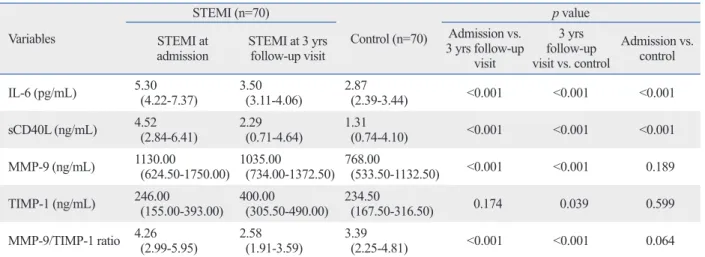
Significant differences in the levels of IL-6, sCD40L, MMP- 2D long-axis view was used to obtain linear measurements
of the LV cavity [LV end-diastolic diameter (LVEDD) and LV end-systolic diameter (LVESD)] and walls. LV mass was estimated using the anatomically validated formula of Devereux. The indices that adjusted LV mass (LVM) was LV mass index (LVMI), defined as LVM/body surface area.
The relative wall thickness was calculated as 2 posterior wall/LVEDD. LV end-systolic volume (LVESV), LV end- diastolic volume (LVEDV), and LV ejection fraction (LVEF) were estimated using the biplanar modified Simp- son’s rule from apical 2 and 4 chamber views. LV diastolic function was evaluated by the following pulse-wave Dop- pler echocardiographic parameters: early (E)/late (A) ratio, indicating the pattern of LV diastolic filling, measured by pulse-wave Doppler flow pattern at mitral anulus traced electronically to measure peak velocities of early and late diastolic LV filling, that is, early (E) and late (A) transmitral peak flow velocities.
Statistical analysis
Data are expressed as means±SD or median and interquar- tile ranges, as appropriate. Unpaired or paired Student’s t- test was used to evaluate differences in normally distributed continuous variables between the two groups. Because of non-normal distribution of inflammatory markers variables, Table 1. Baseline Characteristics of the Study Population
Variables STEMI (n=70) Control (n=70) p value
Male, n (%) 59 (84.3) 60 (85.7) 0.813
Age (yrs) 59.20±11.32 57.89±10.55 0.479
BMI (kg/m2) 25.33±2.70 24.97±3.17 0.473
Waist circumference (cm) 91.62±11.53 88.56±10.17 0.099
Hypertension, n (%) 29 (41.4) 29 (41.4) 1.000
Hypercholesterolemia, n (%) 14 (20.0) 22 (31.4) 0.122
Diabetes mellitus, n (%) 9 (12.9) 9 (12.9) 1.000
Current smokers, n (%) 46 (65.7) 38 (54.3) 0.168
Sedentary lifestyle, n (%) 32 (45.7) 36 (52.2) 0.446
Negative life events occurred in the past 6 months, n (%) 19 (27.1) 18 (25.7) 0.848
Medication -
β blocker use, n (%) 63 (90)
ACE-I use, n (%) 68 (97.1)
Statin use, n (%) 68 (97.1)
Antiplatelet agents, n (%) 70 (100)
Reperfusion therapy, n (%) 56 (80.0) -
Peak CK (u/L) 1514.00 (695.00-3059.00) -
Peak CK-MB (u/L) 121.00 (53.00-210.00) -
Peak TnI (ng/mL) 42.65 (13.18-134.80) -
Killip’s class ≥II, n (%) 15 (21.4) -
BMI, body mass index; ACE-I, angiotensin-converting enzyme inhibitors; CK, creatine kinase; CK-MB, creatine kinase-myocardial fraction; Tn-I, Troponin I;
STEMI, segment elevation myocardial infarction; Age, BMI and waist circumference are presented as mean±standard deviation; Peak CK, CK-MB and TnI are presented as medians (25th-75th percentile); categorical variables are presented as number (%).
p=0.014 and p=0.034, respectively), and LVEF was im- paired (p=0.006), compared with baseline (Table 3).
There was no association between the levels of IL-6, sCD40L, MMP-9, TIMP-1 and echocardiographic markers of LV structure or function at the same time. However, MMP-9 measured at admission showed weak association with great LVEDD and LVEDV (r=0.294, p=0.022 and r=0.269, p=0.036, respectively), and TIMP-1 positively correlated with E/A ratio at follow-up (r=0.278, p=0.044).
DISCUSSION
The main findings of this study are that the levels of serum IL-6, sCD40L and MMP-9 were steadily increased among 9 and TIMP-1 were detected among the three studied
groups, as shown in Table 2. The levels of IL-6 and sCD40L were found to be elevated in acute STEMI as compared to controls (both p<0.001); In patients with remote myocardi- al infarction (at 3-year follow-up visit), the levels of IL-6, sCD40L, MMP-9 and MMP-9/TIMP-1 ratio were signifi- cantly reduced compared admission levels (all p<0.001);
The levels of IL-6, sCD40L, MMP-9, and TIMP-1 were still higher at 3-year follow-up visit in STEMI patients com- pared to controls (p<0.001, p<0.001, p<0.001 and p=0.039, respectively).
Echocardiographic assessment
After 3-years, LVEDD, LVM, LVMI, LVESV and E/A ra- tio were significantly increased (p=0.045, p=0.048, p=0.047, Table 2. Serum Levels of Inflammatory Markers
Variables
STEMI (n=70)
Control (n=70)
p value STEMI at
admission STEMI at 3 yrs follow-up visit
Admission vs.
3 yrs follow-up visit
3 yrs follow-up visit vs. control
Admission vs.
control
IL-6 (pg/mL) 5.30
(4.22-7.37) 3.50
(3.11-4.06) 2.87
(2.39-3.44) <0.001 <0.001 <0.001 sCD40L (ng/mL) 4.52
(2.84-6.41) 2.29
(0.71-4.64) 1.31
(0.74-4.10) <0.001 <0.001 <0.001 MMP-9 (ng/mL) 1130.00
(624.50-1750.00) 1035.00
(734.00-1372.50) 768.00
(533.50-1132.50) <0.001 <0.001 0.189 TIMP-1 (ng/mL) 246.00
(155.00-393.00) 400.00
(305.50-490.00) 234.50
(167.50-316.50) 0.174 0.039 0.599
MMP-9/TIMP-1 ratio 4.26 (2.99-5.95) 2.58
(1.91-3.59) 3.39
(2.25-4.81) <0.001 <0.001 0.064
IL-6, interleukin-6; sCD40L, soluble CD40 ligand; MMP-9, metalloproteinase-9; TIMP-1, tissue inhibitor of metalloproteinase-1; STEMI, segment elevation myocardial infarction.
Data are presented as medians (25th-75th percentile).
Table 3. Changes in Echocardiographic Parameters from Admission to 3 Years Follow-Up Visit in STEMI Patients Variables STEMI (at admission) (n=70) STEMI (at 3 yrs follow-up visit) (n=70) p value
IVSd (cm) 1.02±0.11 0.96±0.16 0.110
LVPWd (cm) 0.98±0.07 0.99±0.12 0.677
LVEDD (cm) 5.15±0.54 5.49±0.69 0.045
LVM (g) 227.38±56.08 261.31±77.32 0.048
LVMI (g/m2) 125.45±30.22 143.54±37.09 0.047
RWT 0.39±0.04 0.36±0.06 0.051
LVEDV (mL) 128.44±32.15 147.83±43.92 0.061
LVESV (mL) 49.54±22.76 64.38±30.78 0.014
LVEF (%) 61.71±10.03 55.61±8.91 0.006
E/A ratio 0.75±0.18 0.96±0.35 0.034
IVSd, interventricular septal thickness in diastole; LVPWd, left ventricular posterior wall thickness in diastole; LVEDD, left ventricular end-diastolic dimen- sion; LVM, left ventricular mass; LVMI, left ventricular mass index; RWT, relative wall thickness; LVEDV, left ventricular end-diastolic volume; LVESV, left ventricular end-systolic volume; LVEF, left ventricular ejection fraction.
E, mitral peak flow velocity of early filling; A, peak flow velocity of late filling.
Data are presented as mean±standard deviation.
remodeling takes place. Previous studes12-15 have reported that patients with ACS exhibited significantly higher levels of MMP-9 and TIMP-1 than those with stable angina and normal control subjects, and the higher levels were related to the presence of plaque rupture in the culprit lesion. In our pervious study,16 we found that acute STEMI patients ex- hibited significantly higher level of MMP-9 and slightly el- evated TIMP-1 as compared to control, therefore, the MMP-9/TIMP-1 ratio was remarkably increased, indicating that an imbalance between MMP-9 and TIMP-1 in the mi- cro-environment of the vulnerability atherosclerotic plaques may be responsible at least in part for plaque disruption eventually leading to occurrence of cardiovascular events.
In the present study, we failed to find any significant differ- ence of the MMP-9 and TIMP-1 levels between acute STE- MI patients and control due to a small sample size. Howev- er, we found that the levels of MMP-9 and the ratio of MMP-9/TIMP-1 were significantly higher in acute STEMI patients than those in remote STEMI, which also add weight to our previous findings.
MMPs and TIMPs participate not only in the process of atherogenesis and atherosclerotic plaque destabilization, they may also influence LV dilation and function impair- ment after myocardial infarction.17-20 In the present study, we found that MMP-9 measured at admission showed a positive association with great LVEDD and LVEDV at 3-year follow-up visit, which add the additional weight to the above-described notion. We also found that TIMP-1 was elevated remarkly in remote myocardial, and that TIMP-1 at admission positively correlated with E/A ratio measured in the subsequent follow-up period. Thus, we speculate that TIMP-1, following the activation of MMP-9, plays a protective role during the phase of LV remodeling and function impairment. Indeed, MMPs inhibition has been postulated as a potential therapeutic intervention in patients with acute myocardial infarction. Beneficial effects of selective MMP inhibition on regional myocardial geom- etry post-myocardial infarction were shown in experiment animal models.21 Miyazaki, et al.22 found that LV remodel- ing might be suppressed in association with MMP-9 sup- pression in acute myocardial infarction patients treated with percutaneous coronary intervention and regular dose or half-dose-combination of renin-angiotensin system inhibi- tors. However, the broad spectrum MMP inhibitor PG- 116800 failed to attenuate adverse LV remodeling after myocardial infarction in prevention of myo cardial infarc- tion early remodeling trial.23 Therefore, selective MMP in- control subjects, remote myocardial infarction and acute
STEMI patients, that TIMP-1 was elevated, and MMP-9/
TIMP-1 ratio decreased markedly in remote STEMI, that MMP-9 measured at admission showed a positive associa- tion with great LVEDD and LVEDV, and that TIMP-1 pos- itively correlated with E/A ratio measured at subsequent follow-up period. The results suggest that elevated levels of IL-6, sCD40L, and MMP-9 and the imbalance of MMP-9/
TIMP-1 system are involved in the process of plaque rup- ture, leading to the incidence of myocardial infarction, and that MMP-9/TIMP-1 may also be linked to the post-infarc- tion remodeling of myocardium, probably leading to heart failure.
Acute coronary syndrome (ACS) is triggered by dys- functional endothelial and atherosclerotic plaque instability.
The stage of plaque instability is that pro-inflammatory cy- tokines and chemoattractants induce leukocyte chemoat- traction to the endothelium, and together with other triggers such as the CD40L-CD40 co-stimulation system activate plaque monocytes (macrophages). The macrophages then produce MMPs that disintegrate extra-cellular plaque ma- trix, causing coronary plaque instability.7 IL-6 is a circulat- ing cytokine known to be secreted from a number of differ- ent cells including activated macrophages and lymphocytes, which could stimulates hepatocytes to synthesize acute phase response proteins such as C-reactive protein and fi- brinogen. The CD40L on T-cells binding to the CD40 re- ceptor on macrophages is involved in the multiple stages of atherosclerosis and ACS, inducing macrophages to secrete tissue factor, up-regulating the expression of adhesion mol- ecules, and activating secretion of MMPs. Previous study8,9 have shown, in agreement with our results, that the concen- tration of IL-6 and sCD40L are significantly elevated from stable CAD to ACS, as compared to control, possibly sup- porting their role in plaque instability and rupture.
MMPs are a tightly regulated group of zinc-dependent peptidases that participate in matrix turnover in both nor- mal and pathological conditions.10 In the present study, we studied one member from the family of gelatinases, such as MMP-9, which has been found highly expressed in the shoulder regions of advanced atherosclerotic lesions and contributes to plaque instability.11 The main endogenous MMPs inhibitors are TIMPs, and TIMP-1 inhibits most MMPs except for MMP-2. Under normal circumstances, the activeity of MMPs and TIMPs maintain a balance be- tween connective tissue synthesis and degradation. When the balance is disturbed, the undesired tissue destruction or
Eur J Clin Invest 2007;37:623-8.
9. Wang J, Zhang S, Jin Y, Qin G, Yu L, Zhang J. Elevated levels of platelet-monocyte aggregates and related circulating biomarkers in patients with acute coronary syndrome. Int J Cardiol 2007;115:
361-5.
10. Lijnen HR. Plasmin and matrix metalloproteinases in vascular re- modeling. Thromb Haemost 2001;86:324-33.
11. Brown DL, Hibbs MS, Kearney M, Loushin C, Isner JM. Identifi- cation of 92-kD gelatinase in human coronary atherosclerotic le- sions. Association of active enzyme synthesis with unstable angi- na. Circulation 1995;91:2125-31.
12. Inokubo Y, Hanada H, Ishizaka H, Fukushi T, Kamada T, Okumu- ra K. Plasma levels of matrix metalloproteinase-9 and tissue in- hibitor of metalloproteinase-1 are increased in the coronary circu- lation in patients with acute coronary syndrome. Am Heart J 2001;141:211-7.
13. Derosa G, D’Angelo A, Scalise F, Avanzini MA, Tinelli C, Peros E, et al. Comparison between metalloproteinases-2 and -9 in healthy subjects, diabetics, and subjects with acute coronary syn- drome. Heart Vessels 2007;22:361-70.
14. Shu J, Ren N, Du JB, Zhang M, Cong HL, Huang TG. Increased levels of interleukin-6 and matrix metalloproteinase-9 are of car- diac origin in acute coronary syndrome. Scand Cardiovasc J 2007;
41:149-54.
15. Fukuda D, Shimada K, Tanaka A, Kusuyama T, Yamashita H, Ehara S, et al. Comparison of levels of serum matrix metallopro- teinase-9 in patients with acute myocardial infarction versus un- stable angina pectoris versus stable angina pectoris. Am J Cardiol 2006;97:175-80.
16. Tan J, Hua Q, Gao J, Fan ZX. Clinical implications of elevated se- rum interleukin-6, soluble CD40 ligand, metalloproteinase-9, and tissue inhibitor of metalloproteinase-1 in patients with acute ST- segment elevation myocardial infarction. Clin Cardiol 2008;31:
413-8.
17. Matsunaga T, Abe N, Kameda K, Hagii J, Fujita N, Onodera H, et al. Circulating level of gelatinase activity predicts ventricular re- modeling in patients with acute myocardial infarction. Int J Cardi- ol 2005;105:203-8.
18. Webb CS, Bonnema DD, Ahmed SH, Leonardi AH, McClure CD, Clark LL, et al. Specific temporal profile of matrix metalloprotein- ase release occurs in patients after myocardial infarction: relation to left ventricular remodeling. Circulation 2006;114:1020-7.
19. Vanhoutte D, Schellings M, Pinto Y, Heymans S. Relevance of matrix metalloproteinases and their inhibitors after myocardial in- farction: a temporal and spatial window. Cardiovasc Res 2006;69:
604-13.
20. Kelly D, Khan SQ, Thompson M, Cockerill G, Ng LL, Samani N, et al. Plasma tissue inhibitor of metalloproteinase-1 and matrix metalloproteinase-9: novel indicators of left ventricular remodel- ling and prognosis after acute myocardial infarction. Eur Heart J 2008;29:2116-24.
21. Apple KA, Yarbrough WM, Mukherjee R, Deschamps AM, Esco- bar PG, Mingoia JT, et al. Selective targeting of matrix metallo- proteinase inhibition in post-infarction myocardial remodeling. J Cardiovasc Pharmacol 2006;47:228-35.
22. Miyazaki S, Kasai T, Miyauchi K, Miyazaki T, Akimoto Y, Takagi A, et al. Changes of matrix metalloproteinase-9 level is associated with left ventricular remodeling following acute myocardial in- farction among patients treated with trandolapril, valsartan or both. Circ J 2010;74:1158-64.
hibition seems to be the better solution.
The limitations of our study should be considered. First, although significant, the associations between MMP-9, TIMP-1 and LV remodeling were relatively weak, which can not be entirely due to relatively small sample size, nev- ertheless, a larger population is needed to confirm our find- ings. Second, we should study the change of the inflamma- tory markers during the period between admission and discharge, and then select the highest levels in the acute state. Ideally, control group should be the patients with sta- ble CAD and healthy subjects, so as to observe the deffer- ent levels of the inflammatory markers, and elucidate the role of inflammation in the different phases of CAD. Third, the development of ventricular remodeling and function impairment after myocardial infarction are strongly affect- ed by the infracted area and drug treatment during follow- up period, and these factors should not be excluded in the study.
In conclusion, the admission levels of serum MMP-9 and TIMP-1 closely correlated with left ventricular structure and function, which may be involved in the process of post- infarction remodeling of myocardium.
REFERENCES
1. Ross R. Atherosclerosis--an inflammatory disease. N Engl J Med 1999;340:115-26.
2. Jugdutt BI. Ventricular remodeling after infarction and the extra- cellular collagen matrix: when is enough enough? Circulation 2003;108:1395-403.
3. Kelly D, Cockerill G, Ng LL, Thompson M, Khan S, Samani NJ, et al. Plasma matrix metalloproteinase-9 and left ventricular re- modelling after acute myocardial infarction in man: a prospective cohort study. Eur Heart J 2007;28:711-8.
4. Kelly D, Khan S, Cockerill G, Ng LL, Thompson M, Samani NJ, et al. Circulating stromelysin-1 (MMP-3): a novel predictor of LV dysfunction, remodelling and all-cause mortality after acute myo- cardial infarction. Eur J Heart Fail 2008;10:133-9.
5. Fisman EZ, Benderly M, Esper RJ, Behar S, Boyko V, Adler Y, et al. Interleukin-6 and the risk of future cardiovascular events in pa- tients with angina pectoris and/or healed myocardial infarction.
Am J Cardiol 2006;98:14-8.
6. Yan JC, Zhu J, Gao L, Wu ZG, Kong XT, Zong RQ, et al. The ef- fect of elevated serum soluble CD40 ligand on the prognostic val- ue in patients with acute coronary syndromes. Clin Chim Acta 2004;343:155-9.
7. Gidron Y, Gilutz H, Berger R, Huleihel M. Molecular and cellular interface between behavior and acute coronary syndromes. Car- diovasc Res 2002;56:15-21.
8. Tousoulis D, Antoniades C, Nikolopoulou A, Koniari K, Vasiliad- ou C, Marinou K, et al. Interaction between cytokines and sCD40L in patients with stable and unstable coronary syndromes.
cardial infarction: results of the PREMIER (Prevention of Myo- cardial Infarction Early Remodeling) trial. J Am Coll Cardiol 2006;48:15-20.
23. Hudson MP, Armstrong PW, Ruzyllo W, Brum J, Cusmano L, Krzeski P, et al. Effects of selective matrix metalloproteinase in- hibitor (PG-116800) to prevent ventricular remodeling after myo-