Clinical Predictors of Incomplete ST-Segment Resolution in the Patients With Acute ST Segment Elevation Myocardial Infarction
7
0
0
전체 글
(2) So Ra Park, et al.·311. angiography after successful PCI.9) Hemodynamic parameters such as coronary flow reserve and index of microcirculatory resistance provide more physiological information concerning microvascular blood flow.10)11) But, while these methods are acceptable for academic assessments, they are impractical for clinical use. Clinically, it is more useful and convenient to monitor STsegment resolution (STR) on electrocardiography (ECG) after successful PCI. Because STR reflects the physiology of cardiac cells, which are the final target of coronary blood flow, it may be a theological physiologic surrogate. Incomplete STR following revascularization therapy is associated with increased mortality and recurrence rate of myocardial infarction (MI), and decreased left ventricular ejection fraction. Incomplete STR also is an independent and poor prognostic factor apart from the restoration of epicardial blood flow following PCI.12)13) In previous studies, the degrees of STR have been estimated in all elevated leads and were expressed as the sum of ST resolution in all leads. However, it is onerous to measure the sum of ST resolution in the clinical practice setting. Fortunately, rapid estimation of maximal ST resolution has no difference from that of sum of ST resolution in all leads.14) Practically, knowledge of the clinical predictors of incomplete recovery of maximal elevation of ST-segment in the early stage would be helpful for screening patients at high risk for microcirculation perfusion failure and for the planning of more aggressive treatment to recover the coronary microcirculation in such patients. Therefore, the aim of the present study was to compare clinical parameters between complete STR and incomplete STR patients that are readily evident in the emergency setting, and to determine clinical predictors in an early clinical setting for incomplete STR.. Subjects and Methods Patients We recruited a consecutive series of 140 STEMI patients who had undergone primary PCI at Gyeongsang National University Hospital between October, 2005 and June, 2007. Acute MI (AMI) was defined as cases in which the typical chest pain was persistently present for more than 30 minutes, elevation of ST-segment >2 mm on the precordial leads and >1 mm on the limb lead on more than two consecutive leads, and the maximal level of the cardiac enzymes creatinin kinase (CK-MB) and troponin I were elevated three times the upper limit of normal. Patients were excluded if they had a previous AMI, >12 hours of symptoms, left bundle branch block, ventricular tachycardia, ventricular fibrillation, paced or ventricular rhythm, negative T waves in fewer than two adjacent leads with maximal ST elevation or an unsatisfactory ECG image. Based on these criteria,. 39 patients were excluded from the analysis. The initial laboratory findings at admission and coronary risk factors were compared between the complete and incomplete STR groups. Systolic and diastolic blood pressure, heart rate at initial admission, pre-infarction angina, door-to-balloon time, pain-to-balloon time, left ventricular ejection fraction on echocardiography during admission, infarct site on ECG, Killip class and ischemic grade of initial ECG were acquired by protocol. Among the clinical factors, “pre-infarction angina” was defined as new onset or worsening of angina within 48 hours before admission. Analysis of electrocardiography findings Twelve-lead ECGs at admission and 15, 30 and 60 minutes after PCI were assessed by the investigators who were blinded to the clinical characteristics and coronary angiography findings of patients. The degrees of ST-elevation were assessed by the maximal ST-segment elevation, which is similar to the sum of STR in all leads.14) Using a caliper, the degree of ST-segment elevation was assessed at the J-point of each lead from the isoelectric line (excluding aVR). STR on the 12-lead ECG at 15, 30 and 60 minutes after PCI was measured at the corresponding lead that was maximally elevated. The previous definition of complete STR as resolution >70% from maximal elevated ST segment15) was used. The ischemia grade of each standard 12-lead ECG at admission were scored by the criteria of Sclarovsky.16) Grade 3 ischemia (G3I) was defined as the absence of a S wave below the TP-PR isoelectric line in ≥2 leads that usually have a terminal S configuration (leads V1V3) or a ST J-point amplitude >50% of the R-wave amplitude measured from the TP-PR baseline in >2 of all other infarct-related leads (Fig. 1). Patients meeting the ST-elevation criteria but not the grade 3 ischemia criteria were classified as having grade 2 ischemia(G2I) (Fig. 2). An assessment of coronary angiography The location and number of lesions with significant extent of stenosis (≥50%) on coronary angiography were compared between complete and incomplete STR. After primary PCI, the blood flow recovery in the infarct-related epicardial artery was assessed by TIMI flow grade.17) Statistical analyses Continuous variables were expressed as mean±standard deviation (SD) and were compared using the unpaired t-test. Categorical variables were expressed as the frequency and were compared using Fisher’s exact or Chi-square tests. To identify the independent factors that could predict incomplete STR, multivariate logistic regression analysis was performed for the following pa-.
(3) 312·Clinical Predictors of Incomplete ST Resolution. Fig. 1. ECGs of a patient with inferior STEMI on admission showing grade 3 ischemia. The J point is above 50% of the R wave amplitude in leads III and aVF. ECG: electrocardiography, STEMI: ST elevation myocardial infarction.. Fig. 2. ECGs of a patient with anterior STEMI on admission showing grade 2 ischemia. A ST-segment elevation in V1-4 is evident. However, the J point is below 50% of the R wave amplitude in all lead and V1 exhibits a S wave. ECG: electrocardiography, STEMI: ST elevation myocardial infarction.. rameters: hypertension, duration of the chest pain, location of myocardial infarction, Killip Class, ischemia grade on ECG at the admission, initial level of troponine I and brain natriuretic peptide (BNP) and TIMI flow grade before PCI. Statistical analysis was performed using Statistical Package for the Social Sciences (SPSS)-PC 12.0 (SPSS, Chicago, IL, USA). A value of p<0.05 was considered statistically significant.. Results Clinical factors Between the two groups, there were no significant differences with respect to age and the male-to-female. ratio. In regard to risk factors of developing coronary artery diseases, hypertension was significantly higher in the incomplete STR group (p<0.04). However, there were no significant differences in numbers of patients with diabetes mellitus, hyperlipidemia and tobacco smokers. No differences were apparent in the numbers of patients with preinfarction angina, mean of systolic and diastolic blood pressure and heart rate at admission. Although the incomplete STR group had a longer tendency in the total duration of chest pain (p=0.08), symptom-to-door time and door-to-balloon time were not different between the two groups. Incomplete STR group showed relatively lower tendency of the left ventricular ejection fraction (p=0.06), higher tendency of Killip class grade (p=0.08) and more frequent anterior.
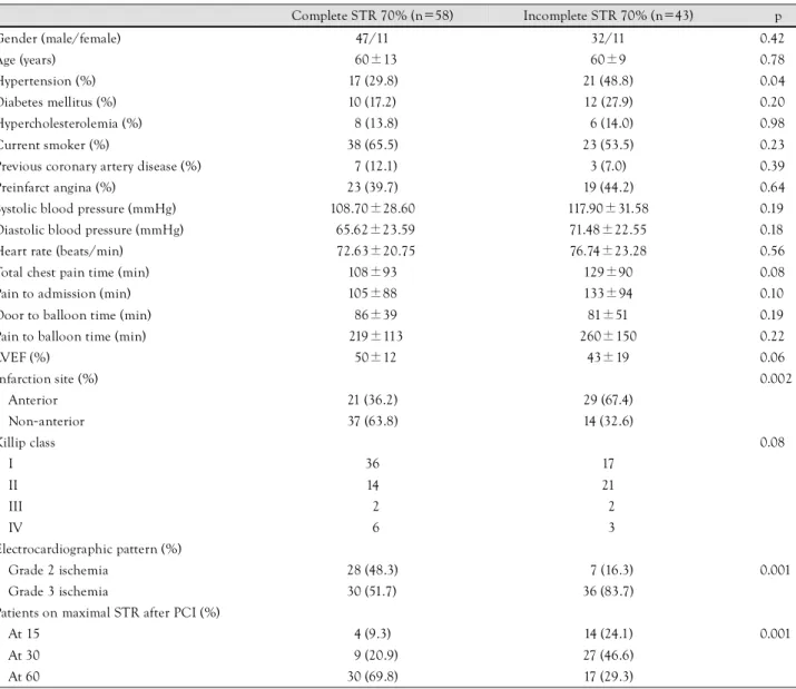
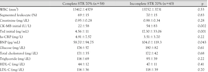
(4) So Ra Park, et al.·313. myocardial infarction (p=0.002). The incomplete STR group also showed more frequent grade 3 ischemia (p=0.001). Resolution time of ST-segment was delayed in the incomplete STR group (p=0.001) (Table 1). Laboratory findings at admission The levels of CK-MB, TnI and BNP were significantly higher in the incomplete STR group (p<0.002, p<0.001 and p<0.043, respectively). However, laboratory markers indicative of the severity of inflammations {white blood cell count (WBC), proportion of segment neutrophils, high sensitivity-C reactive protein (hs-CRP)} did not show a significant difference between the two groups (Table 2). Angiographic findings and clinical outcome TIMI flow grade 3 before PCI was significantly evident in the complete STR group (p=0.029). Successful recovery of TIMI flow grade 3 after PCI was similar in. both groups. The location of culprit lesions was more common in the left anterior descending branch in the incomplete STR group, but more common in the right coronary artery (p=0.002) in the complete STR group. The three deaths that occurred were all in the incomplete STR group (p=0.042) (Table 3). Predictors for incomplete ST-segment resolution TIMI flow grade 2 or less before PCI was the most important independent prognostic factor for incomplete STR. Anterior wall myocardial infarction, grade 3 ischemia on ECG and hypertension were independent predictors for incomplete STR (Table 4).. Discussion Our results indicate that TIMI flow grade 2 or less was the most crucial independent prognostic factor for an incomplete STR as a practical marker representing. Table 1. Clinical characteristics between complete STR and incomplete STR after PCI in patients with acute ST elevation. Gender (male/female) Age (years) Hypertension (%). Complete STR 70% (n=58). Incomplete STR 70% (n=43). p. 47/11. 32/11. 0.420. 60±13. 60±9. 0.780. 17 (29.8). 21 (48.8). 0.040 0.200. Diabetes mellitus (%). 10 (17.2). 12 (27.9). Hypercholesterolemia (%). 08 (13.8). 06 (14.0). 0.980. Current smoker (%). 38 (65.5). 23 (53.5). 0.230. Previous coronary artery disease (%). 07 (12.1). 3 (7.0). 0.390. Preinfarct angina (%). 23 (39.7). 19 (44.2). 0.640. Systolic blood pressure (mmHg). 108.70±28.60. 117.90±31.58. 0.190. Diastolic blood pressure (mmHg). 65.62±23.59. 71.48±22.55. 0.180. Heart rate (beats/min). 72.63±20.75. 76.74±23.28. 0.560. Total chest pain time (min). 108±93. 129±90. 0.080. Pain to admission (min). 105±88. 133±94. 0.100. Door to balloon time (min). 86±39. 81±51. 0.190. Pain to balloon time (min). 219±113. 260±150. 0.220. 50±12. 43±19. LVEF (%) Infarction site (%). 0.060 0.002. Anterior. 21 (36.2). 29 (67.4). Non-anterior. 37 (63.8). 14 (32.6). I. 36. 17. II. 14. 21. Killip class. 0.080. III. 02. 02. IV. 06. 03. Electrocardiographic pattern (%) Grade 2 ischemia. 28 (48.3). 07 (16.3). Grade 3 ischemia. 30 (51.7). 36 (83.7). 0.001. Patients on maximal STR after PCI (%) At 15. 04 (9.3)0. 14 (24.1). At 30. 09 (20.9). 27 (46.6). 0.001. At 60 30 (69.8) 17 (29.3) STR: ST-segment resolution, PCI: percutaneous coronary intervention, LVEF: left ventricular ejection fraction, MI:myocardial infarction.
(5) 314·Clinical Predictors of Incomplete ST Resolution Table 2. Laboratory findings between complete STR and incomplete STR after PCI in patients with STEMI. Complete STR 70% (n=58). Incomplete STR 70% (n=43). p. 13412±4579. 13752±3731. 0.530. 69±15. 70±15. 0.830. Creatinine (mg/dL). 0.95±0.28. 0.98±0.34. 0.280. CK-MB initial (U/L). 22±58. 54±83. 0.002. WBC (mm3) Segmented leukocyte (%). TnI initial (ng/mL). 4.56±11. 17.30±33.06. 0.001. hs-CRP (mg/L). 4.91±7.57. 3.51±3.70. 0.220. 58.70±94.75. 104.0±119.3. 0.043. 174±57. 190±82. 0.610. Total cholesterol (mg/dL). 171±35. 172±42. 0.680. Triglyceride (mg/dL). 114±69. 95±39. 0.220. 44±12. 47±11. 0.410. BNP (pg/mL) Glucose (mg/dL). HDL-C (mg/dL). LDL-C (mg/dL) 114±36 118±39 0.700 All data were expressed as mean±standard deviation. STR: ST-segment resolution, PCI: percutaneous coronary intervention, STEMI: ST elevation myocardial infarction, WBC: white blood count, CK-MB: creatinin kinase-MB, TnI: Troponin I, hs-CRP: high sensitive C-reactive protein, BNP: B-type nitriuretic peptide, HDL-C: high density lipoprotein-cholesterol, LDL-C: low density lipoprotein-cholesterol Table 3. Angiographic parameters and outcome between complete STR and incomplete STR after PCI in patients with STEMI. Complete STR 70% N (%). Incomplete STR <70% N (%). Coronary flow before PCI. p 0.029. TIMI 2 or less. 49 (84.5). 42 (97.7). TIMI 3. 09 (15.5). 01 (2.3)0. TIMI ≤2. 04 (6.9)0. 05 (11.6). TIMI 3. 54 (93.1). 38 (88.4). Coronary flow after PCI. 0.410. No. of diseased vessels 1. 35 (60.3). 26 (60.5). 2. 14 (24.1). 08 (18.6). 3. 09 (15.5). 09 (20.9). Culprit artery. 0.002. Left main. 01 (1.7)0. LAD. 20 (34.5). 27(62.8). RCA. 30 (51.7). 12 (27.9). LCX. 09 (12.1). 02 (4.7)0. 07 (12.1). 10 (23.3). Treatment with tirofiban. 0.840. 02 (4.7)0. 0.130. Death 00 (00.0) 03 (7.0)0 0.042 All data represent mean±standard deviation. STR: ST-segment resolution, STEMI: ST elevation myocardial infarction, TIMI flow: Thrombolysis in Myocardial Infarction flow, TMG grade: Thrombolysis In Myocardial Infarction Perfusion grade, PCI: percutaneous coronary intervention, LAD: left anterior descending artery, RCA: right coronary artery, LCX: left circumflex artery. an incomplete recovery of microvascular flow to cardiac tissue after primary PCI. Anterior wall myocardial infarction showing elevation of ST-segment on the precordial lead, grade 3 ischemia on initial ECG and presence of hypertension were other independent prognostic factors. However, the inflammatory markers assumed to be the crucial for microvascular damages, including WBC counts and hs-CRP showed no difference in incomplete STR group compared to the complete. Table 4. Predictors of incomplete ST segment resolution from multivariate logistic regression analysis. Variable. OR (95% CI). p. Initial TIMI flow grade ≤2. 12.12 (1.23-119.35) 0.032. Anterior MI. 03.93 (1.46-10.57). 0.007. Ischemia grade 3 ECG at admission 03.87 (1.31-11.41). 0.014. Hypertension 03.03 (1.13-8.15) 0.027 OR: odds ratio, CI: confidence Interval, TIMI flow: Thrombolysis In Myocardial Infarction Flow, ECG: electrocardiography. STR group. A previous study reported that 86% of patients showed recovery of TIMI flow grade 3 after pPCI, and old age, lower systolic blood pressure and the presence of blood clots in culprit coronary lesions were independent prognostic factors for recovery of TIMI flow grade 3.18) Our study also showed recovery of TIMI grade 3 in 91% of the patients. However, complete STR in the early stage was achieved only in 57% of total patients. These results confirmed that although PCI was successfully performed, a significant portion of patients with AMI after PCI suffer from inadequate microvascular perfusion to the cardiac tissue in the early stage in incomplete STR. STR has been reported to be a more powerful prognostic factor beyond recovery of epicardial blood flow.19) Therefore, STR in the early stage may be a convenient marker to predict the prognosis of AMI patients after PCI, and must be measured after PCI. Microvascular damage due to the ischemia has been proposed to be classified into two types based on the pathophysiology, time and clinical significance in patients with STEMI.20) One type is the structural microvascular damage present within the necrotized cardiac muscle, which cannot be recovered due to the irreversible damage of microvascular wall. The other type is the functional microvascular damage where the anatomical microvessels are intact, but there is a vascular constric-.
(6) So Ra Park, et al.·315. tion due to endothelial dysfunction or the changes in the sympathetic nervous system, compression of microvessels due to the edema within the tissue and the microvascular obstruction due to platelets or neutrophils. In these cases, recovery of microvascular blood flow could be made by the intensive treatment. Early STR could be an important indicator discriminating between structural and functional microvascular damage. Furthermore, selection of patients judged as incomplete STR by prognostic factors will allow more aggressive treatment and an expected better clinical outcome. TIMI flow before PCI has been significantly correlated with TIMI flow grade 3 and myocardial blush grade 3 after PCI, infarction size, left ventricular ejection fraction prior to the discharge and one-year mortality.21) The same authors reported a significant correlation (p= 0.009) between TIMI flow grade before PCI and recovery of ST-segment.22) Consistent with these studies, we presently confirmed that the maintenance of blood flow prior to the procedure or a spontaneous recovery of ST-segment as powerful independent factors for a recovery of ST-segment. Previous studies have demonstrated that the proportion of cases in which there was a TIMI flow grade 3 prior to PCI was approximately 20%.23)24) Early restoration of coronary blood flow is achieved by the spontaneous dissolution of clots, which can be promoted by drugs as aspirin, other ADP blockers and heparin. Our result also emphasizes that the development of drugs and treatment regimens capable of improving blood flow prior to PCI is most important for reducing microvascular damage in the patients with STEMI. As compared with non-anterior wall MI, the degree of STR was lower in anterior wall MI with precordial ST elevation. Other studies have reported similar results.25)26) To date, little is known about the exact mechanisms. In one study, the degree of correlation between the mortality and incomplete STR in anterior wall MI was found to be lower than that seen in nonanterior wall MI.27) It has been hypothesized that the degree of STR at a precordial lead is, to a lesser extent, due to previous early repolarization or left ventricular hypertrophy. Because patients with anterior wall MI commonly have a greater infarction size than at other sites, it can be inferred that the resulting severe inflammatory responses may contribute to incomplete STR. Presently, the patients with grade 3 ischemia showed agreater size of infarction, higher mortality and higher recurrence rate than those with grade 2 ischemia. “No reflow” phenomenon after PCI is also more frequent in patients with grade 3 ischemia.27)28) This grade of ischemia could be a prognostic factor that predicts incomplete STR and inappropriate reperfusion after PCI.29) Our study confirmed that grade 3 ischemia is also an important prognostic factor for an incomplete STR.. Interestingly, hypertension was also an independent prognostic factor for an incomplete STR. These results support the possibility that microvascular dysfunction is prevalent in patients with hypertension, and that a recovery of ST-segment is measured to a lesser extent in patients with hypertension who had a left ventricular hypertrophy. Left ventricular hypertrophy itself may cause ST elevation. Based on our results, the recovery of epicardial coronary blood flow before PCI is implicated as the most important factor to prevent incomplete STR. Earlier use of anti-platelet agents such as aspirin and platelet IIb/IIIa receptor antagonist must be considered in STEMI patients, especially those at high risk with all predictors for incomplete STR. Adenosine triphosphate (ATP)-sensitive potassium channel openers that act to prevent microvascular constriction can also be useful in this setting. The use of instruments associated with thrombectomy or distal protection during PCI have displayed the more beneficial effect on coronary microvasculature.30) Therefore, these predictors for incomplete STR can be helpful in selecting high risk patients, although PCI itself can produce microvascular damage due to the migration of thrombus or plaque debris. Our study has several limitations. First, because the study was conducted in a single-institution setting, selection bias may influence the results. Secondly, although STR is useful and easy to measure, it can not be a standard and accurate indicator for the microvascular damage unlike invasive hemodynamic parameters. Further studies utilizing hemodynamic factors such as coronary flow reserve and index of microcirculatory resistance or recovery of corresponding wall motion abnormalities as an indicator for microvascular damage are warranted. Studies using these parameters will further our understanding of STR as a indicator of microvascular damage, which has advantage and limitation. REFERENCES 1) Zijlstra F, de Boer MJ, Hoorntje JC, Reiffers S, Reiber JH, Sur-. 2). 3). 4). 5). yapranata H. A comparison of immediate coronary angioplasty with intravenous streptokinase in acute myocardial infarction. N Engl J Med 1993;328:680-4. Keeley EC, Boura JA, Grines CL. Primary angioplasty versus intravenous thrombolytic therapy for acute myocardial infarction: a quantitative review of 23 randomised trials. Lancet 2003; 361:13-20. Boersma E. Does time matter?: a pooled analysis of randomized clinical trials comparing primary percutaneous coronary intervention and in-hospital fibrinolysis in acute myocardial infarction patients. Eur Heart J 2006;27:779-88. Yoon MH, Tahk SJ, Choi SY, et al. Microvascular integrity and ventricular function according to early ST-segment resolution in acute myocardial infarction. Korean Circ J 2003;33:183-95. Choi SY, Tahk SJ, Yoon MH, et al. Comparison of TIMI myocardial perfusion grade with coronary flow reserve for prediction of recovery of LV function and LV remodeling in acute myocardial.
(7) 316·Clinical Predictors of Incomplete ST Resolution infarction. Korean Circ J 2004;34:247-57. 6) Ito H, Okamura A, Iwakura K, et al. Myocardial perfusion pat-. 7). 8) 9). 10). 11). 12). 13). 14). 15). 16). 17) 18). 19). terns related to thrombolysis in myocardial infarction perfusion grades after coronary angioplasty in patients with acute anterior wall myocardial infarction. Circulation 1996;93:1993-9. Kloner RA, Ganote CE, Jennings RB. The ‘no-reflow’ phenomenon after temporary coronary occlusion in dog. J Clin Invest 1974;54:1496-508. Reffelmann T, Kloner RA. The ‘no-reflow’ phenomenon: basic science and clinical correlates. Heart 2002;87:162-8. Gibson CM, Schomig A. Coronary and myocardial angiography: angiographic assessment of both epicardial and myocardial perfusion. Circulation 2004;109:3096-105. Fearon WF, Faroque HM, Balsam LB, et al. Comparison of coronary thermodilution and Doppler velocity for assessing coronary flow reserve. Circulation 2003;108:2198-200. Lim HS, Tahk SJ, Yoon MH, et al. A novel index of microcirculatory resistance for invasively assessing myocardial viability after primary angioplasty for treating acute myocardial infarction. Korean Circ J 2007;37:318-26. Matetzky S, Novikov M, Gruberg L, et al. The significance of persistent ST elevation versus early resolution of ST segment elevation after primary PTCA. J Am Coll Cardiol 1999;34:1932-8. Kobayashi N, Ohmura N, Nakada I, et al. Further ST elevation at reperfusion by direct percutaneous transluminal coronary angioplasty predicts poor recovery of left ventricular systolic function in anterior wall AMI. Am J Cardiol 1997;79:862-6. Schroder R, Wegscheider K, Schroder K, Dissmann R, MeyoSabellek W. Extent of early ST segment elevation resolution: a strong predictor of outcome in patients with acute myocardial infarction and a sensitive measure to compare thrombolytic regimens. J Am Coll Cardiol 1995;26:1657-64. de Lemos JA, Braunwald E. ST segment resolution as a tool for assessing the efficacy of reperfusion therapy. J Am Coll Cardiol 2001;38:1283-94. Sclarovsky S, Mager A, Kusniec J, et al. Electrocardiographic classification of acute myocardial ischemia. Isr J Med Sci 1990; 26:525-31. The TIMI Study Group. The Thrombolysis in Myocardial Infarction (TIMI) trial: phase I findings. N Engl J Med 1985;312:932-6. An SG, Oh JH, Park TI, et al. Risk factors for reflow disturbance phenomenon during percutaneous coronary intervention in patients with acute myocardial infarction. Korean J Med 2008;74: 16-22. Matetzky S, Novikov M, Gruberg L, et al. The significance of. 20). 21). 22). 23). 24). 25). 26). 27). 28). 29). 30). persistent ST elevation versus early resolution of ST segment elevation after primary PTCA. J Am Coll Cardiol 1999;34:1932-8. Galiuto L. Optimal therapeutic strategies in the setting of post-infarct no reflow: the need for a pathogenetic classification. Heart 2004;90:123-5. De Luca G, Ernst N, Zijlstra F, et al. Preprocedural TIMI flow and mortality in patients with acute myocardial infarction treated by primary angioplasty. J Am Coll Cardiol 2004;43:1363-7. De Luca G, Maas AC, Suryapranata H, et al. Prognostic significance of residual cumulative ST-segment deviation after mechanical reperfusion in patients with ST-segment elevation myocardial infarction. Am Heart J 2005;150:1248-54. Stone GW, Brodie BR, Griffin JJ, et al. Prospective, multicenter study of the safety and feasibility of primary stenting in acute myocardial infarction: in-hospital and 30-day results of the PAMI stent pilot trial. J Am Coll Cardiol 1998;31:23-30. Stone GW, Brodie BR, Griffin JJ, et al. Clinical and angiographic follow-up after primary stenting in acute myocardial infarction. Circulation 1999;99:1548-54. McLaughlin MG, Stone GW, Aymong E, et al. Prognostic utility of comparative methods for assessment of ST-segment resolution after primary angioplasty for acute myocardial infarction. J Am Coll Cardiol 2004;44:1215-23. Brodie BR, Stuckey TD, Hansen C, et al. Relation between electrocardiographic ST-segment resolution and early and late outcomes after primary percutaneous coronary intervention for acute myocardial infarction. Am J Cardiol 2005;95:343-8. Birnbaum Y, Herz I, Sclarovsky S, et al. Prognostic significance of the admission electrocardiogram in acute myocardial infarction. J Am Coll Cardiol 1996;27:1128-32. Birnbaum Y, Goodman S, Barr A, et al. Comparison of primary coronary angioplasty versus thrombolysis in patients with STsegment elevation acute myocardial infarction and grade II and grade III myocardial ischemia on the enrollment electrocardiogram. Am J Cardiol 2001;88:842-7. Wolak A, Yaroslavtsev S, Amit G, et al. Grade 3 ischemia on the admission electrocardiogram predicts failure of ST resolution and of adequate flow restoration after primary percutaneous coronary intervention for acute myocardial infarction. Am Heart J 2007;153:410-7. Kunadian B, Dunning J, Vijayalakshmi K, Thornley AR, de Belder MA. Meta-analysis of randomized trials comparing anti-embolic devices with standard PCI for improving myocardial reperfusion in patients with acute myocardial infarction. Catheter Cardiovasc Interv 2007;69:488-96..
(8)
수치

관련 문서
patients who underwent a successful primary percutaneous coronary intervention via the distal radial approach (Snuff- box puncture time was defined as time interval from local
This study aimed to evaluate the effects of percutaneous coronary intervention (PCI) on short- and long-term major adverse cardiac events (MACE) in elderly ( > 75 yr old) acute
It is uncertain that atorvastatin pretreatment can reduce myocardial damage in patients undergoing primary percutaneous coronary intervention (PCI) for ST-segment elevation
Although acute diastolic and systolic dysfunction after AMI may improve with the successful restoration of coronary blood flow by means of a percutaneous coronary intervention, some
► See the article “Effect of Operator Volume on In-Hospital Outcomes Following Primary Percutaneous Coronary Intervention for ST-Elevation Myocardial Infarction: Based on the 2014
Contrast-induced nephropathy (CIN) is a common complication of primary percutaneous coronary intervention (pPCI) and is associated with high mortality and morbidity, long
loon time in patients with suspected STEMI. 3)4) However, the decis- ion to provide immediate reperfusion therapy to patients presenting with chest pain and ST-segment elevation
Our study suggests an association among high plasma le- vels of TnI, NT-proBNP, and hsCRP at admission, incomplete Fig. Prognostic value of a multi-marker approach. The addition
