동맥경화성 관상동맥질환은 선진국에서 성인 사망률을 높이 는 주요한 원인이며, 우리나라에서도 식습관의 서구화와 생활 습관의 변화로 인한 활동량의 감소 등으로 말미암아 이환율 및 사망률이 빠르게 증가하고 있다(1).
죽상동맥경화증은 혈관벽 손상에 대한 일련의 염증반응으 로, 조직학적으로는 혈관벽 내에 지방선조(fatty streak)로 시작해서, 지질 및 괴사조직으로 이루어진 핵(core of lipid and necrotic tissue)과 이를 덮는 섬유성 모자(fibrous cap) 를 갖는 연성 죽상판(soft plaque)을 형성하고, 여기에 석회화 가 일어나면서 병변이 안정화되는 것으로 알려져 있다(2).
최근 급속한 CT 기술의 발달로 새롭게 대두한 관상동맥 CT 혈관촬영술(coronary CT angiography, 이하 CCTA)은 고 식적 혈관조영술에 비해 비침습적 방법으로 관상동맥의 협착 을 진단할 수 있을 뿐 아니라, 죽상판 내 지질이나 석회화와 같 은 조직학적 특성을 알 수 있어 그 유용성이 강조되고 있다(3- 5).
본 연구에서는 CCTA에서 발견된 동맥경화 죽상판의 CT 소견을 분석하여, 혈관 협착 정도에 따른 석회화와 관련된 죽
상판 특성을 분석하였다.
대상과 방법
대상
이 연구는 본원 임상시험심사위원회 승인 하에 시행되었다.
2005년 12월부터 2008년 5월까지 CCTA를 시행한 692명의 환자에서 관상동맥경화증을 보였던 239명(38-88세, 평균 62.1세; 남자 156명, 여자 83명)을 대상으로 후향적 연구를 하였다. CCTA를 촬영한 이유는 흉통(n=115), 흉부 불쾌감 (n=22), 호흡곤란(n=13), 빈맥(n=3), 관상동맥질환 과거 력(n=3), 심전도 이상(n=3), 두통(n=1)이었고, 나머지 79 명은 무증상 환자로 선별검사 목적으로 시행하였다.
관상동맥 CT 혈관촬영술 프로토콜
모든 환자에서 CT를 시행하기 1시간 전에 베타차단제인 propranolol(Pranol, Daewoong, Korea) 20 mg을 경구 투 여하였고, 1시간 후 심박동 수가 계속해서 분당 65회 이상으로 측정되면 20 mg을 추가로 투여한 후 시행하였다. 추가적 투여 후에도 심박동수가 분당 70회 이상으로 높게 측정되거나 부정
관상동맥 CT 혈관촬영술에서 죽상판 특성:
관상 동맥 협착에 미치는 영향
1최연희∙황윤미∙백승연∙김유경
목적: 관상동맥 CT 혈관촬영술에서 발견된 동맥경화 죽상판의 CT 소견을 분석하여, 혈관 협착 정도에 따른 죽상판 특성의 차이를 알아보고자 하였다.
대상과 방법: 2005년 12월부터 2008년 5월까지 관상동맥 CT 혈관촬영술에서 동맥경화성 관상
동맥질환을 보였던 239명에서 죽상판의 위치, 협착 정도(경증, < 75% 면적협착; 중증, >
75%), 죽상판의 유형을 기록하고, 협착 정도에 따른 죽상판 유형 간의 빈도와 협착정도와 석회 수치간의 관계를 분석하였다.
결과: 239명의 환자에서 694개의 죽상판이 발견되었다. 151명의 환자에서(경증 협착 환자의 57.1%; 중증 협착 환자의 87.5%) 다발성 병변을 보였다. 죽상판의 유형은 비석회화형, 혼합 형, 석회화형이 각각 경증 협착 환자에서 26.7%, 22.5%, 50.8%, 중증 협착 환자에서 22.9%, 56.3%, 20.8%였다. 죽상판은 좌전하행동맥 근위부에 가장 자주 생겼다. 죽상판에 의한 혈관 협착 정도와 석회수치는 유의한 상관관계를 보였다.
결론: 죽상판 유형은 중증 협착에서는 혼합형이, 경증 협착에서는 석회화형이 가장 많았고, 중 증 협착을 갖는 환자 대부분이 다발성 병변을 보였다. 석회수치는 중증 협착을 갖는 환자에서 유의하게 높았다.
1이화대학교 의과대학 영상의학과
이 논문은 2008년 12월 22일 접수하여 2009년 3월 2일에 채택되었음.
맥을 보이는 환자에서는 CCTA를 시행하지 않았다. CT 촬영 직전에 nitroglycerin(Nitroglycerin, Myung Moon, Korea) 0.6 mg을 설하 투여하였다.
CT 촬영은 64-절편 다중검출 나선형 전산화단층촬영기 (Somatom Sensation 64, Siemens Medical Solutions, Germany)를 사용하여 영상을 얻었다. 조영제 주입 전에 전향 적 심전도 동기화를 사용하여 120 kV, 40 mAs, 검출기폭조 절(collimation) 30×0.6 mm, 겐트리 회전시간 270 msec, feed 18 mm로 석회화영상을 촬영하였다. CCTA는 후향적 심 전도 동조화를 사용하였고, 120 kV, 750 effective mAs, 검 출기폭조절(collimation) 64×0.6 mm, 겐트리 회전시간 370 msec, pitch 0.24로 촬영하였다. 조영제 주입은 18 게이지 카 테터를 상완 전주와 정맥에 삽입하여 60-80 mL 비이온성 조 영제(iohexol, Omnipaque 300, Nycomed; iopamidol, Iopamiro 370, Bracco)를 초당 4-5 mL 속도로 주입한 후 40-50 mL의 생리식염수를 같은 속도로 주입하였고, 스캔 범 위는 기관분기부(carina)에서 심장이 횡격막과 만나는 부위까 지 포함하였다. 영상재구성은 절편 두께 0.5-0.6 mm로 60- 70% RR 간격에서 영상을 재구성하여 최적의 영상을 선택하 였다.
영상 분석
영상분석은 CT 검사 당시 워크스테이션(workstation) (Leonardo, Siemens Medical Solutions)에서 최대강도투 사(maximum intensity projection, 이하 MIP), 다평면 재 구성(multiplanar reconstruction, 이하 MPR), 곡선 다평 면 재구성(curved MPR) 기법을 이용하여 좌주관상동맥, 좌 전하행동맥, 우측관상동맥, 좌회선동맥에 각각 평행 및 직각 방향으로 영상을 분석하고 판독하였다. 의료영상 저장전송시 스템(Picture Archiving and Communication System, 이 하 PACS) (Infinitt, Korea)으로 영상전송은 병변 부위를 혈 관과 평행 및 수직으로 확대하여 촬영한 영상과 절편 두께 3
mm, 간격 2 mm로 구성한 각각의 관상동맥 혈관의 수직횡단 면 영상을 3차원 volume rendering 영상과 함께 전송하였다.
이번 후향적 연구에서는 PACS에 저장된 영상을 검사 당시 판 독지 결과를 참고로 하여 두 명의 흉부 영상의학과 전문의가 협의로 재분석하였다.
각각의 환자에서 분절 별로 죽상판의 유무, 혈관 협착 정도, 죽상판의 유형을 기록하였다. 죽상판의 위치는 1) 좌주관상동 맥(left main coronary artery, 이하 LM), 2) 좌전하행동맥 의 근위부(proximal left anterior descending artery, 이하 p-LAD), 3) 좌전하행동맥의 중간부, 4) 좌전하행동맥의 원위 부, 5) 좌회선동맥의 근위부(proximal left circumflex artery, 이하 p-LCX), 6) 좌회선동맥의 중간부, 7) 좌회선동 맥의 원위부, 8) 우관상동맥의 근위부(proximal right coronary artery, 이하 p-RCA), 9) 우관상동맥의 중간부, 10) 우관상동맥의 원위부, 11) 기타 분절로 분류하였다.
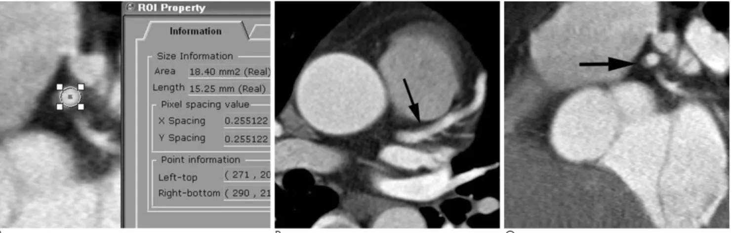
죽상판에 의한 혈관 협착 정도는 면적 협착(area stenosis) 이 혈관 단면적의 75% 미만이면 경증, 75% 이상이면 중증으 로 분류하였다. 면적 협착은 PACS 모니터에서 혈관 협착부와 협착 직상 및 직하부의 혈관강 횡단면적을 관심영역(Region of interest, 이하 ROI)을 이용하여 측정한 후(Fig. 1A), [1- 협착부 혈관강 단면적 × 2 / (협착 직상부 혈관강 단면적 + 협착 직하부 혈관강 단면적)] × 100%로 표시하였다. 협착 직 상 및 직하부의 혈관이 혈관분지 등에 의해 단면적을 정확히 측정하기 어려운 경우에는 둘 중 측정 가능한 부위만을 측정하 여(1- 협착부 혈관강 단면적 / 협착 직상 혹은 직하부 혈관강 단면적) × 100%로 표시하였다.
죽상판의 유형은 석회화 정도에 따라 세 개의 유형으로 분류 하였는데, 1) 석회화가 전혀 없는 비석회화형, 2) 석회화와 비 석회화 부분이 혼재된 혼합형, 3) 완전히 석회화된 석회화형으 로 분류하였다.
각각의 환자에서 가장 심한 협착을 보이는 병변을 대표 병변 으로 하여 경증 및 중증 협착에서 죽상판 유형별 빈도를 비교
A B C
Fig. 1. A 47-year-old man with hypertension who underwent screening coronary CT angiography.
A. Cross-sectional area of vascular lumen was measured by using a region of interest (ROI) on PACS monitor.
B, C. Longitudinal (B) and cross-sectional (C) multiplanar reconstruction image of proximal left anterior descending coronary artery show mild stenosis due to noncalcified plaque (arrows).
하였고, 또 모든 죽상판 병변의 위치, 협착 정도, 죽상판 유형 을 분석하였다.
관상동맥 석회화를 갖는 환자를 두 개의 군: 1) 석회화형 죽 상판 만을 갖는 군; 2) 혼합형 죽상판을 갖거나 비석회화형과 석회화형 죽상판을 함께 갖는 군으로 분류한 후, 각 군에서 경 증과 중증 협착을 보이는 환자들의 평균 석회 수치를 비교하였 다. 여기서 석회화는 130 HU이상으로 정의하였고, 석회수치 를 구하는 방법은 Agatston scoring system을 따랐다. 협착 정 도 와 석 회 수 치 간 의 관 계 는 two-way ANOVA와 Friedman 검정법을 이용하여 분석하였다.
결 과
239명의 환자에서 비석회화형 죽상판 만을 보인 환자가 37 명(Fig. 1), 석회화형 죽상판 만을 보인 환자가 103명, 비석회 화형과 석회화형 죽상판을 함께 갖고 있었던 환자가 21명이었 고, 비석회화형이나 석회화형 죽상판의 유무와 상관없이 혼합 형 죽상판을 갖고 있었던 환자가 78명이었다(Fig. 2). 각각의 환자에서 가장 심한 협착을 보이는 병변을 협착 정도에 따라 분류하면 Table 1과 같다. 151명의 환자에서 2개 이상의 관상 동맥 분절에 죽상판에 의한 협착을 보였는데, 경증, 중증 협착 환자의 각각 57.1%(109/191), 87.5%(42/48)를 차지해서 중 증 협착이 있는 환자일수록 다발성 병변이 있음을 알 수 있었 다(Fig. 2).
환자가 갖는 죽상판 중 가장 심한 협착을 보이는 죽상판을 협착 정도와 죽상판 유형별로 분류했을 때, 비석회화형, 혼합 형, 석회화형의 빈도는 경증 협착을 보이는 환자에서는 각각 51명(26.7%), 43명(22.5%), 97명(50.8%), 중증 협착을 보 이는 환자에서는 11명(22.9%)(Fig. 3), 27명(56.3%), 10명
(20.8%)으로, 경증 협착 환자일수록 석회화형이나 비석회화 형 죽상판의 빈도가 높았고, 중증 협착 환자에서는 혼합형 죽 상판의 빈도가 가장 높았다(Fig. 4).
관상동맥 분절 별로 보면, 죽상판은 총 239명의 환자의 694 개 관상동맥분절에서 발견되었다. 각각의 죽상판의 위치 및 유 형은 Table 2와 같다. 비석회화형, 혼합형, 석회화형 죽상판이 각각 91예(13.1%), 150예(21.6%), 453예(65.3%)였고, 경 증 및 중증 협착이 각각 597예(86.0%), 97예(14.0%)였다.
죽상판의 가장 흔한 위치는 p-LAD(27.7%)였고(Fig. 1), 다 음으로 p-RCA(13.4%) (Fig. 2, 3), p-LCX(11.0%)에 잘 발생하였다.
협착 정도와 석회 수치간의 관계를 보면, 석회화형 죽상판만
Table 1. The Distribution of Each Plaque Type According to the Degree of Stenosis
Degree of Stenosis*
Plaque Type
Mild Severe Total Noncalcified 33 (5)� 4 (2) 37 (7)0
Mixed 48 (31) 30 (26) 78 (57)
Calcified 95 (58) 8 (8) 103 (66)0
Noncalcified and Calcified 15 (15) 6 (6) 21 (21)
Total 191 (109) 48 (42) 239 (151)
Note.─ Noncalcified = patients who had only noncalcified plaques, Mixed = patients who had mixed plaques with or with- out other type plaques, Calcified = patients who had only calci- fied plaques, Noncalcified and Calcified = patients who had non- calcified and calcified plaques.
* Degree of stenosis represents the most severe stenosis in a pa- tient with multiple segment stenosis.
�Figures in parentheses are numbers of patients with multiple segment stenosis.
A B
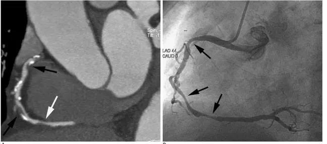
Fig. 2. A 69-year-old man with chest pain and family history of coronary artery disease.
A. Multiplanar reconstruction image of the right coronary artery shows multiple severe stenosis due to mixed (black arrows) or noncalcified plaque (white arrow).
B. Coronary angiography reveals multiple severe stenoses through the right coronary artery (arrows), corresponding to the stenoses on CT angiography.
을 갖는 환자군에서 경증 및 중증 협착을 보이는 환자들의 평 균 석 회 수 치 는 각 각 322.9+315.9(mean+SD), 2582+1025.1로, 협착 정도가 높은 환자일수록 유의하게 높 은 수치를 보였다(p-value < 0.0001). 혼합형 죽상판을 갖거 나 비석회화형과 석회화형 죽상판을 함께 갖는 환자군에서도 경증 및 중증 협착을 보이는 환자들의 평균 석회 수치는 각각 142.1+122.4, 459.9+498.4로 중증 협착 환자에서 유의하게 높았다(p-value < 0.0001) (Fig. 5).
고 찰
죽상동맥경화증의 병태생리가 아직 명확히 밝혀진 것은 아 니지만, 현재로는 혈관손상에 대한 일련의 염증반응으로 설명 되고 있다. 흡연, 고혈압, 당뇨병, 저밀도 지단백의 증가와 같 은 여러 가지 원인으로 혈관에 손상이 일어나면 내피세포에 기 능장애가 일어나면서 염증반응이 일어나게 되는데, 초기에는 혈중 지단백이 혈관 내피세포에 침착하면서 지방 선조를 만들 고, 염증반응이 진행되면서 평활근의 이주와 증식으로 죽상판
의 외벽을 형성하면서 지질과 괴사조직으로 이루어진 핵과 이 를 덮는 섬유성 모자로 구성된 연성 죽상판을 형성한다. 죽상 판이 점차 진행되면서 평활근 세포에 의한 골기질 형성으로 석 회화가 이루어지게 되는데, 이는 진행의 마지막 단계로 지질이 풍부해 불안정했던 부위에 혈전 형성에 의한 상처치유 반응으 로 석회화되면서 죽상판은 안정성 만성병변이 된다(2). 본 연 구에서 비석회화형과 혼합형 죽상판의 빈도를 비교하였을 때 경증 협착을 보이는 환자에서는 비석회화형 죽상판이 높게 나 타나고, 중증 협착 환자에서는 혼합형 죽상판이 높게 나타나는 것은 이러한 죽상판 진행에 따른 석회화 증가를 반영한다고 할 수 있겠다.
저자들의 연구에서 경증 협착을 갖는 환자들에서 석회화형 죽상판의 빈도가 다른 유형에 비해 가장 높게 나타났는데, 여 기에 속한 대부분의 환자는 크기가 작은 석회화형 죽상판을 갖 고 있어 아마도 죽상판의 크기가 중증 협착을 일으킬 정도로 커지기 전에 석회화를 통하여 안정화된 것으로 생각된다. 또 중증 협착 환자의 8.3%는 비석회화형 죽상판에 의한 협착이 었다는 점을 고려할 때, 죽상판 내 석회화가 일어나는 시점이
Table 2. The Location and Plaque Type in Patients with Coronary Artery Disease on Coronary CT Angiography Coronary Artery Segments
LM pLAD mLAD dLAD pLCX mLCX dLCX pRCA mRCA dRCA Others Total
Plaques type
Noncalcified 08 040 05 03 08 03 00 08 06 04 06 091
Mixed 07 054 08 02 14 04 02 21 11 17 10 150
Calcified 38 098 37 13 54 29 09 64 44 38 29 453
Total 53 192 50 18 76 36 11 93 61 59 45 694
Note.─ LM = left main coronary artery, pLAD = proximal segment of left anterior descending artery, mLAD = middle segment of left anterior descending artery, dLAD = distal segment of left anterior descending artery, pLCX = proximal segment of left circumflex artery, mLCX = middle segment of left circumflex artery, dLCX = distal segment of left circumflex artery, pRCA = proximal segment of right coronary artery, mRCA = middle segment of right coronary artery, dRCA = distal segment of right coronary artery, Others = Other seg- ment
A B
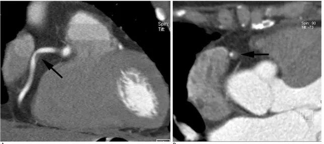
Fig. 3. A 63-year-old woman with hypertension and diabetes mellitus who underwent coronary CT angiography for screening.
A, B. Longitudinal (A) and cross-sectional (B) multiplanar reconstruction image of the middle right coronary artery show severe stenosis due to noncalcified plaque (arrows).
꼭 병변의 크기와 관계가 있는 것은 아닐 것으로 생각된다.
중증 협착에서는 병변의 진행에 따른 석회화 증가를 고려할 때 석회화형 죽상판의 빈도가 가장 높을 것으로 예상함에도 불 구하고, 본 연구에서는 오히려 석회화형 죽상판의 빈도가 20.8%로 다른 유형에 비해 가장 낮고 혼합형의 빈도가 56.3%로 가장 높게 나타났다. Feuchtner 등(6)의 관상동맥 질환이 의심되는 다양한 임상증상을 보이는 환자들(무증상, 비 전형적 흉통, 안정성 및 불안정성 협심증, 급성관상동맥증후 군)을 대상으로 한 CCTA 연구에서도 77.5%가 중증 협착인 전체 죽상판에서 비석회화형이 24%, 석회화형이 31%, 혼합 형이 45%으로 나타나 중증 협착에서 혼합형의 빈도가 가장 높고, 석회화형의 빈도는 낮은 저자들의 연구와 유사한 결과를 보였다.
이러한 분포를 보이는 이유로는 대상 환자의 구성을 생각해 볼 수 있다. 안정성 협심증(stable angina pectoris) 환자가 불안정성 협심증이나 급성관상동맥 증후군 환자들에 비해 죽 상판 내 석회화 양이 많고(7, 8) 주로 만성 석회화형 죽상판에 의한다는 점 등을 고려할 때(6), 연구 대상에 안정성 협심증 환자들이 많이 포함될수록 석회화형 죽상판의 빈도도 늘어날 것으로 예상한다. 또 반대로 저자와 Feuchtner 등(6)의 연구 결과를 고려할 때 중증 협착성 관상동맥질환을 갖는 전체 환자 에서 만성 석회화 병변에 의한 안정성 협심증이 차지하는 비율 이 높지 않고, 중증 협착을 일으킬 정도의 큰 혼합형이나 비석 회화형 죽상판이 완전히 석회화되기 까지는 매우 오랜 기간이 필요하다는 것을 의미할 수 도 있다. 또 다른 이유로는 죽상판 의 석회화가 진행됨에 따라 죽상판 크기가 줄어들어 협착의 정 도가 감소하였을 가능성도 생각해 볼 수 있다. 죽상판을 형성 하는 과정에서 초기에는 보상적인 혈관 확장(positive remodeling)이 일어나 죽상판에 의한 혈관 면적 협착이 약
40% 정도에 이를 때까지도 혈관 내경이 감소되지 않다가, 후 기로 가면 동맥이 더 이상 확장에 의해 보상하지 못하고 죽상 판이 내강 내로 함입되어 혈관이 좁아지는 것으로 알려져 있다 (9). 그러나 죽상판이 만성 석회화 병변으로 진행되면서 혈관 내경에 어떤 변화가 오는지는 아직 알려진 바가 없고, 이를 증 명하려면 혼합형 혹은 비석회화 죽상판을 갖는 환자들에서 CCTA 혹은 혈관초음파(intravascular ultrasound)를 이용 한 오랜 기간의 추적 검사가 필요할 것으로 보인다.
Choi 등(10)은 관상동맥질환 위험인자를 갖는 무증상 환자 의 4%에서, Hausleiter 등(11)은 비전형적 증상을 보이는 관 상동맥 질환이 의심되는 환자의 6.2%에서 비석회화형 죽상판 이 있었다고 보고하였다. 본 연구에서도 CCTA를 시행한 주로 무증상 고위험군 혹은 비전형적 증상을 보인 환자의 5.3%에 서 비석회화형 죽상판을 가지고 있었고, 이 중 소수에서는 중 증 협착을 보였다. 비석회화형 죽상판은 그 크기와 관계없이 파열과 혈전 형성에 의해 급성관상동맥증후군을 일으킬 수 있 으므로 경증 협착을 갖는 무증상 환자일 지라도 그 진단이 중 요하다(6). 이러한 점들을 고려할 때 현재 사용되고 있는 관상 동맥 석회수치는 선별 검사로서 제한점이 있고, 앞으로 CCTA 가 선별 검사로 유용할 것으로 생각한다.
관상동맥 석회수치는 그 환자가 가진 죽상판 전체 양과 밀접 한 상관관계를 보이고, 관상동맥 협착의 정도와도 관련이 있는 것으로 알려져 있다(12-14). 본 연구에서도 석회화형군 및 혼 합형군 모두에서 석회수치는 경증 협착보다 중증 협착에서 유 의한 차이를 보이며 증가하는 것을 볼 수 있었다. 그러나 주로 석회화형 죽상판을 갖는 환자는 경증 혈관 협착이 있더라도 높 은 석회수치를 보일 수 있고, 반대로 주로 비석회화형 혹은 혼 합형 죽상판을 갖는 환자는 중증 협착이 있더라도 상대적으로 낮은 석회수치를 보일 수 있으므로, 모든 환자에서 석회수치가 꼭 죽상판 양이나 혈관 협착과 비례하는 것은 아니다.
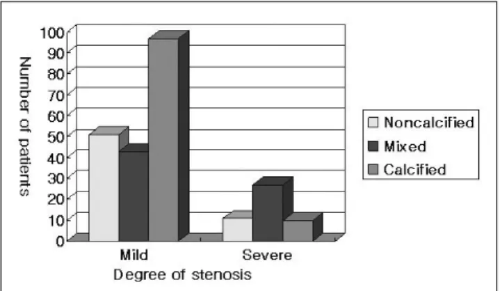
Fig. 4. The distribution of each plaque type according to the stenosis degree. In patients with mild degree stenosis, noncal- cified or calcified plaques are much more common than mixed plaque, but in patients with severe stenosis mixed plaque is the most common type.
Noncalcified = patients with a maximum stenosis due to non- calcified plaque, Mixed = patients with a maximum stenosis due to mixed plaque, Calcified = patients with a maximum stenosis due to calcified plaque
Fig. 5. The relationship between calcium score and degree of coronary artery stenosis in patients with atherosclerotic coro- nary disease. The mean calcium score in each group of steno- sis shows significant correlation with degree of stenosis in both calcified and mixed group (p < 0.0001).
Mixed = the group of patients with mixed plaques or with noncalcified and calcified plaques, Calcified = the group of pa- tients who had only calcified plaques
본 연구의 제한점은 첫째, 본 연구의 대상이 전형적인 증상 을 보이는 관상동맥 질환 환자가 아니고 주로 CCTA의 적응증 인 무증상 고위험군 환자 및 비전형적 흉통을 보이는 환자들이 어서 협착 정도나 죽상판의 유형별 분포가 전형적인 증상을 보 이는 관상동맥 질환 환자들과는 차이가 있을 수 있다. 둘째, CCTA에서 석회화형 죽상판에 의한 협착은 과대평가될 수 있 으므로(15) 본 연구 결과에서 중증 협착으로 분류된 석회화 죽 상판의 일부는 경증 협착일 가능성이 있다. 그러나 석회화형 죽상판의 빈도가 경증협착에서 가장 높고, 중증협착에서 가장 낮다는 저자들의 연구결과에는 변화가 없을 것으로 생각된다.
결론적으로, CCTA를 시행한 환자들에서 경증 협착에서는 석회화형 죽상판의 빈도가 가장 높았고, 비석회화형이 혼합형 에 비해 많았으며, 중증 협착에서는 혼합형 죽상판의 빈도가 가장 높았다. 중증 협착을 갖는 환자 대부분이 다발성 병변을 보였고, 석회수치는 같은 협착 정도에서는 혼합형에 비해 석회 화형 죽상판 환자들에서 높았지만, 혼합형 및 석회화형 모두에 서 경증 협착에 비해 중증 협착에서 높은 수치를 보였다.
참 고 문 헌
1. Beaglehole R. Global cardiovascular disease prevention: time to get serious. Lancet 2001;358:661-663
2. Falk E, Shah P, Fuster V. Coronary plaque disruption. Circulation 1995;92:657-671
3. Achenbach S, Moselewski F, Ropers D, Ferencik M, Hoffmann U, MacNeill B, et al. Detection of calcified and noncalcified coronary atherosclerotic plaque by contrast-enhanced, submillimeter multi- detector spiral computed tomography: a segment-based compari- son with intravascular ultrasound. Circulation 2004;109:14-17 4. Leber A, Knez A, Becker A, Becker C, von Ziegler F, Nikolaou K,
et al. Accuracy of multidetector spiral computed tomography in identifying and differentiating the composition of coronary athero- sclerotic plaques: a comparative study with intracoronary ultra- sound. J Am Coll Cardiol 2004;43:1241-1247
5. Myerburg RJ, Interian A Jr, Mitrani RM, Kessler KM, Castellanos A. Frequency of sudden cardiac death and profiles of risk. Am J Cardiol 1997;80:10F-19F
6. Feuchtner G, Postel T, Weidinger F, Frick M, Alber H, Dichtl W, et al. Is there a relation between non-calcifying coronary plaques and acute coronary syndromes? A retrospective study using multi- slice computed tomography. Cardiology 2008;110:241-248 7. Motoyama S, Kondo T, Sarai M, Sugiura A, Harigaya H, Sato T et
al. Multislice computed tomographic characteristics of coronary le- sions in acute coronary syndromes. J Am Coll Cardiol 2007;50:319- 326
8. Shemesh J, Stroh C, Tenenbaum A, Hod H, Boyko V, Fisman EZ.
Comparison of coronary calcium in stable angina pectoris and in first acute myocardial infarction utilizing double helical computer- ized tomography. Am J Cardiol 1998;81:271-275
9. Glagov S, Weisenberg E, Zarins CK, Stankunavicius R, Kolettis GJ.
Compensatory enlargement of human atherosclerotic coronary ar- teries. N Engl J Med 1987;316:1371-1375
10. Choi EK, Choi SI, Rivera JJ, Nasir K, Chang SA, Chun EJ, et al.
Coronary computed tomography angiography as a screening tool for the detection of occult coronary artery disease in asymptomatic individuals. J Am Coll Cardiol 2008;52:357-365
11. Hausleiter J, Meyer T, Hadamitzky M, Kastrati A, Martinoff S, Scho¨mig A. Prevalence of noncalcified coronary plaques by 64- slice computed tomography in patients with an intermediate risk for significant coronary artery disease. J Am Coll Cardiol 2006;48:
312-318
12. Rumberger J, Simons D, Fitzpatrick L, Sheedy P, Schwartz R.
Coronary artery calcium area by electron-beam computed tomog- raphy and coronary atherosclerotic plaque area. A histopathologic correlative study. Circulation 1995;92:2157-2162
13. Sangiorgi G, Rumberger J, Severson A, Edwards WD, Gregoire J, Fitzpatrick LA, et al. Arterial calcification and not lumen stenosis is highly correlated with atherosclerotic plaque burden in humans: a histologic study of 723 coronary artery segments using nondecalci- fying methodology. J Am Coll Cardiol 1998;31:126-133
14. Detrano R, Hsiai T, Wang S, Puentes G, Fallavollita J, Shields P, et al. Prognostic value of coronary calcification and angiographic stenoses in patients undergoing coronary angiography. J Am Coll Cardiol 1996;27:285-290
15. Zhang S, Levin DC, Halpern EJ, Fischman D, Savage M, Walinsky P. Accuracy of MDCT in assesing the degree of stenosis caused by calcified coronary artery palques. AJR Am J Roentgenol 2008;191:
1676-1683
J Korean Soc Radiol 2009;60:411-417
Address reprint requests to : Yookyung Kim, M.D., Department of Radiology, Ewha Womans University Mokdong Hospital 911-1, Mokdong, Yancheon-gu, Seoul 158-710, Korea.
Tel. 82-2-2650-5380 Fax. 82-2-2650-5302 E-mail: [email protected]
Plaque Characteristics on Coronary CT Angiography:
Effects on the Degree of Coronary Artery Stenosis
1Yeon Hee Choi, M.D., Yunmi Hwang, M.D., Seung Yon Baek, M.D., Yookyung Kim, M.D.
1Department of Radiology, School of Medicine, Ewha Womans University
Purpose: We evaluated the CT characteristics of atherosclerotic coronary plaques in relation to the degree of stenosis.
Materials and Methods: We analyzed coronary CT angiography of 239 patients showing plaques obtained from December 2005 to May 2008, in terms of degree of stenosis (mild < 75% area stenosis; severe >75%), location of plaque, plaque types, and calcium score.
Results: Total 694 segments were found to have plaque in 239 patients. Multiple lesions were found in 151 pa- tients (57.1% of patients with mild stenosis; 87.5% with severe stenosis). The type of plaque was classified into noncalcified, mixed, and calcified in 26.7%, 22.5%, 50.8% of patients with mild stenosis and 22.9%, 56.3%, 20.8% with severe stenosis, respectively. The most common plaque location was the proximal left anterior de- scending artery. In patients with calcified or mixed plaques, there was a significant correlation between degree of stenosis and calcium score.
Conclusion: Mixed plaque was the most common type in severe stenosis, while calcified plaque was the most common in mild stenosis. Multiple lesions were found in most patients with severe stenosis. The calcium score was significantly higher in patients with severe stenosis.
Index words :Coronary artery diseases Tomography, X-ray computed