D I A B E T E S & M E T A B O L I S M J O U R N A L
This is an Open Access article distributed under the terms of the Creative Commons At- tribution Non-Commercial License (http://creativecommons.org/licenses/by-nc/3.0/) which permits unrestricted non-commercial use, distribution, and reproduction in any medium, provided the original work is properly cited.
Diagnostic Accuracy of 64-Slice MDCT Coronary Angiography for the Assessment of Coronary Artery Disease in Korean Patients with Type 2 Diabetes
Jun Sung Moon1, Ji Sung Yoon1, Kyu Chang Won1, Ihn-Ho Cho2, Hyoung Woo Lee1
Departements of 1Internal Medicine, 2Nuclear Medicine, Yeungnam University College of Medicine, Daegu, Korea
Background: A 64-slice multidetector computed tomography (MDCT) is well known to be a useful noninvasive form of angiog- raphy for the general population, but not for certain patients with diabetes. The aim of this study was to investigate the diagnostic accuracy and usefulness of 64-slice MDCT coronary angiography for detecting coronary artery disease in Korean patients with type 2 diabetes mellitus (T2DM).
Methods: A total of 240 patients were included, 74 of whom had type 2 diabetes (M:F=40:33; 41.8±9.5 years). We compared sig- nificant coronary stenosis (>50% luminal narrowing) in MDCT with invasive coronary angiography (ICA) by segment, artery, and patient. We also evaluated the influence of obesity and coronary calcium score on MDCT accuracy.
Results: Of the 4,064 coronary segments studied, 4,062 segments (T2DM=1,109) were assessed quantitatively by both MDCT and ICA, and 706 segments (T2DM=226) were detected as a significant lesion by ICA in all patients. Sensitivity, specificity, as well as positive and negative predictive values for the presence of significant stenosis in T2DM were: by segment, 89.4%, 96.4%, 85.8%, and 97.4%, respectively; by artery (n=222), 95.1%, 92.9%, 94.4%, and 93.8%, respectively; by patients (n=74), 98.4%, 100.0%, 98.4%, and 90.0%, respectively. Regardless of presence of diabetes, there was no significant difference in diagnostic accu- racy. Obesity (≥25 kg/m2) and coronary calcium score did not also affect the diagnostic accuracy of MDCT.
Conclusion: The 64-slice MDCT coronary angiography was found to have similar diagnostic accuracy with ICA, regardless of diabetes. These results suggest MDCT may be helpful to reduce unnecessary invasive studies for patients with diabetes.
Keywords: Accuracy; Coronary artery disease; Diabetes mellitus, type 2; Multidetector computed tomography
Corresponding author: Hyoung Woo Lee
Department of Internal Medicine, Yeungnam University College of Medicine, 170 Hyeonchung-ro, Nam-gu, Daegu 705-717, Korea
E-mail: [email protected]
INTRODUCTION
In Korea, the prevalence of diabetes and its complications is rapidly increasing, thereby leading to a significant rise in health care expenditures [1]. It is well known that cardiovas- cular disease is the most common cause of death in type 2 dia- betes patients. Moreover, about 79% of patients with diabetes die of cardiac complications after a myocardial infarction [2,3]. Cardiovascular disease (30.6%) is the most common cause of mortality in patients with type 2 diabetes in Korea [1].
The cost of treating coronary artery disease (CAD) is reported
to be US $14,680 per patient, and holds the rank of second highest in economic burden for diabetic complications [4].
Compared to the nondiabetic population, diabetic patients have distinguished characteristics of CAD: higher prevalence [5], more extensive, diffuse, calcified coronary lesions, in- creased rates of left ventricular dysfunction, and silent isch- emia [6]. Although invasive coronary angiography (ICA) is known as the ‘gold standard’ for detection of CAD, it is not ap- plicable in every CAD candidate due to its several limitations such as expense, invasive nature and complications. So several noninvasive diagnostic tools were suggested for substituting http://dx.doi.org/10.4093/dmj.2013.37.1.54
pISSN 2233-6079 · eISSN 2233-6087
ICA; however, which tool should replace invasive angiography still remains controversial.
A 64-slice multidetector computed tomography (MDCT) is the current technique of choice for noninvasive angiography because it is simple, fast, and reproducible [6]. Several obser- vational studies [7,8] in the general population have shown that MDCT is a safe and feasible modality for evaluating acute chest pain. Moreover, in low-risk chest pain patients, MDCT is at least as good as a nuclear stress imaging study [9]. How- ever, few studies have reported the diagnostic accuracy of MDCT in patients with diabetes compared to the nondiabetic population, especially among Asians such as Koreans who have distinctive features different from western patients [10].
The aim of this study is to evaluate the usefulness of MDCT coronary angiography by comparing it with conventional cor- onary angiography in Korean patients with type 2 diabetes.
METHODS
Study population
From September 2007 to May 2008, MDCT was performed within 30 days of catheterization in 240 patients (147 male) at Yeungnam University Hospital who were scheduled to have conventional coronary angiography for evaluation of chest pain. Seventy-four patients had type 2 diabetes (40 male), and the other 166 were nondiabetes as the control group (107 male).
Criteria for the diagnosis of diabetes, recommended by the American Diabetes Association [11], are as follows: 1) symp- toms of diabetes plus spontaneous plasma glucose concentra- tion ≥200 mg/dL (11.1 mmol/L), or 2) fasting plasma glucose level ≥126 mg/dL (7.0 mmol/L). The control group was de- fined as nondiabetic patients who have taken previously oral hypoglycemic agents or insulin injection, or individuals that did not meet the diagnostic criteria for diabetes. There were 15 patients (8 type 2 diabetes) who had coronary stents placed previously. Exclusion criteria consisted of atrial fibrillation, renal insufficiency (serum creatinine ≥120 mmol/L), iodine contrast allergy, severe claustrophobia, and pregnancy. All pa- tients provided informed consent, and the study protocol was approved by our local ethics committee, as well as the Institu- tional Review Board of Yeungnam University Hospital.
CT protocols
The following scanning parameters were used for 64-slice MDCT (DVCT; GE Healthcare, Milwaukee, WI, USA): elec-
trocardiogram-triggered X-ray tube modulation, 64-channel detectors along the z-axis, a tube voltage of 120 kV, a tube cur- rent of 350 to 650 mA (depending on patient size), a scan field view of 25 cm, a gantry rotation of 0.35 seconds per rotation, a matrix of 512×512, a slice width of 0.625 mm, and a helical pitch of 0.16. A single oral dose of 25 to 50 mg metoprolol tar- trate (Betaloc®; Yuhan Corp., Seoul, Korea) was administrated 1 hour before CT angiography (CTA) if the patient’s heart rate was >65 beats per minute. Prior to scanning, patients fasted, were placed in a supine position, and received 1.25 to 2.5 mg of sublingual isosorbid dinitrate so long as they were asymptom- atic (Isoket®, Spray; Schwarz Pharma, Munchenstein, Switzer- land). After 2 localization scans, a low-dose native scan of the heart was performed for coronary calcium detection and scor- ing. A bolus of 80 mL of iodixanol (Visisense®, 320 mgI/mL;
Taejoon Pharma., Seoul, Korea) was injected into an antecubi- tal vein via an 18-gauge catheter at a flow rate of 4 mL/sec fol- lowed by 50 mL saline solution. Bolus tracking was performed with a region of interest placed into the ascending aorta.
Coronary artery calcium scoring
Coronary artery calcium scoring was performed using gated noncontrast images acquired prior to CTA. Images were ana- lyzed using GE Advantage Workstation 4.4 (Advantage Work- station 4.4; GE Healthcare, Milwaukee, WI, USA) with Smart- score software. Using the Agatston method, calcium detection and quantification was performed by an electrocardiographi- cally triggered, sequential step-and-shoot acquisition mode, with 120 kV tube voltage, 430 mA tube current, and 2.5 mm slice thickness.
MDCT data analysis
Stenosis evaluation was done using a modified American Col- lege of Cardiology/American Heart Association(ACC/AHA) segmentation model [12]. All CTA images were evaluated on a 3D image analysis workstation (Advantage Workstation 4.4).
Images were analyzed by 2 experienced observers blinded to the catheterization results. First, each segment was evaluated for whether it was interpretable or not. We defined significant CAD as vessels with ≥50% reduction in lumen diameter. In the case of multiple lesions in a given segment, the segment was classified according to the worst segment.
ICA
ICA was performed using standard techniques. Vascular ac-
cess was obtained using a femoral approach with the Seldinger technique. Coronary angiograms were visually evaluated by 2 experienced observers blinded to the MDCT data. Similar to MDCT, a significant lesion was defined as a ≥50% luminal narrowing.
Statistical analysis
To evaluate the diagnostic accuracy of MDCT, we defined
“positive” if there was the discrepancy between MDCT and ICA for results from the same segment. If either observer be- tween two tests was not able to assess the segment, the case was also deemed “positive” for a discrepancy. Sensitivity, spec- ificity, positive predictive values (PPV), and negative predic- tive values (NPV) for MDCT were calculated based on ICA as the “gold standard.” Statistical analysis was done using SPSS version 13.0 for windows (SPSS Inc., Chicago, IL, USA). Re- sults were expressed as a mean value±standard deviation for continuous variables. Qualitative data are presented in num- bers (%). To determine the significant differences between MDCT and ICA, a McNemar test was used and a P value of less than 0.05 was considered to be statistically significant.
Agreement for significant stenosis was expressed by a kappa index(k). A k value of 0 represents poor agreement, 0.01 to 0.20 represents slight agreement, 0.21 to 0.40 is fair agreement, 0.41 to 0.60 moderate agreement, 0.61 to 0.80 good agreement, and >0.8 excellent agreement.
RESULTS
Patient characteristics
Patient characteristics are summarized in Table 1. Patients with type 2 diabetes had a significantly higher coronary calcium score (type 2 diabetes vs. control: mean 658.6 vs. 426.4 Agatston score), creatinine (114.9±88.4 μmol/L vs. 88.4±17.7 μmol/L), a lower total bilirubin (11.3±4.5 μmol/L vs. 12.0±5.0 μmol/
L), and direct bilirubin (3.4±1.6 μmol/L vs. 4.5±1.7 μmol/L) (P<0.05). Lipid profiles and C-reactive protein were not dif- ferent from the control group. There was also no significant difference in body mass index (BMI) between both groups.
Final clinical diagnoses of patients are shown in Fig. 1 with chronic stable angina pectoris as the most common diagnosis for both diabetics (48.6%) and the control group (52.4%).
Quality of MDCT image analysis
We classified image quality into three categories: good, ade-
quate and poor. The good quality (219, 91.3%) was defined as a clear image with no difficulties reading. Adequate quality (7, 2.9%) was defined as an interpretable image despite some in- terruption, whereas poor quality (14, 5.8%) included images have artifacts hard to interpret accurately. We identified sever- al reasons for poor image quality including: multiple factors being the largest contributor (6, 42.9%), followed by irregular Table 1. Baseline clinical characteristics of subjects
Characteristic T2DM (n=74) Control (n=166) Male sex 40 (54.1) 107 (64.5)
Age, yr 41.8±9.5 42.2±10.3
SBP, mm Hg 123.4±18.6 121.9±17.4
DBP, mm Hg 72.8±12.2 73.5±10.9
DM duration, yr 9.5±8.7 -
HbA1c, % 8.1±1.7 6.1±1.2
BMI, kg/m2 23.5±3.0 23.8±3.5
CCSa, Agatston score 658.6±91.8 426.4±54.0
Hba, g/dL 12.3±2.2 13.0±1.7
ESRa, mm/H 37.3±35.5 -
hs-CRP, mg/dL 1.3±3.3 1.0±2.0
ASTa, IU/L 23.1±10.2 28.6±20.3
ALT, IU/L 24.0±15.0 29.2±28.2
GGT, IU/L 59.0±120.4 44.9±52.4
ALPa, mg/dL 219.8±107.3 189.3±61.8
T-bila, μmol/L 11.3±4.5 12.0±5.0
D-bila, μmol/L 3.4±1.6 4.5±1.7
BUNa, mmol/L 7.6±4.2 5.8±2.0
Cra, μmol/L 114.9±88.4 88.4±17.7
TC, g/L 1.4±0.9 1.4±0.9
Triglyceride, mmol/L 1.9±0.5 1.9±1.0
HDL-C, mmol/L 1.3±0.3 1.3±0.3
LDL-C, mmol/L 2.8±0.9 2.9±0.9
Values are presented as mean±standard deviation or number (%).
T2DM, type 2 diabetes; SBP, systolic blood pressure; DBP, diastolic blood pressure; BMI, body mass index; CCS, coronary calcium score;
Hb, hemoglobin; ESR, erythrocyte sedimentation rate; hs-CRP, high- sensitivity C-reactive protein; AST, aspartate aminotransferase; ALT, alanine aminotransferase; GGT, gamma-glutamyl transpeptidase;
ALP, alkaline phosphatase; T-bil, total bilirubin; D-bil, direct biliru- bin; BUN, blood urea nitrogen; Cr, creatinine; TC, total cholesterol;
HDL-C, high density lipoprotein cholesterol; LDL-C, low density li- poprotein cholesterol.
aP<0.05.
heart rhythm (4, 28.6%), calcified atherosclerosis (3, 21.4%), and respiratory artifact (1, 7.1%).
Diagnostic accuracy of 64-slice MDCT between nondiabetes and diabetes
MDCT results are summarized in Table 2. Of the 4,064 coro- nary artery segments, 4,062 segments were candidates for both MDCT and ICA analysis, and 706 segments were detect- ed to be significant ICA lesions in all patients. In patients with type 2 diabetes, there were 226 significant stenotic portions of 1,109 segments in ICA. On a per-segment basis, MDCT had a sensitivity of 89.4% in detecting significant lesions in ICA, and a specificity of 96.4% (Table 3). The PPV was 85.8%, NPV was 97.4%, and accuracy was 95.0%. In the control group, sensitiv- ity, specificity, PPV, and NPV were 83.8%, 97.6%, 88.0%, and 96.6%, respectively.
On a per-vessel basis, sensitivity, specificity, PPV, and NPV for MDCT were 95.1%, 92.9%, 94.4%, and 93.8%, respectively in patients with diabetes. The nondiabetes group showed high sensitivity, specificity, PPV, and NPV with 95.2%, 96.4%, 96.4%,
Table 2. Diagnostic accuracy of 64-slice MDCT to ICA in all patients
Coronary segment Sensitivity, % Specificity, % PPV, % NPV, % Accuracy, % k McNemar
All (n=4,064) 99.0 97.1 99.5 94.3 98.8 0.86 NS
Lt. main (n=240) 100.0 100.0 100.0 100.0 100.0 1.00 NS
LAD (n=240) 95.7 98.7 99.4 91.7 96.7 0.92 NS
Proximal 100.0 99.3 99.0 100.0 99.6 0.99 NS
Mid 98.9 99.3 99.0 100 98.6 0.98 NS
Distal 84.4 100.0 100.0 100.0 97.7 0.90 NS
D1 61.2 98.3 93.2 86.7 87.9 0.66 P<0.05
D2 25.0 94.3 18.8 96.0 90.8 0.17 NS
LCX (n=240) 97.9 95.1 93.1 98.6 96.3 0.92 NS
Proximal 93.3 99.5 96.6 99.1 98.8 0.94 NS
Distal 98.6 92.4 84.2 99.4 94.2 0.86 P<0.05
OM 96.8 99.0 93.8 99.5 98.8 0.94 NS
RI 75.0 99.1 75.0 99.1 98.3 0.74 NS
RCA (n=240) 85.3 94.8 86.6 94.2 92.1 0.84 NS
Proximal 98.6 100.0 100.0 99.4 99.6 0.99 NS
Mid 93.6 96.6 90.6 97.7 95.8 0.88 NS
Distal 69.4 99.0 92.6 94.8 94.6 0.74 P<0.05
PDA 33.3 91.5 9.1 98.2 90.0 0.11 P<0.05
PL 66.7 95.9 40.0 98.6 94.8 0.50 P<0.05
MDCT, multidetector computed tomography; ICA, invasive coronary angiography; PPV, positive predictive value; NPV, negative predictive value; NS, not significant; LAD, left anterior descending artery; LCX, left circumflex artery; OM, obtuse marginal branch; RCA, right coronary artery; PDA, posterior descending artery; PL, postero-lateral branch.
Fig. 1. We clinically classified patients with chest pain as hav- ing myocardial infarction, angina pectoris, or other according to changes in cardiac enzymes and electrocardiogram. Clini- cally, we classified unstable angina pectoris in patients as chest pain aggravated by exercise/exertion; otherwise, it was consid- ered chronic stable angina pectoris. Variant angina pectoris and myocardial bridging were diagnosed based on character- istic findings on invasive coronary angiography. T2DM, type 2 diabetes; STEMI, ST elevation myocardiac infarction; NSTE- MI, non-ST elevation myocardiac infarction; USAP, unstable angina pectoris; CSAP, chronic stable angina pectoris; VAP, variant angina pectoris.
60 50 40 30 20 10
0 All patients T2DM Control
Noncardiac origin STEMI NSTEMI USAP CSAP VAP
Myocardial bridging
and 95.2%, respectively. Accuracy of both diabetes and nondi- abetes group were 94.1% and 95.8%. On a per-patient basis of diabetes, MDCT correctly detected significant CAD in 63 of 64 cases for an overall sensitivity of 98.4%. MDCT had a speci- ficity, PPV, and NPV of 100%, 98.4%, and 90%, respectively.
Accurate determination of the presence or absence of signifi- cant CAD was made in 72 of 74 (97.3%) in patients with diabe- tes, and the accuracy of the control group was 98.8%. Through a McNemar test, all segment, vessel, and patient based analyses showed no significant differences between MDCT and ICA regardless of being diabetic or not.
Influence of coronary calcium score (CCS) on MDCT accuracy
We evaluated the accuracy of MDCT in detecting significant
CAD according to CCS. In the type 2 diabetes group, the sen- sitivity, specificity, PPV, and NPV were not influenced by the degree of calcium score (Table 4). The sensitivity, specificity, PPV, and NPV remained 100% when calcium scores were moderate (100≤CCS<400) or high (CCS≥400). Also, there were no differences between MDCT and ICA irrespective of diabetes.
Influence of BMI on MDCT accuracy
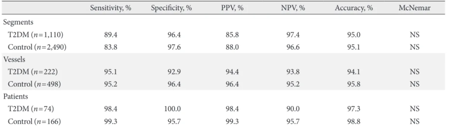
We examined the accuracy of MDCT in detecting significant stenosis in a given patient using BMI (Table 5). According to the definition of obesity (BMI ≥25 kg/m2) for Koreans [13], we classified patients into two groups: normal/overweight (nonobese) vs. obese. Although NPV of the diabetic group slightly dropped to 85.7%, sensitivity, specificity and PPV re- Table 3. Comparison of diagnostic accuracy between nondiabetes and diabetes
Sensitivity, % Specificity, % PPV, % NPV, % Accuracy, % McNemar
Segments
T2DM (n=1,110) 89.4 96.4 85.8 97.4 95.0 NS
Control (n=2,490) 83.8 97.6 88.0 96.6 95.1 NS
Vessels
T2DM (n=222) 95.1 92.9 94.4 93.8 94.1 NS
Control (n=498) 95.2 96.4 96.4 95.2 95.8 NS
Patients
T2DM (n=74) 98.4 100.0 98.4 90.0 97.3 NS
Control (n=166) 99.3 95.7 99.3 95.7 98.8 NS
PPV, positive predictive value; NPV, negative predictive value; T2DM, type 2 diabetes; NS, not significant.
Table 4. The effect of coronary calcium score on diagnostic accuracy of 64-slice MDCT
Sensitivity, % Specificity, % PPV, % NPV, % Accuracy, % McNemar
CCS <100
All patients (n=91) 97.1 100.0 100.0 91.7 97.8 NS
T2DM (n=24) 94.1 100.0 100.0 85.7 95.7 NS
Control (n=67) 98.0 100.0 98.0 94.1 98.5 NS
100≤CCS<400
All patients (n=58) 100.0 83.3 97.9 100.0 98.1 NS
T2DM (n=14) 100.0 100.0 100.0 100.0 100.0 NS
Control (n=44) 100.0 75.0 97.3 100.0 97.5 NS
CCS ≥400
All patients (n=91) 100.0 80.0 98.8 100.0 98.9 NS
T2DM (n=36) 100.0 100.0 100.0 100.0 100.0 NS
Control (n=55) 100.0 75.0 98.1 100.0 98.2 NS
MDCT, multidetector computed tomography; PPV, positive predictive value; NPV, negative predictive value; CCS, coronary calcium score; NS, not significant; T2DM, type 2 diabetes.
mained at 97.2%, 100%, and 100%, respectively in patients with a BMI <25 kg/m2. MDCT remained highly accurate in the presence of obesity, with a sensitivity, specificity, PPV, and NPV of 100% for all patients. There were no significant differ- ences between MDCT and ICA with or without diabetes.
DISCUSSION
As the prevalence of diabetes continues to rise rapidly in Korea, diabetes and its many complications have become a major contributor to increased morbidity and mortality. According to Kim et al. [1], diabetes is the fifth-leading cause of death in Korea, with a two-fold increased death rate among diabetics compared to nondiabetics; moreover, diabetes-related mortal- ity was found to be 19.6 per 100,000 persons in 2009. It is well known that cardiovascular disease is the most common cause of death (30.6%); therefore, the need for appropriate diagnosis and cost-effective management of CAD has become a rising concern. Although the gold standard for CAD detection is known to be coronary angiography, there are several limita- tions to this procedure: 1) invasiveness, 2) expense, 3) small but definite risk of complications, and 4) the need for a skillful operator and special facilities. Given these limitations, multi- ple noninvasive methods have been developed and are now available. These can be divided into functional and anatomical imaging. Functional imaging examines the hemodynamic changes, whereas anatomical imaging permits direct visualiza- tion of the coronary arteries [6]. There are widely used func- tional imaging techniques to detect CAD such as positron emission tomography or single photon emission computed to- mography and myocardial contrast or stress echocardiography [6]. These tests are useful in defining ischemic territories and
predicting myocardium reversibility, but they have several limitations: That methods have some risks because of using pharmacologic stress inducing perfusion defects manner.
Moreover, they do not permit direct visualization of the coro- nary arteries. Specificity of such tests tends to be lower, there- by occasionally requiring other tests for further evaluation.
A 64-slice MDCT is a newer, more widely available method to assess coronary artery disease more directly irrespective of symptoms [9]. Previous studies in the general population re- ported MDCT sensitivity and specificity as 95% to 99% (95%
credible interval [Crl]) and 83% to 94% (95% Crl), respectively [14]. Miller et al. [15] provided a diagnostic performance of a 64-slice MDCT with 291 symptomatic patients (23% of whom have diabetes), and its sensitivity, specificity, PPV, and NPV were 85%, 90%, 91%, and 83%, respectively in a patient-based analysis. They concluded that although MDCT cannot be used as a replacement for ICA, it may help guide clinician decisions as to whether a patient needs further invasive studies [16]. In our study, 64-slice MDCT demonstrated high quality nonin- vasive coronary angiograms that accurately detected signifi- cant lesions of symptomatic patients with type 2 diabetes. Only a few studies on 64-slice MDCT performance in diabetic pa- tients have been done; one of them by Schuijf et al. [17] re- ported that MDCT is a feasible noninvasive test in type 2 dia- betes patients with both a 95% sensitivity and specificity of in- terpretable segments (220/256, 86%). When uninterpretable segments were included, sensitivity and specificity dropped to 81% and 82%, respectively. The present study shows relatively higher quality image (93.4% above adequate image), with 98.4% sensitivity and 100% specificity for diabetic patients re- gardless of imaging quality. This is consistent with meta-anal- yses results, which showed that the sensitivity of a 64-slice CT Table 5. The effect of obesity on diagnostic accuracy of 64-slice MDCT
Sensitivity, % Specificity, % PPV, % NPV, % Accuracy, % McNemar
BMI <25 kg/m2
All patients (n=141) 98.4 94.4 99.2 89.5 97.9 NS
T2DM (n=42) 97.2 100.0 100.0 85.7 97.6 NS
Control (n=99) 98.9 91.7 98.9 91.7 97.8 NS
BMI ≥25 kg/m2
All patients (n=74) 100.0 100.0 100.0 100.0 100.0 NS
T2DM (n=23) 100.0 100.0 100.0 100.0 100.0 NS
Control (n=51) 100.0 100.0 100.0 100.0 100.0 NS
MDCT, multidetector computed tomography; PPV, positive predictive value; NPV, negative predictive value; BMI, body mass index; NS, not significant; T2DM, type 2 diabetes.
(98%) was significantly higher than that of a 16-slice CT (95%) in a patient-based analysis [18]. Because a 64-slice MDCT has increased slices per gantry rotation (64 vs. 16) and a faster gantry speed (330 vs. 375 ms/rotation), the quality of image is increased; therefore, we were able to identify more accurate images compared with former studies. At the bedside, these results may have important clinical implications. Patients with diabetes can be delay proper diagnosis and management be- cause they present atypical chest pain often confused with gas- trointestinal or pulmonary symptoms, so high specificity and NPV of MDCT could be useful in reducing unnecessary inva- sive studies. MDCT could be also beneficial in the preopera- tive assessment of a noncardiac surgery, rapid triage in an emergency center or outpatient department, and evaluation of equivocal stress test results. Therefore, the risks and costs of invasive coronary angiogram could be avoided in a substantial number of diabetic patients by ruling out significant CAD with MDCT.
It is well known that patients with type 2 diabetes experi- ence more diffuse, calcified, and extensive CAD, with signifi- cantly higher coronary calcium scores compared to controls.
Calcified atherosclerosis has been identified as the main rea- son for poor image quality (46%, included multiple factors).
However, there were no significant differences between sensi- tivity and specificity in both groups, probably due to small voxel size effect. Calcium deposits form a metal density that overwhelms the density of other tissues (calcium blooming ef- fect), leading to misinterpreted adjacent plaques through arti- facts attenuating low-energy X-rays (beam-hardening arti- facts). Because small voxel size reduces and modifies these ar- tifacts [19], 64-slice MDCT ameliorates imaging challenges and is helpful in detecting CAD regardless of high coronary calcium scores in diabetic groups.
Raff et al. [19] pointed out that factors related to poor image quality included not only calcium scores over 400, but also obesity (BMI ≥30 kg/m2) and heart rates over 70 beats/min.
In a recent study, Dewey et al. [20] also reported that increased BMI, heart rate, and the presence of breathing artifact were as- sociated with worse image quality.
Our study found that the aforementioned factors are not as- sociated with imaging quality and diagnostic accuracy. Be- cause BMI is a distinctive characteristic in this study com- pared with Caucasian-based research, we suggest that MDCT may be favorable in far-east Asians including Koreans. Asia- Oceanian use different BMI cutoffs for overweight (23 kg/m2)
and obese (25 kg/m2) patients compared to Western measures of BMI and commonly tend to have a more lean body shape than Caucasians [13]. Koreans generally have a higher total body fat content and higher abdominal fat distribution than Caucasians. It is well known that they develop obesity-related complications at a lower BMI level [21]. This point implies that previous surveys about the applicability of MDCT for Caucasians cannot be applied to all ethnicities. In fact, because obesity is common in Western patients with diabetes, ade- quate adjustments such as scanner settings of kV and mA must be considered [20]. Therefore, we believe that the ethnic differences in obesity, for example, are the reason for different image quality and accuracy. Dewey et al. [20] showed that Asians have a tendency for better imaging quality compared to Caucasians (odds ratio, 2.68; P=0.06) and significantly better quality than African Americans. Therefore, Koreans may be in a more favorable group with lesser variables affected by body size in comparison to Caucasians.
It is widely accepted that the accuracy of distal segments is lower than that of proximal segments because less blood flow due to small diameters reduces contrast-to-noise, making it difficult to interpret peripheral lesions [22-26]. This study was consistent with prior results. The sensitivity of 2nd diagnostic and posterior descending artery (PDA) branches dropped sharply to 25% and 33.3%, respectively. Namgung et al. [27]
had reported conflicting results with higher sensitivity in dis- tal segments (95%) compared to proximal segments (89%), as well as a greater degree of calcification and motion artifact at the proximal segment. According to Yun et al. [28], diameters of the distal segment of left anterior descending artery and PDA of right coronary artery were 2.16±0.39 mm (median, 2.09 mm) and 2.09±0.48 mm (median, 2.02 mm), respectively for Koreans. Although it is believed that coronary plaques are more prevalent in the proximal segments of each vessel re- gardless of diabetes [29], diabetic patients often present with equivocal symptoms that could lead to physician misdiagnosis.
Additionally, this raises the concern that perhaps radiologists should be more thorough in interpreting peripheral lesions and selecting patients for further study.
Our study has several limitations. Given this is a cross-sec- tional, single-center study, the diagnostic accuracy of MDCT may not represent the true accuracy in all Koreans with diabe- tes. Since including all patients had chest pain possible to in- fluence the results, thus selection bias may be not excluded in this study. Limitations of coronary 64-slice MDCT include ra-
diation-exposure [30], the use of contrast, the need for heart rate control, and anatomical imaging not directly indicative of ischemic changes.
In conclusion, our results demonstrate that there is no sig- nificant difference in accuracy between invasive coronary an- giogram and MDCT regardless of diabetes. Thus, MDCT may be helpful in reducing unnecessary invasive studies for diabet- ic patients with chest pain, and may facilitate more reliable de- cisions for physicians in Korea.
CONFLICTS OF INTEREST
No potential conflict of interest relevant to this article was re- ported.
REFERENCES
1. Kim JH, Kim DJ, Jang HC, Choi SH. Epidemiology of micro- and macrovascular complications of type 2 diabetes in Korea.
Diabetes Metab J 2011;35:571-7.
2. Morrish NJ, Wang SL, Stevens LK, Fuller JH, Keen H. Mortali- ty and causes of death in the WHO Multinational Study of Vascular Disease in Diabetes. Diabetologia 2001;44 Suppl 2:
S14-21.
3. Jacoby RM, Nesto RW. Acute myocardial infarction in the dia- betic patient: pathophysiology, clinical course and prognosis. J Am Coll Cardiol 1992;20:736-44.
4. Lee KW. Costs of diabetes mellitus in Korea. Diabetes Metab J 2011;35:567-70.
5. Hammoud T, Tanguay JF, Bourassa MG. Management of coro- nary artery disease: therapeutic options in patients with diabe- tes. J Am Coll Cardiol 2000;36:355-65.
6. Bax JJ, Inzucchi SE, Bonow RO, Schuijf JD, Freeman MR, Bar- rett EJ. Cardiac imaging for risk stratification in diabetes. Dia- betes Care 2007;30:1295-304.
7. White CS, Kuo D, Kelemen M, Jain V, Musk A, Zaidi E, Read K, Sliker C, Prasad R. Chest pain evaluation in the emergency de- partment: can MDCT provide a comprehensive evaluation?
AJR Am J Roentgenol 2005;185:533-40.
8. Rubinshtein R, Halon DA, Gaspar T, Jaffe R, Karkabi B, Flugel- man MY, Kogan A, Shapira R, Peled N, Lewis BS. Usefulness of 64-slice cardiac computed tomographic angiography for di- agnosing acute coronary syndromes and predicting clinical outcome in emergency department patients with chest pain of uncertain origin. Circulation 2007;115:1762-8.
9. Gallagher MJ, Ross MA, Raff GL, Goldstein JA, O’Neill WW, O’Neil B. The diagnostic accuracy of 64-slice computed tomog- raphy coronary angiography compared with stress nuclear im- aging in emergency department low-risk chest pain patients.
Ann Emerg Med 2007;49:125-36.
10. Park JY, Lee KU, Kim CH, Kim HK, Hong SK, Park KS, Lee HK, Min HK. Past and current obesity in Koreans with non- insulin-dependent diabetes mellitus. Diabetes Res Clin Pract 1997;35:49-56.
11. American Diabetes Association. Diagnosis and classification of diabetes mellitus. Diabetes Care 2004;27 Suppl 1:S5-10.
12. Austen WG, Edwards JE, Frye RL, Gensini GG, Gott VL, Griffith LS, McGoon DC, Murphy ML, Roe BB. A reporting system on patients evaluated for coronary artery disease. Re- port of the Ad Hoc Committee for Grading of Coronary Ar- tery Disease, Council on Cardiovascular Surgery, American Heart Association. Circulation 1975;51(4 Suppl):5-40.
13. Bassett J; International Diabetes Institute; World Health Orga- nization Regional Office for the Western Pacific; International Association for the Study of Obesity; International Obesity Task Force. The Asia-Pacific perspective: redefining obesity and its treatment. Sydney: Health Communications Australia; 2000.
14. Mowatt G, Cook JA, Hillis GS, Walker S, Fraser C, Jia X, Waugh N. 64-Slice computed tomography angiography in the diagno- sis and assessment of coronary artery disease: systematic review and meta-analysis. Heart 2008;94:1386-93.
15. Miller JM, Rochitte CE, Dewey M, Arbab-Zadeh A, Niinuma H, Gottlieb I, Paul N, Clouse ME, Shapiro EP, Hoe J, Lardo AC, Bush DE, de Roos A, Cox C, Brinker J, Lima JA. Diagnos- tic performance of coronary angiography by 64-row CT. N Engl J Med 2008;359:2324-36.
16. McCulloch AC. Coronary angiography by 64-row CT. N Engl J Med 2009;360:2027.
17. Schuijf JD, Bax JJ, Jukema JW, Lamb HJ, Vliegen HW, Salm LP, de Roos A, van der Wall EE. Noninvasive angiography and as- sessment of left ventricular function using multislice comput- ed tomography in patients with type 2 diabetes. Diabetes Care 2004;27:2905-10.
18. Stein PD, Yaekoub AY, Matta F, Sostman HD. 64-slice CT for diagnosis of coronary artery disease: a systematic review. Am J Med 2008;121:715-25.
19. Raff GL, Gallagher MJ, O’Neill WW, Goldstein JA. Diagnostic accuracy of noninvasive coronary angiography using 64-slice spiral computed tomography. J Am Coll Cardiol 2005;46:552-7.
20. Dewey M, Vavere AL, Arbab-Zadeh A, Miller JM, Sara L, Cox
C, Gottlieb I, Yoshioka K, Paul N, Hoe J, de Roos A, Lardo AC, Lima JA, Clouse ME. Patient characteristics as predictors of image quality and diagnostic accuracy of MDCT compared with conventional coronary angiography for detecting coro- nary artery stenoses: CORE-64 Multicenter International Tri- al. AJR Am J Roentgenol 2010;194:93-102.
21. Kim DM, Ahn CW, Nam SY. Prevalence of obesity in Korea.
Obes Rev 2005;6:117-21.
22. Vogl TJ, Abolmaali ND, Diebold T, Engelmann K, Ay M, Dogan S, Wimmer-Greinecker G, Moritz A, Herzog C. Techniques for the detection of coronary atherosclerosis: multi-detector row CT coronary angiography. Radiology 2002;223:212-20.
23. Kuettner A, Kopp AF, Schroeder S, Rieger T, Brunn J, Meisner C, Heuschmid M, Trabold T, Burgstahler C, Martensen J, Schoebel W, Selbmann HK, Claussen CD. Diagnostic accuracy of multidetector computed tomography coronary angiography in patients with angiographically proven coronary artery dis- ease. J Am Coll Cardiol 2004;43:831-9.
24. Kuettner A, Trabold T, Schroeder S, Feyer A, Beck T, Brueck- ner A, Heuschmid M, Burgstahler C, Kopp AF, Claussen CD.
Noninvasive detection of coronary lesions using 16-detector multislice spiral computed tomography technology: initial clinical results. J Am Coll Cardiol 2004;44:1230-7.
25. Martuscelli E, Romagnoli A, D’Eliseo A, Razzini C, Tomassini M, Sperandio M, Simonetti G, Romeo F. Accuracy of thin-slice
computed tomography in the detection of coronary stenoses.
Eur Heart J 2004;25:1043-8.
26. Gaudio C, Mirabelli F, Alessandra L, Nguyen BL, Di Michele S, Corsi F, Tanzilli G, Mancone M, Pannarale G, Francone M, Carbone I, Catalano C, Passariello R, Fedele F. Noninvasive as- sessment of coronary artery stenoses by multidetector-row spiral computed tomography: comparison with conventional angiography. Eur Rev Med Pharmacol Sci 2005;9:13-21.
27. Namgung J, Choe H, Kwon SU, Doh JH, Lee SY, Hur G, Lee WR. Diagnostic accuracy of 64-slice multidetector CT coro- nary angiography for the evaluation of coronary artery disease.
Korean J Med 2008;75:42-53.
28. Yun YK, Park KH, Choi YS, Kim KM, Jun TG, Kim JG. Com- puterized quantative analysis of cornary angiogram in patients without coronary pathology. Korean J Thorac Cardiovasc Surg 1998;31:488-93.
29. Chu ZG, Yang ZG, Dong ZH, Zhu ZY, Deng W, Peng LQ, Shao H, Tang SS, Chen J. Comparative analysis of coronary artery disease assessed by coronary CT angiography between pa- tients with type 2 diabetes mellitus and non-diabetic patients.
Int J Cardiol 2011;147:178-81.
30. Einstein AJ, Henzlova MJ, Rajagopalan S. Estimating risk of cancer associated with radiation exposure from 64-slice com- puted tomography coronary angiography. JAMA 2007;298:
317-23.