Validation of the diagnostic
performance of ‘HeartMedi V.1.0’, a
novel CT- derived fractional flow reserve measurement, for patients with coronary artery disease: a study protocol
Soo- Hyun Kim,
1Si- Hyuck Kang,
1Woo- Young Chung,
2Chang- Hwan Yoon,
1Sang- Don Park,
3Chang- Wook Nam,
4Ki- Hwan Kwon,
5Joon- Hyung Doh,
6Young- Sup Byun,
7Jang- Whan Bae,
8Tae- Jin Youn ,
1In- Ho Chae
1To cite: Kim S- H, Kang S- H, Chung W- Y, et al. Validation of the diagnostic performance of
‘HeartMedi V.1.0’, a novel CT- derived fractional flow reserve measurement, for patients with coronary artery disease:
a study protocol. BMJ Open 2020;10:e037780. doi:10.1136/
bmjopen-2020-037780
►Prepublication history for this paper is available online.
To view these files, please visit the journal online (http:// dx. doi.
org/ 10. 1136/ bmjopen- 2020- 037780).
So- HK and Si- HK contributed equally.
Received 25 February 2020 Revised 12 May 2020 Accepted 05 June 2020
For numbered affiliations see end of article.
Correspondence to Dr Tae- Jin Youn;
ytjmd@ snubh. org
© Author(s) (or their employer(s)) 2020. Re- use permitted under CC BY- NC. No commercial re- use. See rights and permissions. Published by BMJ.
ABSTRACT
Introduction Coronary CT angiography (CCTA) is widely used for non- invasive coronary artery evaluation, but it is limited in identifying the nature of functional characteristics that cause ischaemia. Recent computational fluid dynamic (CFD) techniques applied to CCTA images permit non- invasive computation of fractional flow reserve (FFR), a measure of lesion- specific ischaemia. However, this technology has limitations, such as long computational time and the need for expensive equipment, which hinder widespread use.
Methods and analysis This study is a prospective, multicentre, comparative and confirmatory trial designed to evaluate the diagnostic performance of HeartMedi V.1.0, a novel CT- derived FFR measurement for the detection of haemodynamically significant coronary artery stenoses identified by CCTA, based on invasive FFR as a reference standard. The invasive FFR values ≤0.80 will be considered haemodynamically significant. The study will enrol 184 patients who underwent CCTA, invasive coronary angiography and invasive FFR. Computational FFR (c- FFR) will be analysed by CFD techniques using a lumped parameter model based on vessel length method.
Blinded core laboratory interpretation will be performed for CCTA, invasive coronary angiography, invasive FFR and c- FFR. The primary objective of the study is to compare the area under the receiver–operator characteristic curve between c- FFR and CCTA to non- invasively detect the presence of haemodynamically significant coronary stenosis. The secondary endpoints include diagnostic accuracy, sensitivity, specificity, positive predictive value, negative predictive value and correlation of c- FFR with invasive FFR.
Ethics and dissemination The study has ethic approval from the ethics committee of Seoul National University Bundang Hospital (E-1709/420-001) and informed consent will be obtained for all enrolled patients. The result will be published in a peer- reviewed journal.
Trial registration number KCT0002725; Pre- results.
INTRODUCTION
Fractional flow reserve (FFR) has become the standard of care for functional assess- ment of the extent and severity of coronary disease.
1 2Recent advances in CT and compu- tational fluid dynamics (CFD) have enabled estimation of FFR with routine CT angiog- raphy acquired at rest. Haemodynamics of the aorta and coronary arteries calculated using CFD are coupled with parameter models of the cardiovascular system. The current tech- nology has shown acceptable diagnostic accu- racy compared with invasive FFR.
3–5However, there are several limitations with it, such as prolonged time for calculation and the need for high- performance computational power;
these hamper its widespread use in clinical practice.
Recently, a novel simulating method for predicting FFR with coronary CT angiography
Strengths and limitations of this study
► The non- invasive measurement of fractional flow reserve (FFR) can improve diagnostic performance for the severity of coronary disease and enhance the quality of patient outcomes.
► The novel simulating method for predicting FFR in the study allows shorter time and easier access us- ing an on- site personal computer.
► The study aims to reduce the biases associated with selection and referrals through a multicentre, pro- spective study design.
► The study excludes patients with acute myocardial infarction, previous percutaneous coronary inter- vention or coronary artery bypass graft, so there is a limitation that the generalisation potential of computed FFR for the overall patients with coronary artery disease is unknown.
on January 28, 2021 by guest. Protected by copyright.http://bmjopen.bmj.com/BMJ Open: first published as 10.1136/bmjopen-2020-037780 on 20 July 2020. Downloaded from
(CCTA) has been developed.
6 7It represents a simple simulation method using a personal computer to estimate FFR values. CT images of coronary arteries and basal phys- iological data of patients are the only requirements for patient- specific simulation model.
6For the construction of
a patient- specific CFD model, a fast segmentation system of CT images is used, which enables the on- site solution of computational FFR (c- FFR). The lumped parameter model used to reflect the effect of microvasculature and veins adopts only the coronary circulation rather than
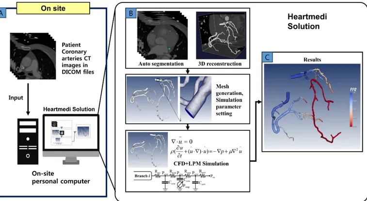
Figure 1 Process of computational fractional flow reserve (FFR) calculation. (A) Digital imaging and communicationsin medicine (DICOM) data sets from segmented coronary CT angiography image and physiological data required for
haemodynamic calculations. (B) FFR is calculated through computational fluid dynamic (CFD) technique and three- dimensional (3D) vascular modelling. Blood flow is calculated using coronary artery length instead of the volume of myocardium based on lumped parameter model (LPM) resistance. (C) Visualised results are derived based on the computed FFR.
Figure 2 An example case of computed and invasive FFR. (A) 3D model reconstruction derived from CCTA image
segmentation and FFR simulated using the novel methods. (B) Coronary angiography shows significant stenosis at the proximal LAD, an intermediate lesion at the distal LCX and an insignificant lesion at the mid- RCA. The arrow indicates the position of the pressure sensor when measuring FFR. The measured FFR was 0.79 for LAD, 0.87 for LCX and 0.99 for RCA, respectively. (C) The computed FFR at the corresponding point was 0.77, 0.90 and 0.96. 3D, three- dimensional; CCTA, coronary CT angiogram;
FFR, fractional flow reserve; LAD, left anterior descending; LCX, left circumflex; RCA, right coronary artery.
on January 28, 2021 by guest. Protected by copyright.http://bmjopen.bmj.com/BMJ Open: first published as 10.1136/bmjopen-2020-037780 on 20 July 2020. Downloaded from
using the entire cardiovascular system (figures 1 and 2).
Potential advantages include shorter computational time and no need for supercomputers.
A recent retrospective analysis demonstrated accept- able diagnostic performance of the simulation method.
8In this study, we will prospectively perform a trial to confirm the diagnostic performance of c- FFR. c- FFR esti- mated using the routine CCTA images will be compared with anatomical assessment alone with invasive FFR as the reference standard.
METHODS Study aim
This study will be a prospective, multicentre, comparative and confirmatory trial. The primary objective of the trial is to assess the diagnostic performance of c- FFR based on routinely acquired CCTA (HeartMedi V.1.0, Silicon- Sapiens, Korea) in patients with coronary artery disease.
The invasive FFR acquired during invasive coronary angi- ography (ICA) will be the reference standard, and non- invasive CCTA will be the comparator diagnostic method.
The primary endpoint is to test the superiority of c- FFR compared with CCTA alone in terms of identification of significant haemodynamic stenosis validated by invasive FFR with ICA.
Study population
Patients with coronary artery disease undergoing non- emergent ICA and invasive FFR will be eligible for inclu- sion in the trial. All study subjects will provide written informed consent. CCTA with ≥64 multidetector slices needs to be taken within 90 days before enrolment. ICA and invasive FFR will be performed with a clinical indi- cation that will be left on physicians’ discretion. Patients will be enrolled after completion of ICA and invasive FFR if he/she provides informed consent (figure 3). The key inclusion criteria include the presence of CCTA within 90 days, available ICA and invasive FFR measurements.
Key exclusion criteria include resting anginal symptom,
chronic kidney disease, tachycardia, hypotension and high coronary artery calcium score. The inclusion and exclu- sion criteria are detailed in table 1. The study subjects will be enrolled from 12 medical centres in Korea.
Study process
Study images such as CCTA, ICA and invasive FFR will be transferred to blinded independent core laboratories where study images of CCTA and invasive FFR will be interpreted independently. CCTA images will be anony- mised and sent to the vendor (c- FFR core laboratory), where measurements of c- FFR will be performed. All study processes will be blinded, and measurements will be conducted independently. All measurement data will be recorded in electronic case report forms, which will be blinded to other participants of the study. Specifically, the c- FFR core laboratory of SiliconSapiens will be completely blinded to the findings of the CCTA, quantitative coro- nary angiography and invasive FFR core laboratories. The independent statistical core laboratory will collect the data after completion of the trial.
Coronary CT angiography
Patients who underwent CCTA as part of routine clinical care will be enrolled in the study. The minimum require- ment for CCTA includes ≥64 multidetector slices and a row width of ≤0.75 mm. CT angiography scanning proto- cols in the participating centres are consistent with the quality standards by the Society of Cardiac Computed Tomography.
9The quality of CCTA images of each partic- ipating centre has been confirmed by the core laboratory before the trial initiation. Study CCTA images will be transmitted to the core laboratory, where the characteris- tics and severity of coronary atherosclerotic lesions will be quantified by two independent, blinded radiologists. Any disagreement between the two radiologists will be resolved by discussion. The coronary system will be divided into the left anterior descending artery, left circumflex artery and right coronary artery (RCA) and then further into 15 segments according to the American Heart Association (AHA) classification guidelines.
10 11Using a semiauto- mated dedicated three- dimensional workstation (Intellis- pace Portal, Philips Healthcare, Cleveland, Ohio, USA), curved multiplanar reformatted images will be recon- structed for assessment. The degree of stenosis of the vessels will be measured around the narrowest area at a rate based on the average of the normal coronary arteries above and below the stenosis site. Quantitative analysis of stenosis grade will be classified as normal (0%), minimal (1%–24%), mild (26%–49%), moderate (50%–69%), severe (70%–99%) and occlusion (100%) according to Society of Cardiovascular Computed Tomography guidelines.
9Coronary angiography
ICA and invasive FFR procedures will be performed according to the American College of Cardiology/
AHA for guidelines for coronary angiography and
Figure 3 Study flow. CCTA, coronary CT angiography; FFR,fractional flow reserve.
on January 28, 2021 by guest. Protected by copyright.http://bmjopen.bmj.com/BMJ Open: first published as 10.1136/bmjopen-2020-037780 on 20 July 2020. Downloaded from
intervention.
12Intracoronary nitroglycerine (100–
200 mg) will be administered in the coronary arteries before initial cine angiograms unless contraindicated.
Coronary arterial images will be obtained with selective catheterisation of the left coronary artery and RCA. The coronary angiography images will be analysed using an automated edge- detection system (Cardiovascular Angi- ography Analysis Systems, Maastricht, the Netherlands) at the core laboratory by an experienced technician who is blinded to the study. After calibration with the outer diameter of the coronary catheter, the minimal lumen diameter, reference vessel diameter, % diameter stenosis, will be measured. If there are two or more stenosed vessels over 2.0 mm, the most severe lesion will be chosen as the index lesion.
Invasive FFR
The invasive FFR measurements performed in the coro- nary arteries with a diameter of ≥2 mm will be included in the study. The invasive FFR should be measured using a sensor- tipped 0.014- inch guidewire (PressureWire;
St. Jude Medical, St. Paul, Minnesota, USA or Verrata Wire; Philips, Eindhoven, the Netherlands) through a 5–7 Fr guiding catheter. Pressure calibration should be confirmed as zero at the ascending aorta or proximal
segment of the coronary arteries. The location of the pres- sure wire distal to the index lesion should be recorded on a coronary angiographic image. Maximal myocardial hyperaemia should be induced by continuous intravenous adenosine infusion via a central or peripheral vein with an infusion rate of 140 mg/kg/min. The invasive FFR will be calculated as the mean distal coronary pressure divided by the mean aortic pressure during hyperaemia. A pull- back recording should be performed and recorded. The absence of pressure signal drift (0.97‒1.03) needs to be confirmed at the distal end of the guiding catheter. The raw data of the invasive FFR measurements will be sent to the invasive FFR core laboratory, where potential bias such as maximum hyperaemia and pressure drift will be confirmed, and the measurements will be validated. The validated invasive FFR values will be transmitted to the statistical core laboratory.
Computational FFR
The vendor (c- FFR core laboratory) will receive the segmented CCTA images from the CCTA core laboratory.
The invasive FFR core laboratory will demarcate the loca- tion of invasive FFR measurement on the reconstructed CCTA, which will be transmitted to SiliconSapiens via the study coordinator. SiliconSapiens will analyse c- FFR
Table 1 Inclusion and exclusion criteriaInclusion criteria Exclusion criteria
1. Men and women age ≥19
2. Voluntary agreement to a written consent
3. 64 Multidetector row CCTA taken within 90 days of coronary angiography
4. Subjects who need a preliminary test for FFR during coronary angiography
1. Needs for emergency procedures
2. Difficult cooperating with medical staff for reasons such as cognitive impairment
3. Experienced acute myocardial infarction within the last 30 days
4. Report of chest pain during rest (CCS class IV)
5. Impaired chronic renal function (serum creatinine >2.0 mg/
dL)
6. Heart rate ≥100 beats/min 7. Systolic BP ≤90 mm Hg 8. CAC ≥1000
9. Pregnancy
10. Body mass index >35 kg/m2
11. Prior PCI or CABG in the subject blood vessel 12. Previous valvular surgery
13. Complicated congenital heart disease 14. Acute pulmonary oedema
15. Unstable haemodynamics including cardiogenic shock, abrupt chest pain
16. Pacemaker or internal defibrillator leads implanted 17. Known hypersensitivity or contraindication to β-blocker,
nitroglycerin, adenosine 18. History of contrast dye allergy
19. Significant arrhythmia including complete AV block, ventricular arrhythmia
20. Subjects who are currently participating in other clinical trials or have participated in other clinical trials within 30 days before screening
21. Others who are inappropriate subject judged by clinician AV, atrioventricular; BP, blood pressure; CABG, coronary artery bypass graft; CAC, coronary artery calcium; CCS, Canadian Cardiovascular Society; CCTA, coronary CT angiography; FFR, fractional flow reserve; PCI, Percutaneous coronary intervention.
on January 28, 2021 by guest. Protected by copyright.http://bmjopen.bmj.com/BMJ Open: first published as 10.1136/bmjopen-2020-037780 on 20 July 2020. Downloaded from
according to the method of using medical devices for clinical trials. The simulations use a three- dimensional model of epicardial coronary arteries derived from CCTA image segmentation, and the estimation is based on vessel lengths but not on myocardial volume. The parameters will be assigned by physiological data customised to each individual patient- specific model. Coronary blood flow will be simulated under conditions that mimic maximal hyperaemia. For suboccluded or chronically occluded arteries by CCTA (ie, stenosis >90%), default c- FFR values of 0.50 will be assigned to that vessel.
Primary efficacy analysis
The primary measure of performance will be the area under the receiver–operator characteristic curve (AUC) to detect haemodynamically significant stenosis. The gold standard for significant stenosis will be defined as inva- sive FFR ≤0.80. The measurements will be % stenosis for CCTA and simulated FFR based on CT for c- FFR. Sensi- tivity will be plotted against (1−specificity) for different cut- off points of the study measurements. The AUC, SE and 95% CIs will be presented. Delong’s test will be used to compare two correlated C- statistics.
13Secondary efficacy analysis
Diagnostic accuracy, sensitivity, specificity, positive predic- tive value (PPV), negative predictive value (NPV) and correlation will be presented as secondary analyses. The cut- off for significant obstruction of CCTA will be defined as a diameter stenosis of ≥50%. The cut- off for c- FFR will the simulated c- FFR measured by the software HeartMedi V.1.0 of ≤0.8. Each value will be calculated as shown below.
Predictive accuracy = TP+FP+ FN+TNTP+TN
Sensitivity = TP+FNTP
Specificity = TN+FPTN
Positive predictive value = TP+FPTP
Negative predictive value = TN+FNTN
(TP, True positive; TN, True negative; FP, False positive;
FN, False negative.)
Each value and 95% CI will be presented. The perfor- mance will be compared using McNemar’s test. Correla- tion will be assessed with the use of Pearson or Spearman Correlation, whereby coefficient (r) and p values will be determined.
Statistical hypotheses and sample size calculation
The study hypothesis is that the AUC of c- FFR would be greater than that of CCTA. The NXT (HeartFlow anal- ysis of coronary blood flow using CT angiography: NeXt sTeps) trial previously reported the AUC of FFR
CT(Heart- Flow) and CCTA to be 0.90 and 0.81, respectively.
3 14The NOVEL- FLOW (Diagnostic Performance of a Novel Method for Fractional Flow Reserve Computed From Noninvasive Computed Tomography Angiography) study also showed similar discriminatory functions (AUC, 0.93 for CT- FFR and 0.74 for CCTA).
8In the present study,
the AUC of c- FFR and CCTA was assumed to be 0.90 and 0.81, respectively. We expected the prevalence of haemo- dynamically significant stenosis to be 31.5% based on the previous studies.
3 15–17The assumptions included 0.6 of the correlation coefficient of AUCs between c- FFR and CCTA and the attrition rate of 15%. With these assump- tions, 184 study participants would be required to achieve a one- sided significance level of 0.025 and power of 80%.
Patient and public involvement
There was no patient or public involvement in the design of the present study, and there is no planned patient or public involvement to recruit and conduct the study.
DISCUSSION
FFR- guided coronary revascularisation has shown clinical benefits over angiography guidance alone.
18 19CCTA is currently the most widely used imaging modality for non- invasive coronary evaluation.
20 21Recent advances in CT imaging enabled high diagnostic accuracy for detecting obstructive coronary artery disease.
22 23The combination of high image quality of CCTA and functional assessment of FFR has the potential to improve diagnostic perfor- mance and enhance the quality of patient outcomes.
It allows the functional assessment of coronary stenosis without invasive catheterisation, which inevitably is associ- ated with complications.
Previous studies have proven the benefit of such approaches, including FFR
CTdeveloped by Heart- Flow.
24–26The limitations of this technology include the need for high- performance computing power, long computation time and potential simulation errors.
The novel simulation method tested in this study (CT- FFR, HeartMedi V.1.0) has several advantages over the previous methods. The previous methods use CFD that requires myocardial mass estimation based on the whole cardiac anatomy coupled with lumped parameter models (volume- based method).
3 4 27In contrast, CFD used in the novel c- FFR technology calculates vessel length and three- dimensional coronary artery geometry, which is combined with coronary circulation of lumped param- eter models (length- based method). A previous study demonstrated no significant difference in haemody- namic simulation between the two estimation methods.
7The feature is translated into less need for computational power. Functional assessment can be performed on- site with a personal computer environment without transfer- ring large volume CT images to central laboratories. In addition, this method excludes the possibility of errors due to the segmentation of left ventricular muscle.
One previous study retrospectively analysed 218 vessels from 117 patients to validate the c- FFR method compared with invasively measured FFR.
8The accuracy, sensitivity, specificity, PPV and NPV of c- FFR were shown to be 85.8%, 86.2%, 85.5%, 79.8% and 90.3%, respec- tively. The diagnostic performance measured by the AUC was significantly higher for c- FFR than those for CCTA.
on January 28, 2021 by guest. Protected by copyright.http://bmjopen.bmj.com/BMJ Open: first published as 10.1136/bmjopen-2020-037780 on 20 July 2020. Downloaded from
c- FFR showed a slight underestimation of the functional severity of the lesions. The present study is designed to prospectively validate the performance of the novel simu- lation method. Eligible subjects who have coronary artery disease with CCTA and invasive FFR available will be prospectively enrolled. The sample size is planned based on the statistical power calculation.
In conclusion, the present study will prospectively assess the diagnostic performance of c- FFR. The values will be compared with that of CCTA with invasive FFR as the gold standard.
Limitations
Since this study excludes patients with acute myocardial infarction, previous percutaneous coronary intervention or coronary artery bypass graft, there is a limitation that the generalisation potential of computed FFR for the overall patients with coronary artery disease is unknown.
However, the novel method in this study can be easily applied to these cases, and further study will attempt to include them.
Another limitation of the present study is that, although prospective, we are recruiting patients following the performance of CCTA and invasive FFR. This may lead to selection bias. Finally, while the technology was developed for on- site usage, the measurements will be performed in a core laboratory. This was included in the study design to ensure adequate blinding of investigators to the refer- ence values and hence to minimise the study bias.
Protocol amendments
All changes in the study protocol were reviewed by the ethics committee of Seoul National University Bundang Hospital and reported to the sponsor and funder. Signifi- cant protocol changes were recorded in Clinical Research Information Service.
Ethics and dissemination
This study was approved by the institutional review board of Seoul National University Bundang Hospital (E-1709/420-001). Written informed consent will be obtained for all enrolled patients. The result will be published in a peer- reviewed journal.
Author affiliations
1Division of Cardiology, Department of Internal Medicine, Seoul National University Bundang Hospital, Seongnam- si, Gyeonggi- do, The Republic of Korea
2Department of Internal Medicine, Seoul Metropolitan Boramae Hospital, Dongjak- gu, Seoul, The Republic of Korea
3Division of Cardiology, Department of Internal Medicine, Inha University Hospital, Incheon, The Republic of Korea
4Department of Medicine, Keimyung University Dongsan Medical Center, Daegu, The Republic of Korea
5Division of Cardiology, Department of Internal Medicine, Ewha Womans University School of Medicine, Seoul, The Republic of Korea
6Department of Internal Medicine, Inje University Ilsan Paik Hospital, Goyang- si, Gyeonggi- do, The Republic of Korea
7Division of Cardiology, Department of Internal Medicine, Inje University Sanggye Paik Hospital, Seoul, The Republic of Korea
8Department of Internal Medicine, College of Medicine, Chungbuk National University, Cheongju, The Republic of Korea
Contributors Si- HK and T- JY conceived and designed the study. So- HK and Si- HK wrote the draft of the paper. W- YC, C- HY, S- DP, C- WN, K- HK, J- HD, Y- SB and J- WB will conduct screening and data collection. Analysis will be performed by Si- HK, So- HK and T- JY. T- JY and I- HC were involved in critical revision of the study for important intellectual content. All authors contributed to revision and approved the final version of the manuscript.
Funding This research was supported by a grant of the Korea Health Technology R&D Project through the Korea Health Industry Development Institute (KHIDI) funded by the Ministry of Health & Welfare, Republic of Korea (grant number: HI17C2006).
The sponsor, SiliconSapiens, due to the nature of the study, will only provide data that independently analyse computational FFR. The protocol design or the implementation, management, data collection and analysis of the study is entirely independent of the vendor company (SiliconSapiens) or funder.
Competing interests None declared.
Patient and public involvement Patients and/or the public were not involved in the design, or conduct, or reporting, or dissemination plans of this research.
Patient consent for publication Not required.
Provenance and peer review Not commissioned; externally peer reviewed.
Open access This is an open access article distributed in accordance with the Creative Commons Attribution Non Commercial (CC BY- NC 4.0) license, which permits others to distribute, remix, adapt, build upon this work non- commercially, and license their derivative works on different terms, provided the original work is properly cited, appropriate credit is given, any changes made indicated, and the use is non- commercial. See: http:// creativecommons. org/ licenses/ by- nc/ 4. 0/.
ORCID iDs
Tae- Jin Youn http:// orcid. org/ 0000- 0001- 9957- 4204 In- Ho Chae http:// orcid. org/ 0000- 0003- 1644- 2105
REFERENCES
1 Neumann F- J, Sousa- Uva M, Ahlsson A, et al. 2018 ESC/
EACTS guidelines on myocardial revascularization. Eur Heart J 2019;40:87–165.
2 Levine GN, Bates ER, Blankenship JC, et al. 2011 ACCF/AHA/
SCAI guideline for percutaneous coronary intervention: a report of the American College of cardiology Foundation/American heart association Task force on practice guidelines and the Society for cardiovascular angiography and interventions. Circulation 2011;124:e574–651.
3 Nørgaard BL, Leipsic J, Gaur S, et al. Diagnostic performance of noninvasive fractional flow reserve derived from coronary computed tomography angiography in suspected coronary artery disease: the NXT trial (analysis of coronary blood flow using CT angiography: next steps). J Am Coll Cardiol 2014;63:1145–55.
4 Koo B- K, Erglis A, Doh J- H, et al. Diagnosis of ischemia- causing coronary stenoses by noninvasive fractional flow reserve computed from coronary computed tomographic angiograms. results from the prospective multicenter DISCOVER- FLOW (diagnosis of Ischemia- Causing stenoses obtained via noninvasive fractional flow reserve) study. J Am Coll Cardiol 2011;58:1989–97.
5 Min JK, Leipsic J, Pencina MJ, et al. Diagnostic accuracy of fractional flow reserve from anatomic CT angiography. JAMA 2012;308:1237–45.
6 Kwon S- S, Chung E- C, Park J- S, et al. A novel patient- specific model to compute coronary fractional flow reserve. Prog Biophys Mol Biol 2014;116:48–55.
7 Lee KE, Kwon S- S, Ji YC, et al. Estimation of the flow resistances exerted in coronary arteries using a vessel length- based method.
Pflugers Arch 2016;468:1449–58.
8 Chung J- H, Lee KE, Nam C- W, et al. Diagnostic performance of a novel method for fractional flow reserve computed from noninvasive computed tomography angiography (NOVEL- FLOW study). Am J Cardiol 2017;120:362–8.
9 Leipsic J, Abbara S, Achenbach S, et al. SCCT guidelines for the interpretation and reporting of coronary CT angiography: a report of the Society of cardiovascular computed tomography guidelines Committee. J Cardiovasc Comput Tomogr 2014;8:342–58.
10 Austen WG, Edwards JE, Frye RL, et al. A reporting system on patients evaluated for coronary artery disease. Report of the AD hoc Committee for grading of coronary artery disease, Council on cardiovascular surgery, American heart association. Circulation 1975;51:5–40.
on January 28, 2021 by guest. Protected by copyright.http://bmjopen.bmj.com/BMJ Open: first published as 10.1136/bmjopen-2020-037780 on 20 July 2020. Downloaded from
11 Hausleiter J, Meyer T, Hadamitzky M, et al. Prevalence of noncalcified coronary plaques by 64- slice computed tomography in patients with an intermediate risk for significant coronary artery disease. J Am Coll Cardiol 2006;48:312–8.
12 Levine GN, Bates ER, Blankenship JC, et al. 2011 ACCF/AHA/
SCAI guideline for percutaneous coronary intervention: a report of the American College of cardiology Foundation/American heart association Task force on practice guidelines and the Society for cardiovascular angiography and interventions. Catheter Cardiovasc Interv 2013;82:E266–355.
13 DeLong ER, DeLong DM, Clarke- Pearson DL. Comparing the areas under two or more correlated receiver operating characteristic curves: a nonparametric approach. Biometrics 1988;44:837–45.
14 Gaur S, Achenbach S, Leipsic J, et al. Rationale and design of the HeartFlowNXT (HeartFlow analysis of coronary blood flow using CT angiography: next sTeps) study. J Cardiovasc Comput Tomogr 2013;7:279–88.
15 Park S- J, Kang S- J, Ahn J- M, et al. Visual- functional mismatch between coronary angiography and fractional flow reserve. JACC Cardiovasc Interv 2012;5:1029–36.
16 Tesche C, De Cecco CN, Caruso D, et al. Coronary CT angiography derived morphological and functional quantitative plaque markers correlated with invasive fractional flow reserve for detecting hemodynamically significant stenosis. J Cardiovasc Comput Tomogr 2016;10:199–206.
17 Wu J, Barton D, Xie F, et al. Comparison of fractional flow reserve assessment with demand stress myocardial contrast echocardiography in angiographically intermediate coronary stenoses. Circulation 2016;9.
18 Tonino PAL, De Bruyne B, Pijls NHJ, et al. Fractional flow reserve versus angiography for guiding percutaneous coronary intervention.
N Engl J Med 2009;360:213–24.
19 De Bruyne B, Fearon WF, Pijls NHJ, et al. Fractional flow reserve- guided PCI for stable coronary artery disease. N Engl J Med 2014;371:1208–17.
20 , Montalescot G, Sechtem U, et al, Task Force Members. 2013 ESC guidelines on the management of stable coronary artery disease: the task force on the management of stable coronary artery disease of the European Society of cardiology. Eur Heart J 2013;34:2949–3003.
21 National Institute for Health and Clinical Excellence: Guidance. Chest pain of recent onset: assessment and diagnosis of recent onset chest pain or discomfort of suspected cardiac origin. London: National Institute for Health and Clinical Excellence, 2010.
22 Min JK, Shaw LJ, Berman DS. The present state of coronary computed tomography angiography a process in evolution. J Am Coll Cardiol 2010;55:957–65.
23 Budoff MJ, Dowe D, Jollis JG, et al. Diagnostic performance of 64- multidetector row coronary computed tomographic angiography for evaluation of coronary artery stenosis in individuals without known coronary artery disease: results from the prospective multicenter accuracy (assessment by coronary computed tomographic angiography of individuals undergoing invasive coronary angiography) trial. J Am Coll Cardiol 2008;52:1724–32.
24 Coenen A, Lubbers MM, Kurata A, et al. Fractional flow reserve computed from noninvasive CT angiography data: diagnostic performance of an on- site clinician- operated computational fluid dynamics algorithm. Radiology 2015;274:674–83.
25 Renker M, Schoepf UJ, Wang R, et al. Comparison of diagnostic value of a novel noninvasive coronary computed tomography angiography method versus standard coronary angiography for assessing fractional flow reserve. Am J Cardiol 2014;114:1303–8.
26 Kruk M, Wardziak Łukasz, Demkow M, et al. Workstation- Based calculation of CTA- Based FFR for intermediate stenosis. JACC Cardiovasc Imaging 2016;9:690–9.
27 Thompson AG, Raju R, Blanke P, et al. Diagnostic accuracy and discrimination of ischemia by fractional flow reserve CT using a clinical use rule: results from the determination of fractional flow reserve by anatomic computed tomographic angiography study. J Cardiovasc Comput Tomogr 2015;9:120–8.
on January 28, 2021 by guest. Protected by copyright.http://bmjopen.bmj.com/BMJ Open: first published as 10.1136/bmjopen-2020-037780 on 20 July 2020. Downloaded from