J Korean Soc Radiol 2015;73(1):1-10 http://dx.doi.org/10.3348/jksr.2015.73.1.1
INTRODUCTION
Hypertrophic cardiomyopathy (HCM) is a complex and rela- tively common genetic cardiac disease with an estimated preva- lence of 0.2% (1). Frequently, patients with HCM have chest pain suggestive of angina pectoris, and the electrocardiogram (ECG) can resemble that of a myocardial infarction (MI) in the
absence of coronary artery disease (CAD) (2, 3). However, adult patients with HCM may also develop atherosclerotic CAD, and patients with HCM and coexistent CAD have an increased rate of morbidity compared to patients with HCM without CAD (4, 5). Therefore, early detection of coexistent CAD in HCM is of high importance.
Myocardial bridging (MB) is more commonly associated with
Coexistent Coronary Artery Disease or Myocardial Bridging in Patients with Hypertrophic Cardiomyopathy Using Coronary CT Angiography
비후성 심근증을 가진 성인 환자에서 동반되는 관상동맥질환과 심근교락:
다중검출기 심장 CT를 통한 연구
Jae Hwan Lee, MD
1, Eun Ju Chun, MD
1, Yeo Koon Kim, MD
1, Jin Young Yoo, MD
1, Sang Il Choi, MD
1, Dong-Ju Choi, MD
2Departments of 1Radiology, 2Internal Medicine, Seoul National University Bundang Hospital, Seongnam, Korea
Purpose: To evaluate the prevalence of coexistent coronary artery disease (CAD) or myocardial bridging (MB) in patients with hypertrophic cardiomyopathy (HCM) using coronary CT angiography (CCTA) and assess the role of CCTA.
Materials and Methods: The prevalence of obstructive CAD (> 50% luminal reduc- tion) and MB (partial and full encasement) were assessed in 150 patients with HCM diagnosed by clinical findings, electrocardiography, and echocardiography of 19588 consecutive patients who underwent CCTA for suspected CAD.
Results: The overall feasibility of coronary artery visualization was 98.9% with CCTA.
In patients with HCM, the prevalence of obstructive CAD and MB (14.7% partial and 28.0% full encasement) were 23.3% and 42.7%, respectively. Age, hypertension, fam- ily history of premature CAD, Framingham risk score and severe chest pain were as- sociated with CAD, whereas male gender and septal type were associated with MB (all p < 0.05). In comparison to invasive coronary angiography (n = 37), the diagnos- tic accuracy of CCTA for the detection of CAD and full encasement MB was 89.2%
and 86.5%, respectively.
Conclusion: One-quarter of patients with HCM had coexistent obstructive CAD or full encasement MB. CCTA can be a feasible and accurate noninvasive imaging modality for the detection of CAD and MB in patients with HCM.
Index terms
Hypertrophic Cardiomyopathy Coronary Artery Disease Myocardial Bridging
Coronary Computed Tomography Angiography
Received February 23, 2015 Accepted March 22, 2015
Corresponding author: Eun Ju Chun, MD Department of Radiology, Seoul National University Bundang Hospital, 82 Gumi-ro 173beon-gil, Bundang-gu, Seongnam 463-707, Korea.
Tel. 82-31-787-7618 Fax. 82-31-787-4011 E-mail: [email protected]
This is an Open Access article distributed under the terms of the Creative Commons Attribution Non-Commercial License (http://creativecommons.org/licenses/by-nc/3.0) which permits unrestricted non-commercial use, distri- bution, and reproduction in any medium, provided the original work is properly cited.
This work was supported by the grant (11-2009-035) from the Seoul National University Bundang Hospital (SNUBH) Research Fund and by the National Research Foundation (NRF) grant funded by the Korea government (MEST) (NRF-2010-0023504).
HCM than with non-HCM (6). Although most occurrences of MB are thought to be innocent, in some cases, they may cause angina pectoris, MI, life-threatening ventricular arrhythmias, or even sudden cardiac death (6-8).
Traditionally, coexistent CAD or MB in HCM was diagnosed with two different methods; the magnitude of myocardial hy- pertrophy was usually assessed with 2-dimensional echocardiog- raphy, while coexistent CAD or MB was evaluated with invasive coronary angiography (ICA) (5, 7). Although pharmacologic stress echocardiography was used as a reasonable method for diagnosis of the occurrence of both coexistent CAD and HCM at the same time, the feasibility of dipyridamole-stress echocar- diography was often limited, especially in patients with poor echo windows or chronic theophylline therapy (9). Currently, coronary computed tomography angiography (CCTA) with the ECG-gated technique is an attractive noninvasive imaging meth- od for the evaluation of coronary arteries and myocardial hy- pertrophy at the same time (10). However, there is little data re- garding the role of CCTA in the evaluation of patients with HCM.
Therefore, the purpose of this study was to investigate the prevalence and risk factors associated with coexistent CAD or MB in patients with HCM using CCTA. In addition, we assessed the feasibility and diagnostic accuracy of CCTA for the detection of coexistent CAD or MB in patients with HCM in comparison to ICA.
MATERIALS AND METHODS
Study Population
The Institutional Review Board approved the study protocol, and informed consent was waived. Among a registry of 19588 patients who underwent CCTA between January 2008 and De- cember 2012 in Seoul National University Bundang Hospital, we retrospectively enrolled 150 adults with HCM [112 men;
mean age ± standard deviation (SD): 62.4 ± 11.0 years; range:
36–90 years]. The diagnosis of HCM was based on clinical and ECG findings and echocardiographic features, with left ventric- ular hypertrophy (LVH), in the absence of another cardiac or systemic disease, known to cause hypertrophied myocardium (1). Clinical data including family history and ECG findings were retrieved through medical record review. LVH was as-
sessed in the end-diastolic stage with M-mode and 2-dimen- sional transthoracic echocardiography by standard techniques.
According to morphological phenotype, septal HCM was de- fined as a septal thickness greater than 15 mm or a septal thick- ness to inferior LV wall thickness ratio of greater than 1.5 at the mid-ventricular level, and apical HCM was defined as an abso- lute apical wall thickness of greater than 15 mm or an apical to basal LV wall thicknesses ratio of 1.3–1.5 showing characteristic
“spade-like configuration”. The other types of HCM were named according to the hypertrophied location, such as midventricu- lar type or concentric type HCM (10). The combination of two or more phenotypes was classified as combined or mixed type HCM.
Clinical Symptoms and Risk Factors
Clinical symptoms, basic demographic data, and clinical risk factors of each patient were obtained through the physician’s interview and medical record. According to the Canadian Car- diac Society (CCS) angina classification, CCS III or higher was defined as severe chest pain (11). Body weight, height, and blood pressure were also measured at the time of CT scan. The traditional risk factors for CAD, such as hypertension, diabetes, smoking or hypercholesterolemia, and family history of prema- ture coronary heart disease (FHx-CHD, CHD in a male first- degree relative less than 55 years of age; CHD in a female first- degree relative less than 65 years of age), were evaluated on the basis of the physician’s interview and laboratory findings, and the details were published previously (12). The Framingham risk score (FRS) was calculated using risk factors including sex, age, total cholesterol, high-density lipoprotein cholesterol, smok- ing, systolic blood pressure, and antihypertensive medications and estimated to a 10-year risk of CAD (13).
Scan Protocol and Image Reconstruction of CCTA Patients with a heart rate greater than 70 beats/min received intravenous esmolol 10–30 mg (Jeil Pharm. Co., Ltd., Seoul, Ko- rea) before CCTA imaging. The cardiac CT examinations were performed using a 64-slice multidetector CT scanner (Brilliance 64, Philips Medical Systems, Best, the Netherlands) with 64 × 0.625 mm section collimation, 420-ms rotation time, 120-kV tube voltage, and 800-mA tube current. All scans were performed with ECG-gated dose modulation. A bolus of 80 mL iomeprol
(Iomeron 400, Bracco, Milan, Italy) was intravenously injected (4 mL/s) followed by a 50-mL saline chaser. A region of interest (ROI) was placed in the descending thoracic aorta, and image ac- quisition was automatically initiated once a selected threshold (150 Hounsfield units) had been reached with bolus tracking.
The patient’s ECG was simultaneously recorded to allow for ret- rospective segmental data reconstruction. Images were initially reconstructed at the mid-diastolic phase (75% of R-R interval) of the cardiac cycle. If motion artifacts were present, additional reconstructions were performed for the motion-free phase.
CCTA Image Analysis
Two expert radiologists (SIC 10 years; EJC 8 years for interpre- tation of cardiac imaging) analyzed the CT images with a 3-di- mensional workstation (Brilliance, Philips Medical Systems, Best, the Netherlands). They were blinded to the clinical information.
After making independent evaluations, a consensus interpreta- tion was achieved to obtain a final CCTA diagnosis. The con- trast-enhanced portion of the coronary lumen was semi-auto- matically traced at the maximal stenotic site and compared with the mean value of the proximal and distal reference site. The se- verity of diameter stenosis was evaluated on a per-segment ba- sis according to a 16-segment model (14), and graded as normal nonobstructive CAD (< 50% luminal stenosis), and obstructive CAD (≥ 50% luminal reduction). Image quality was evaluated on a per-segment basis using a 3-point grading scale (good, no ar- tifacts; adequate, mild artifacts but fully evaluable; poor, nonin- terpretable). No segment was excluded from analysis.
MB was diagnosed when one of the vascular segments tunnels through the myocardium, causing the segment to be in contact with the left ventricular myocardium, without intervening fat (15). We divided instances of MB into two groups: 1) partial en- casement, which was defined as the vascular segment partially embedded within the myocardium and 2) full encasement, which was defined as the vascular segment surrounded by the myocar- dium with measureable overlying muscle (15).
Invasive Coronary Angiography
ICA was performed in 37 of 150 patients within 1 month after the CCTA scan.
ICA was determined with a consideration of each patient’s symptoms or subsequent diagnostic test such as single-photon
emission computed tomography. ICA was performed using 5-French high-flow Judkins catheters (Cordis, Miami, FL, USA), and images were acquired in multiple projections.
An experienced cardiologist, blinded to the multidetector CT results, analyzed the coronary angiograms using a validated quantitative coronary angiographic system for determining the degree of coronary artery stenosis (Philips H5000, Philips Medi- cal Systems, Andover, MA, USA; or Allula DCI program, Philips Medical Systems, Best, the Netherlands). The severity of coro- nary stenosis was quantified in two orthogonal views, and ob- structive CAD was classified as significant if the lumen diameter reduction was greater than 50%. MB was diagnosed as a change in luminal narrowing that was more pronounced than that in neighboring normal vessels during systole (15).
Statistical Analysis
Continuous variables are expressed as mean ± SD, while cate- gorical variables are presented using absolute value and percent- age. Between two or more groups according to degree of steno- sis (normal, nonobstructive, or obstructive CAD) or MB (none, partial encasement MB, or full encasement MB), differences in continuous variables were analyzed using the one-way analysis of variance test. Differences in categorical variables were ana- lyzed using the chi-square test. A p value of < 0.05 was consid- ered statistically significant, and all analyses were performed with the Statistical Package for the Social Sciences (SPSS) 20.0 statis- tical package (SPSS Inc., Chicago, IL, USA).
For determining the accuracy of CCTA to detect obstructive CAD or full encasement MB compared with ICA, estimations of sensitivity, specificity, positive predictive value (PPV), and neg- ative predictive value (NPV) were calculated.
RESULTS
Baseline Characteristics of the Study Population Table 1 demonstrates the baseline characteristics of the study population. In total, 56.7% of HCM patients complained of chest pain. Among them, 19 patients (12.7%) had severe chest pain with CCS III or higher. Although 36 patients (24.0%) with HCM were asymptomatic, we performed CCTA on these pa- tients due to the presence of at least one risk factor such as dia- betes, hypertension, or FHx-CHD.
Among the various morphological phenotypes, the septal type (38.0%) was most common, followed by the apical type (32.7%).
Among 32 patients with combined or mixed type HCM, 25 pa- tients (78.1%) had combined apical and septal type HCM.
Prevalence of CAD and MB in Patients with HCM Ninety-six patients with HCM had coronary plaque (64.0%).
Among them, 35 patients (23.3%) had obstructive CAD (Fig.
1). Table 2 demonstrates a comparison of the clinical character- istics and morphological phenotype of HCM according to ste- nosis degree of coexistent CAD. Increasing age and FRS showed a significant correlation to degree of stenosis in a stepwise fash-
ion (p < 0.001). The prevalence of hypertension (p = 0.001) and FHx-CHD (p = 0.034) also tended to increase as severity of ste- nosis progressed. Although overall chest pain did not signifi- cantly differ according to stenosis degree, the prevalence of se- vere chest pain (CCS III–IV) was significantly higher in patients with obstructive CAD (p = 0.039).
The prevalence of coexistent MB was 42.7% (64 of 150 pa- tients). Partial encasement MB and full encasement MB were noted in 22 patients (14.7%) and 42 patients (28.0%), respec- tively. There were no significant differences in clinical charac- teristics among the degrees of MB besides male sex (p < 0.001).
Among the morphological phenotypes of HCM, septal type HCM was significantly higher in full encasement MB (p = 0.001) (Fig. 2). A comparison of clinical characteristics according to the degree of the MB is summarized in Table 3.
Feasibility and Diagnostic Accuracy of CCTA in Comparison to ICA for Coexistent CAD and MB
The image quality of coronary arteries in 150 adults with HCM was classified as good in 2202 segments (91.8%), adequate in 171 segments (7.1%), and poor in 27 segments (1.1%). There- fore, overall feasibility of coronary artery visualization was 98.9%. Poor image quality was attributed to the following rea- sons: motion artifacts (12 segments), blooming artifacts related to calcification (11 segments), or small vessels (4 segments). In- terobserver agreement for the degree of stenosis (Cohen’s kappa
= 0.89) was very good, and those of partial and full encasement MB (Cohen’s kappa = 0.92 and 0.95, respectively) were excellent.
Among 37 patients who underwent ICA, 22 patients (59.5%) had obstructive CAD and 5 patients had MB. Three patients had both CAD and MB. With CCTA, per-patient sensitivity, speci- ficity, PPV, and NPV for obstructive CAD were 86.4%, 93.3%, 95.0%, and 82.4%, respectively. Per-segment sensitivity, speci- ficity, PPV, and NPV were 88.0%, 99.0%, 92.2%, and 98.9%, re- spectively. Among 37 patients with ICA, MB was found in 13 patients with CCTA (partial encasement MB and full encase- ment MB in 3 patients and 10 patients, respectively). However, five patients, all of whom had full encasement MB by CCTA, were finally confirmed by ICA. Therefore, the sensitivity, speci- ficity, PPV, and NPV of CCTA for full encasement MB were 100%, 84.4%, 50.0%, and 100%, respectively.
Table 1. Baseline Characteristics of the Study Population
Characteristics Study Population (n = 150)
Age (years) 62.4 ± 11.0 (range, 36–90)
Male 112 (74.7%)
BMI (kg/m2) 25.4 ± 2.8 (range, 15.3–31.7)
Diabetes mellitus 39 (26.0%)
Hypertension 105 (70.0%)
Hypercholesterolemia 57 (38.0%)
Current smoker 28 (18.7%)
Family history of premature CHD 14 (9.3%)
Framingham risk score (%) 12.7 ± 8.0
Medication
Statin 51 (34.2%)
Aspirin 66 (44.1%)
Antithrombotic drug 21 (14.5%)
Clinical symptom
Chest pain 85 (56.7%)
CCS I–II 66 (44.0%)
CCS III–IV 19 (12.7%)
Dyspnea 13 (8.7%)
Palpitation 9 (6.0%)
Syncope 3 (2.0%)
Nonspecified symptom* 4 (2.7%)
Asymptomatic, with risk factors for CAD 36 (24.0%) Phenotype
Septal type HCM 57 (38.0%)
Apical type HCM 49 (32.7%)
Concentric type HCM 8 (5.3%)
Midventricular type HCM 4 (2.7%)
Combined, mixed type HCM 32 (21.3%)
Data are expressed as number (%) or mean ± standard deviation.
*Defined as numbness or dizziness (without evidence of syncope), head- ache, etc.
BMI = basal metabolic index, CAD = coronary artery disease, CCS = Cana- dian Cardiac Society, CHD = coronary heart disease, HCM = hypertrophic cardiomyopathy
DISCUSSION
The main findings of this study were as follows. 1) The preva- lence of coexistent obstructive CAD and full encasement MB in
patients with HCM were 23.3% and 28.0%, respectively. 2) Age, hypertension, FHx-CHD, FRS and severe chest pain were asso- ciated with CAD, whereas male gender and septal type were as- sociated with MB. 3) In comparison to ICA, CCTA was a feasi-
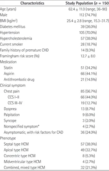
Fig. 1. A 66-year-old male with hypertrophic cardiomyopathy.
A. Serial short axis reformatted images of multidetector-row CT (MDCT) images show ventricular hypertrophy at midventricular anteroseptal wall (dashed arrows). Note the calcified plaque at mid left anterior descending artery (LAD, arrow).
B. 2-chamber view demonstrates significant stenosis at proximal to mid LAD (arrow) with mixed plaques.
C. Invasive angiography shows significant stenosis (arrow) at proximal LAD which corresponded area on MDCT.
A
B C
ble and accurate noninvasive imaging modality for the evaluation of coexistent CAD or MB in patients with HCM.
Because the most common presentation of HCM is chest pain, which mimics angina, the diagnosis of coexistent CAD is very difficult (16, 17). The ECG pattern is also diverse and can resem- ble that of MI even in the absence of obstructive CAD (18). How- ever, adult patients with HCM are known to have a higher inci- dence and severity of coexisting CAD as age increases (19). Ac- cording to earlier observations, the prevalence of CAD con- firmed by ICA is approximately 11–26% in patients with HCM (5, 19, 20), which corresponds with the result of our investiga- tion (23.3%).
In our study, age, hypertension, FHx-CHD, FRS and severe chest pain were associated with CAD in patients with HCM.
FRS provided a 10-year risk of cardiovascular events regarding multiple clinical risk factors including age, sex, smoking history, blood pressure, and lipid profiles, which have been most widely used and validated as a prediction model for CAD (21, 22).
Therefore, FRS may be a good screening tool for CAD in pa- tients with HCM. Numerous studies reported that FHx-CHD had been associated with a persistent increase in mortality risk
across long-term follow-ups (23). FHx-CHD was reported to be associated with enhanced development and progression of subclinical disease, independent of other risk factors, in a mul- tiethnic, population-based study (24).
MB is a well-recognized phenomenon that has a prevalence of 1–3% in the general population (25), though it occurs far more frequently in patients with HCM with variable prevalence rates between 15 and 41% (6, 8, 15, 25). In the present study, the prev- alence of coexistent MB was 42.7%, including partial encase- ment (14.7%) and full encasement (28.0%), in agreement with previous studies. The detection rate of MB by CCTA was signif- icantly higher than that by CAG, due to the ability of CCTA to clearly show the anatomical structure and intramyocardial lo- cation of the involved coronary arterial segment, and to detect partial encasement MB, which cannot be detected by ICA (15).
In our results, MB was frequently found in septal type HCM, indicating that hypertrophied septal myocardia have a higher chance of surrounding the mid portion of the left anterior de- scending artery. Whether MB in HCM is related to poor clini- cal outcomes has been controversial (25, 26). In studies of pedi- atric patients with HCM, MB has been associated with cardiac Table 2. Comparison of the Clinical Characteristics and Morphological Phenotype of HCM According to the Stenosis Degree of Coexistent CAD and Degree MB
Normal Coronary (n = 54)
Non-Obstructive CAD (n = 61)
Obstructive CAD
(n = 35) p-Value
Age (years)† 57.1 ± 9.3 63.1 ± 10.4 69.1 ± 10.4 < 0.001*
Male‡ 41 (75.9%) 45 (73.8%) 26 (74.3%) 0.841
BMI (kg/m2)† 24.9 ± 2.8 26.0 ± 2.7 25.1 ± 3.0 0.092
Diabetes mellitus‡ 12 (22.2%) 14 (23.0%) 13 (37.1%) 0.147
Hypertension‡ 29 (53.7%) 45 (73.8%) 30 (85.7%) 0.001*
Hypercholesterolemia‡ 15 (27.8%) 27 (44.3%) 15 (42.9%) 0.111
Current smoker‡ 10 (18.5%) 9 (14.8%) 9 (25.7%) 0.484
Family history of premature CHD‡ 3 (5.6%) 4 (6.6%) 7 (20.0%) 0.034*
Framingham risk score (%)† 9.6 ± 7.1 12.6 ± 7.8 17.7 ± 7.4 < 0.001*
Chest pain‡ 28 (51.9%) 32 (52.5%) 25 (71.4%) 0.093
CCS I–II‡ 23 (42.6%) 27 (44.3%) 14 (40.0%) 0.638
CCS III–IV‡ 5 (9.3%) 5 (8.2%) 9 (25.7%) 0.039*
Morphological phenotype of HCM
Septal type (vs. non-septal type)‡ 31 (57.4%) 29 (47.5%) 13 (37.1%) 0.061
Apical type (vs. non-apical type)‡ 29 (53.7%) 30 (49.2%) 17 (48.6%) 0.611
Data are expressed as number (%) or mean ± standard deviation.
*p < 0.05.
†Statistical significance of continuous values was tested using one-way analysis of variance among groups.
‡Statistical significance of categorical values was tested using the chi-square test among groups.
BMI = basal metabolic index, CAD = coronary artery disease, CCS = Canadian Cardiac Society, CHD = coronary heart disease, HCM = hypertrophic cardio- myopathy, MB = myocardial bridging
disease severity and an increased risk of sudden cardiac death, while no significant correlation was found in adult patients (6, 25). In our study, the presence of MB was not associated with
specific symptoms in adults with HCM. These results might re- flect the generally benign nature of MB in adults, but future stud- ies will be required to ascertain the correct prognosis of MB in
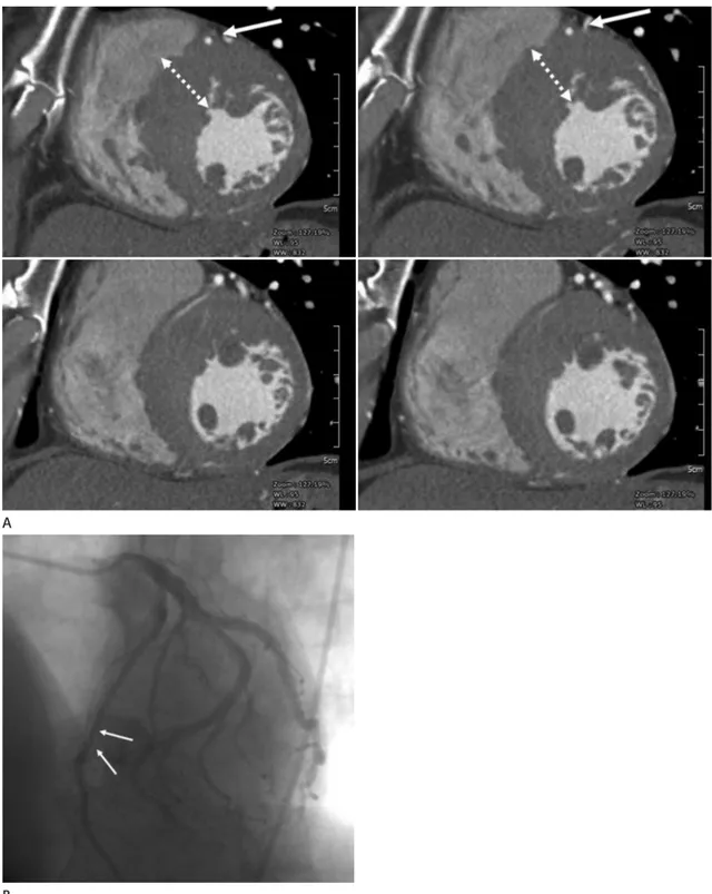
Fig. 2. A 55-year-old male with hypertrophic cardiomyopathy.
A. Serial short axis multidetector-row CT images show full encasement of mid left anterior descending artery (LAD, arrows) within hypertrophied midventricular anteroseptal wall (dashed arrows).
B. Invasive angiography demonstrates milking effect with systolic compression of LAD (arrows).
A
B
HCM.
Although ICA is generally accepted as the gold standard for diagnosing CAD (17), it is difficult to use ICA as a screening test in patients with HCM because ICA is highly invasive. Stud- ies of other noninvasive diagnostic tools, including pharmaco- logical stress echocardiography or stress thallium myocardial perfusion scan, revealed relatively high false positive rates, pos- sibly because of the innate vulnerability of hypertrophied myo- cardium for myocardial ischemia without significant CAD (1, 17). With the advantage of detailed morphological assessment with 3-dimensional views, CCTA has been an effective alterna- tive to ICA in patients with HCM, because it can evaluate both the coronary artery and the myocardium at the same time.
There were several limitations in this study. First, our study population was retrospectively analyzed and subject to bias. Sec- ond, as the decision for ICA was based on clinical reasons, not all patients with HCM underwent ICA. Thus, the diagnostic accuracy of CCTA for the evaluation of coexistent CAD or MB was not validated in all populations of our study, although CCTA showed high diagnostic accuracy in selected patients who underwent both CCTA and ICA. Finally, the lack of long- term follow-up results limited the clinical relevance of coexistent CAD or full encasement MB in patients with HCM. Further
long-term follow-up studies will be needed to determine a more concrete clinical implication.
In conclusion, approximately one-quarter of patients with HCM had coexistent obstructive CAD or full encasement MB.
CCTA can be a feasible and accurate noninvasive method for identification of coexistent CAD or MB in patients with HCM.
REFERENCES
1. Maron BJ. Hypertrophic cardiomyopathy: a systematic re- view. JAMA 2002;287:1308-1320
2. Nishimura RA, Holmes DR Jr. Clinical practice. Hypertrophic obstructive cardiomyopathy. N Engl J Med 2004;350:
1320-1327
3. Sutton MG, Tajik AJ, Smith HC, Ritman EL. Angina in idio- pathic hypertrophic subaortic stenosis. A clinical correlate of regional left ventricular dysfunction: a videometric and echocardiographic study. Circulation 1980;61:561-568 4. Lazzeroni E, Rolli A, Aurier E, Botti G. Clinical significance of
coronary artery disease in hypertrophic cardiomyopathy.
Am J Cardiol 1992;70:499-501
5. Sorajja P, Ommen SR, Nishimura RA, Gersh BJ, Berger PB, Tajik AJ. Adverse prognosis of patients with hypertrophic Table 3. Comparison of the Clinical Characteristics and Morphological Phenotype of HCM According to the Degree of the MB
None (n = 86) Partial encasement MB (n = 22) Full encasement MB (n = 42) p-Value
Age (years)† 63.9 ± 11.0 58.1 ± 11.1 61.5 ± 10.3 0.072
Male‡ 57 (66.3%) 15 (68.2%) 40 (95.2%) 0.001*
BMI (kg/m2)† 25.3 ± 2.9 25.4 ± 2.5 25.5 ± 2.9 0.953
Diabetes mellitus‡ 19 (22.1%) 5 (22.7%) 15 (35.7%) 0.115
Hypertension‡ 62 (72.1%) 12 (54.5%) 30 (71.4%) 0.764
Hypercholesterolemia‡ 35 (40.7%) 6 (27.3%) 16 (38.1%) 0.663
Current smoker‡ 14 (17.7%) 4 (18.2%) 10 (23.8%) 0.315
Family history of premature CHD‡ 11 (12.8%) 1 (4.5%) 2 (4.8%) 0.118
Framingham risk score (%)† 12.7 ± 8.1 10.3 ± 9.3 13.9 ± 7.0 0.229
Chest pain‡ 49 (57.0%) 11 (50.0%) 25 (59.5%) 0.861
CCS I–II‡ 40 (46.5%) 9 (40.9%) 17 (40.5%) 0.341
CCS III–IV‡ 9 (10.5%) 2 (9.1%) 8 (19.0%) 0.201
Morphological phenotype of HCM
Septal type (vs. non-septal type)‡ 33 (38.4%) 11 (50.0%) 29 (69.0%) 0.001*
Apical type (vs. non-apical type)‡ 46 (53.5%) 11 (50.0%) 19 (45.2%) 0.382
Data are expressed as number (%) or mean ± standard deviation.
*p < 0.05.
†Statistical significance of continuous values was tested using one-way analysis of variance among groups.
‡Statistical significance of categorical values was tested using the chi-square test among groups.
BMI = basal metabolic index, CCS = Canadian Cardiac Society, CHD = coronary heart disease, HCM = hypertrophic cardiomyopathy, MB = myocardial bridging
cardiomyopathy who have epicardial coronary artery dis- ease. Circulation 2003;108:2342-2348
6. Tio RA, Van Gelder IC, Boonstra PW, Crijns HJ. Myocardial bridging in a survivor of sudden cardiac near-death: role of intracoronary doppler flow measurements and angiogra- phy during dobutamine stress in the clinical evaluation.
Heart 1997;77:280-282
7. Angelini P, Trivellato M, Donis J, Leachman RD. Myocardial bridges: a review. Prog Cardiovasc Dis 1983;26:75-88 8. Alegria JR, Herrmann J, Holmes DR Jr, Lerman A, Rihal CS.
Myocardial bridging. Eur Heart J 2005;26:1159-1168 9. Lazzeroni E, Picano E, Dodi C, Morozzi L, Chiriatti GP, Lu C,
et al. Dipyridamole echocardiography for diagnosis of co- existent coronary artery disease in hypertrophic cardiomy- opathy. Echo-Persantine International Cooperative (EPIC) Study Group--Subproject Hypertrophic Cardiomyopathy.
Am J Cardiol 1995;75:810-813
10. Chun EJ, Choi SI, Jin KN, Kwag HJ, Kim YJ, Choi BW, et al.
Hypertrophic cardiomyopathy: assessment with MR imaging and multidetector CT. Radiographics 2010;30:1309-1328 11. Campeau L. Letter: Grading of angina pectoris. Circulation
1976;54:522-523
12. Kim KJ, Choi SI, Lee MS, Kim JA, Chun EJ, Jeon CH. The pre- valence and characteristics of coronary atherosclerosis in asymptomatic subjects classified as low risk based on tradi- tional risk stratification algorithm: assessment with coro- nary CT angiography. Heart 2013;99:1113-1117
13. Greenland P, LaBree L, Azen SP, Doherty TM, Detrano RC.
Coronary artery calcium score combined with Framingham score for risk prediction in asymptomatic individuals. JAMA 2004;291:210-215
14. Austen WG, Edwards JE, Frye RL, Gensini GG, Gott VL, Gri- ffith LS, et al. A reporting system on patients evaluated for coronary artery disease. Report of the Ad Hoc Commit- tee for Grading of Coronary Artery Disease, Council on Car- diovascular Surgery, American Heart Association. Circula- tion 1975;51(4 Suppl):5-40
15. Kim PJ, Hur G, Kim SY, Namgung J, Hong SW, Kim YH, et al.
Frequency of myocardial bridges and dynamic compression of epicardial coronary arteries: a comparison between com- puted tomography and invasive coronary angiography.
Circulation 2009;119:1408-1416
16. Maron BJ, Bonow RO, Cannon RO 3rd, Leon MB, Epstein SE. Hypertrophic cardiomyopathy. Interrelations of clinical manifestations, pathophysiology, and therapy (1). N Engl J Med 1987;316:780-789
17. Harjai KJ, Cheirif J, Murgo JP. Ischemia and atherosclerotic coronary artery disease in patients with hypertrophic car- diomyopathy: a review of incidence, pathophysiological mechanisms, clinical implications and management strat- egies. Coron Artery Dis 1996;7:183-187
18. Luzza F, Carerj S, Oreto G. Hypertrophic cardiomyopathy with persistent ST segment elevation simulating acute myo- cardial infarction. Heart 2004;90:380
19. Cokkinos DV, Krajcer Z, Leachman RD. Hypertrophic cardio- myopathy and associated coronary artery disease. Tex Heart Inst J 1985;12:147-151
20. Walston A 2nd, Behar VS. Spectrum of coronary artery dis- ease in idiopathic hypertrophic subaortic stenosis. Am J Car- diol 1976;38:12-16
21. D’Agostino RB Sr, Grundy S, Sullivan LM, Wilson P; CHD Risk Prediction Group. Validation of the Framingham coronary heart disease prediction scores: results of a multiple ethnic groups investigation. JAMA 2001;286:180-187
22. Wilson PW, D’Agostino RB, Levy D, Belanger AM, Silbershatz H, Kannel WB. Prediction of coronary heart disease using risk factor categories. Circulation 1998;97:1837-1847 23. Bachmann JM, Willis BL, Ayers CR, Khera A, Berry JD. Asso-
ciation between family history and coronary heart disease death across long-term follow-up in men: the Cooper Cen- ter Longitudinal Study. Circulation 2012;125:3092-3098 24. Pandey AK, Blaha MJ, Sharma K, Rivera J, Budoff MJ, Blank-
stein R, et al. Family history of coronary heart disease and the incidence and progression of coronary artery calcifica- tion: Multi-Ethnic Study of Atherosclerosis (MESA). Athero- sclerosis 2014;232:369-376
25. Sorajja P, Ommen SR, Nishimura RA, Gersh BJ, Tajik AJ, Holmes DR. Myocardial bridging in adult patients with hy- pertrophic cardiomyopathy. J Am Coll Cardiol 2003;42:
889-894
26. Yetman AT, McCrindle BW, MacDonald C, Freedom RM, Gow R. Myocardial bridging in children with hypertrophic cardiomyopathy--a risk factor for sudden death. N Engl J Med 1998;339:1201-1209
비후성 심근증을 가진 성인 환자에서 동반되는 관상동맥질환과 심근교락: 다중검출기 심장 CT를 통한 연구
이재환
1· 전은주
1· 김여군
1· 유진영
1· 최상일
1· 최동주
2목적: 심장 전산화단층촬영을 통해 비후성 심근증을 가진 성인 환자에서 동반되는 관상동맥질환과 심근교락의 빈도를 알아보고, 심장 전산화단층촬영의 역할을 알아보고자 한다.
대상과 방법: 관상동맥질환이 의심되어 심장 전산화단층촬영을 시행한 19588명의 환자 중 임상양상, 심전도 및 심에코로 비후성 심근증이 확인된 150명의 환자를 대상으로, 협착성 관상동맥질환(50% 이상의 협착)과 심근교락(부분 및 완전 포 위)의 빈도를 조사하였다.
결과: 심장 전산화단층촬영을 통해 전체 환자의 98.9%에서 관상동맥의 관찰이 가능하였다. 비후성 심근증 환자에서 협 착성 관상동맥과 심근교락의 빈도는 각각 23.3%와 42.7%(부분 및 완전포위 심근교락의 빈도는 14.7%와 28.0%)였다.
연령, 고혈압, 심장질환에 의한 가족력, 프래밍험 위험지수 및 심한 흉통 등이 협착성 관상동맥과 관련되며, 남성과 격벽형 비후가 완전포위의 심근교락과 유의하게 관련이 있었다. 침습성 관상동맥조영술(37예)과 비교할 때 협착성 관상동맥과 완전포위 심근교락의 심장 전산화단층촬영의 진단의 정확도는 각각 89.2%와 86.5%였다.
결론: 비후성 심근증 환자의 약 1/4에서 협착성 관상동맥과 완전포위 심근교락이 동반된다. 심장 전산화단층촬영은 비후 성 심근증 환자에서 동반되는 협착성 관상동맥과 심근교락을 진단하는 데 가능성 있는 정확한 비침습적 방법으로 사료된다.
분당서울대학교병원 1영상의학과, 2심장내과