137
<증례보고>
Ocular coherence tomography를 이용한 개의 망막변성증 평가
지동범1·유석종2·서강문3·정만복3,*,†
1지동범동물병원, 2유림동물병원, 3서울대학교 수의과대학
(접수: 2013년 3월 11일, 수정: 2013년 4월 30일, 게재승인: 2013년 5월 15일)
Assessment of retinal degeneration with optical coherence tomography in a dog
Dongbeom Ji1, Sukjong Yoo2, Kangmoon Seo3, Manbok Jeong3,*,†
1Jidongbeom Animal Clinic, Busan 609-310, Korea
2Yoolim Animal Clinic, Seoul 137-909, Korea
3Department of Veterinary Clinical Science, College of Veterinary Medicine, Seoul National University, Seoul 151-742, Korea
(Received: March 11, 2013; Revised: April 30, 2013; Accepted: May 15, 2013)
Abstract : To report retinal image of in a 6-year-old male castrated poodle dog with a 2-month history of nyctalopia using optical coherence tomography (OCT). Ocular reflexes were present in both eyes and slit lamp examination showed anterior subscapular cataract in the right eye. There were fundus abnormalities in both eyes similar to retinal degeneration. Scotopic electroretinograms (ERGs) revealed significantly subabnormal amplitudes and prolonged implicit time, whereas photopic ERGs were better maintained, although far from normal. OCT of affected dogs revealed generalized retinal thinning much more than a normal age-matched dog. Therefore, OCT scanning is considered to be a useful method for retinal evaluation in dogs with retinal degeneration.
Keywords : dogs, electroretinography, nyctalopia, optical coherence tomography, retinal degeneration
Ocular coherence tomography(OCT)는 비침습적, 비접촉성 방법으로 동공을 통하여 망막을 고해상도로 촬영하여 망막 과 시신경판의 구조를 정량적으로 평가할 수 있는 진단 장 비이다 [6]. 이 기술은 의학과 수의학에서 망막연구자나 임 상가들이 시간의 흐름에 따라 망막과 시신경 질환을 모니터 링하고 진단하는 데 이용하고 있다. 이와 같이 망막 내층 구 조를 평가하는 고해상도 기법은 개에서 진행성 망막위축증 (progressive retinal atrophy)과 녹내장, 사람에서 황반부종, 상망막증, 초자체-황반당김증 및 망막하액 등과 같은 다양한 망막질환에 대한 새로운 치료법의 효과를 평가하는데 이용 되고 있다 [4, 8, 9]. 최근에 개에서 OCT를 이용한 개의 정상 망막의 두께에 관한 연구 및 개의 진행성 망막위축증 과 같은 유전성 망막질환의 특성과 새로운 치료법에 관한 연 구가 보고 되고 있다 [7, 9-11]. 그러나 소동물 안과에서 OCT를 이용한 개의 망막질환의 임상증례에 대한 연구는 저 조한 실정이다. 그러므로 본 연구의 목적은 OCT를 이용하 여 개의 망막변성증의 평가에 대하여 보고하고자 한다.
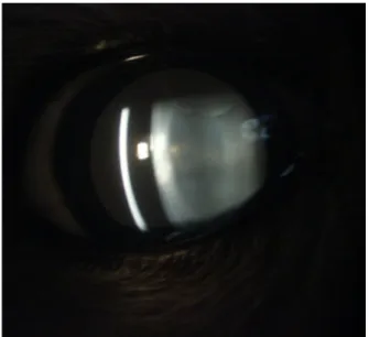
6살 중성화 수컷 Poodle이 2개월 전부터 어두운 곳에서만 사물에 부딪히는 행동양상으로 내원하였다. 어두운 곳에서 실시한 장애물 검사에서 시각이상을 나타냈다. 양안의 위협 반응, 기타 안신경 검사 및 눈물량과 안압(TonoVet; iCare, Espoo, Finland)은 정상이었다. 전안부 검사를 위한 세극등현 미경(BQ900; HAAG-STREIT, Switzerland) 검사에서 우안 의 전낭하 백내장이 관찰되었으며 기타 각막, 전안방 및 홍 채는 정상이었다(Fig. 1). 산동제(Mydrin-P; Taejoon Pharm, Korea) 투여 20분 후에 도상검안경(Omega 500; Heine, Germany) 검사를 실시하고 안저 사진(VX-10α; Kowa, Japan)을 촬영하였다. 양안 반사판의 과반사, 혈관 협소화, 반 사판 주변부의 변색, 비반사판의 탈색소화 및 시신경판 주위 의 위축이 관찰되었다(Fig. 2).
도상검안경 검사 1시간 후에 isoflurane을 이용한 전신마취 상태에서 우안의 망막전위도(RETI-Port; Roland Consult, Germany) 검사를 실시하였다. 망막전위도 검사는 크게 암순 응시(암소시, scotopic vision)과 명순응시(명소시, photopic
*Corresponding author
Tel: +1-515-708-8432, Fax: +1-515-294-7520 E-mail: [email protected]
†Present address: Department of Veterinary Clinical Sciences, College of Veterinary Medicine, Iowa State University, Ames, IA 50011, USA
vision) 망막전위도 검사로 나누어진다 [13]. 암순응시 망막전 위도 검사는 간상세포반응(rod response)과 간상세포-원추세 포혼합 반응(mixed rod-cone response)으로 나누어진다. 먼 저 간상세포반응은 20분 동안 암순응 상태에서 0.025 cd·s/m2 의 빛 강도로 암순응 1분, 5분, 10분, 15분, 20분에 간상세 포 기능을 평가하였다. 간상세포-원추세포 혼합반응은 2.5 cd·s/m2 의 빛 강도로 그 기능을 평가하였다. 명순응시 망막전위도 검사는 원추세포반응(single flash cone response) 과 원추세포 명멸반응(cone flicker response)으로 나누어진다.
10분 동안 25 cd/m2의 빛 강도로 명순응 하고 그 빛 강도 하에서 두 검사를 실시하였다. 원추세포반응은 2.5 cd·s/m2의 빛 강도로 검사하며 원추세포 명멸반응은 2.5 cd·s/m2의 빛 강도를 31 Hz로 명멸하여 검사하였다.
암순응시와 명순응시 망막전위도에 기록된 파형은 모든 반 응에서 정상이었다(Fig. 3). 암순응시 망막전위도 검사의 간
상세포반응에서 b파 진폭(amplitude)과 함의점 시간(implicit time)은 암순응 1분 후에 18.5 µV와 40.0 ms, 5분 후에 11.7 µV과 41.0 ms, 10분 후에 15.7 µV과 44.0 ms, 15분 후에 24.4 µV와 39.0 ms, 20분 후에 11.6 µV과 48.0 ms이 었다. 간상세포-원추세포혼합반응에서 a파 진폭과 함의점 시 간은 8.4 µV와 16.0 ms이었으며 b파 진폭과 함의점 시간은 23.7 µV과 37.0 ms이었다. 원추세포반응에서 a파 진폭과 함 의점 시간은 7.3 µV과 11.0 ms이었으며 b파 진폭과 함의점 시간은 12.1 µV과 30.0 ms이었다. 원추세포 명멸반응에서 b 파 진폭과 함의점 시간은 15.5 µV과 28.0 ms이었다.
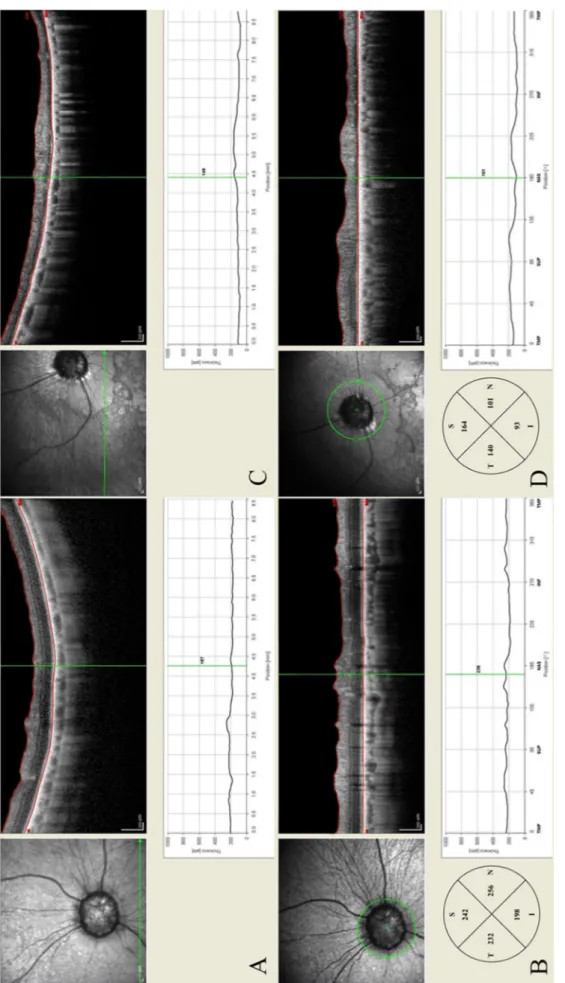
망막전위도 측정 직후에 전신마취를 유지하면서 OCT (Spectralis; Heidelberg Engineering, Germany)를 이용하여 병변이 가장 명확한 시신경판 주위(peripapillary circle scan) 와 비반사판의 망막두께를 측정하였다. 나이가 유사한 정상 Shih Tzu의 망막은 모든 층을 정확히 구별할 수 있을 정도 로 명확하였으나 환자의 망막은 각 층을 구별 할 수 없을 정도로 각 세포층이 변성되었다(Fig. 4). 정상견과 환자의 비 반사판 망막두께는 각각 187.0 µm와 149.0 µm, 시신경판 주 위 망막두께는 코쪽 256.0 µm와 101.0 µm, 외측 232.0 µm 와 140.0 µm, 등쪽 242.0 µm와 164.0 µm, 배쪽 198.0 µm 와 93.0 µm이었다(Fig. 4). 이러한 안검사를 바탕으로 본 증 례의 환자는 양측성 망막변성증으로 진단되었다.
OCT는 안저 사진촬영과 형광 안저촬영 등과 같은 진단법 을 보완하여 망막질환 진단에 우수한 정보를 제공한다 [6].
그러므로 본 연구에서는 빠르고 비침습적이며 고해상도의 망 막구조 사진을 제공하여 살아있는 생명체의 안저 구조를 평 가하는 OCT와 망막의 생리적 기능을 평가하는 망막전위도 를 함께 활용하여 망막질환을 보다 더 정확하게 진단하였다.
본 연구의 환자는 야맹증이 발생하였으나 위협반응은 정 상이었다. 위협반응을 비롯한 다른 안신경 검사는 환자의 민 감도, 검사자의 숙련도, 진료실의 환경 및 광원 등과 같은 여러 가지 요인에 영향을 받는다. 그러므로 일반적인 안신경 검사를 바탕으로 정확하게 시력여부를 평가하기는 어렵다.
그러나 본 환자는 각막에서 초자체까지 빛이 통과하는 것을 Fig. 1. Slit lamp biomicroscopic image of right eye in a 6-
year-old Poodle. Note increase in lens opacity on the anterior subcapsular area.
Fig. 2. Fundus photographs of right eye with retinal degeneration in a 6-year-old Poodle. A: Diffuse hyper-reflectivity, vascular attenuation, severe peripapillary atrophic ring, mild optic disc atrophy and non-tapetal depigmentation were shown. B: Tapetum.
Note the distinct discoloration of peripheral tapetum.
방해하는 요인으로 우안에 전낭하 백내장이 관찰되었으나 이 것은 시력에 영향을 미칠 만큼 심하지 않았고 주간에 밝은 곳에서는 시력이 존재하였으므로 위협반응이 있었을 가능성 이 매우 크다. 본 증례에서 도상검안경을 이용하여 반사판의 과반사, 혈관협소화, 반사판 주변부의 변색, 비반사판의 탈색 소화 및 시신경판 주위의 위축 등과 같은 망막변성을 관찰 할 수 있었다. 예전 연구에 의하면 대체로 망막변성증은 반 사판의 중간과 주변부분, 비반사판의 경계 부분에서 색깔이 회색으로 변한다. 그 이후에 혈관의 협소화로 진행되고 질환 이 심해지면 비반사판의 색소가 손상되고 과색소화 되는 안 저 변화를 나타낸다 [14]. 본 증례에서 비반사판의 안저 변 화가 기존 연구의 후기 단계 망막변성의 안저 변화와 유사하 게 나타났다.
이와 같이 안저 변화가 있으며 안구 내 매질에 빛 통과를 방해하는 요소가 없고 명확한 실명의 원인을 찾아내기 어려 운 경우에 망막전위도를 이용하여 망막의 기능을 평가한다 [5]. 본 연구에서 암순응시 망막전위도 검사의 간상세포반응 에서 b파 진폭은 매우 심하게 감소되었으며 함의점 시간은 지연되었다. 특히 20분 암순응 하는 동안에 b파 진폭이 전혀 증가하지 않았다. 이것은 간상세포와 망막색소상피세포 사이 에서 일어나는 간상세포 시각경로(rod visual cycle)가 비정 상적으로 작동하여 간상세포의 기능이 거의 상실된 것을 의 미한다 [16]. 원추세포 명멸반응을 포함한 명순응시 망막전
위도 검사에서 진폭은 매우 감소하고 함의점 시간은 지연되 었지만 간상세포에 비하여 감소 정도가 매우 낮고 파형이 정 확하게 기록되었다. 본 환자의 임상적 야맹증과 망막전위도 결과를 종합하여 분석하면 기능을 하는 원추세포의 수가 많 이 감소하였지만 어느 정도 기능이 가능한 세포는 존재한다 는 것을 의미한다. 그러므로, 본 환자는 주간에 밝은 곳에서 시력이 있었을 것이다.
오래 전에 Poodle에서 유전성 망막질환인 진행성 망막위 축증의 한 부류인 진행성 간상-원추세포 변성증(progressive rod-cone degeneration)이 보고되었다 [1-3, 17]. 이것은 시각 세포가 정상적으로 분화한 직후에 광수용체 외분절막의 손 상 및 파괴를 의심할 수 있는 층판 해체와 소포가 형성되어 망막이 변성하는 유전성 질환이다. 시간이 지나면서 원추세 포 보다 간상세포가 더 광범위하며 빠르게 퇴행된다 [1].
Poodle에 대한 예전 연구 보고에 의하면 망막전위도 기능 이상은 생후 9.5주에 나타났다 [2]. 다른 연구에서는 생후 28주부터 나타나기 시작하여 18개월에 간상세포와 원추세포 에 대한 반응에서 단지 b파 진폭이 낮게 기록되었으며 암순 응이 진행되면서도 진폭이 상승하지 않았다 [1]. 또 다른 연 구는 Poodle의 진행성 간상-원추세포 변성증을 3단계로 나누 어 실험을 진행하였다 [17]. 3단계에 속하는 Poodle에서 간 상세포의 진폭이 매우 심하게 감소하였거나 파형이 기록되 지 않았으며 원추세포는 진폭이 감소되었다. 본 연구의 간상 Fig. 3. Scotopic and photopic electroretinogram (ERG) waveforms obtained from the right eye in a 6-year-old Poodle with retinal degeneration. Scotopic ERGs (A: rod response and B: mixed rod-cone response) and photopic ERGs (C: single flash cone response and D: 31 Hz cone flicker response).
Fig. 4. OCT scan images. A and B: a 6-year-old normal Shih Tzu. C and D: a 6-year-old Poodle with retinal degeneration in the study. Note significant thinning of both peripapillary and non-tapetal retinal areas in the case (C and D) compared to A and B. The green line in the left fundus image shows the scanned area depicted on the right side of the figure. N (NAL): nasal, S (SUP): superior, I (INF): inferior, T (TEM): temporal.
세포반응 망막전위도 결과는 예전 연구의 3단계의 안저 변 화를 가진 개체의 망막전위도 결과와 동일하다. 또한 여러 연구간의 망막전위도 차이는 이 질환이 동일한 질병이지만 한 품종에서도 질환의 발생 양상이 다양하게 진행되는 이질 성(heterogeneity) 때문에 나타난다.
OCT를 이용하여 시신경판을 포함한 망막의 구조적인 특 징과 질환의 진행, 치료 및 예후를 평가할 수 있다. 이 검사 방법은 망막 전체 두께를 측정할 수 있을 뿐만 아니라 망막 신경절 세포(retinal ganglion cell) 같이 특정 세포층의 두께 를 정밀하게 측정할 수 있다 [9]. 본 증례 환자는 정상견보 다 비반사판 망막두께는 20.4%, 시신경판 주위의 코쪽 60.6%, 배쪽 53.1%, 외측 39.7%, 등쪽 32.3% 감소하여 코 쪽에서 가장 많이 감소하였다. 그러나 이러한 두께 감소보다 더 중요한 것은 정상견의 OCT영상과 비교하여 본 증례는 비반사판과 시신경판 주위 망막의 각 세포층을 전혀 구별할 수 없을 정도로 망막세포가 완전히 변성되었다. 본 증례 망 막의 조직병리학적 소견과 OCT결과를 비교하지 못하였지만 예전 연구에 의하면 10세 Miniature Longhaired Dachshund 의 진행성 망막위축증의 조직병리 소견과 OCT영상이 일치 하는 것을 확인하였다 [12]. 본 연구에서 정상견 망막두께는 예전 연구에서 측정된 망막두께 보다 더 두꺼웠다 [9, 10, 15]. 이것은 품종 혹은 연령간의 차이에 의해 발생했을 것으 로 판단된다.
본 증례에서는 야맹증이 발생한 Poodle의 도상검안경을 이 용한 안저 변화, 망막전위도 검사를 이용한 생리적 검사 및 OCT를 이용한 망막의 구조적 변화를 정상견과 비교하여 보 다 체계적인 정밀 진단 과정에 대하여 보고하였다. 특히 수 의안과학에서 OCT는 일반 안과검사를 통하여 망막질환의 정 확한 진단이 어려울 때 망막의 각 세포층 두께를 측정하여 그 변화 여부를 제시함으로써 망막질환의 임상적 진단, 치료 의 예후 평가 및 망막 연구에 유용하게 이용할 수 있을 것 이다.
References
1. Aguirre G, Alligood J, O'Brien P, Buyukmihci N.
Pathogenesis of progressive rod-cone degeneration in miniature poodles. Invest Ophthalmol Vis Sci 1982, 23, 610- 630.
2. Aguirre GD, Rubin LF. Progressive retinal atrophy in the miniature poodle: an electrophysiologic study. J Am Vet Med Assoc 1972, 160, 191-201.
3. Barnett KC. Canine retinopathies. II. The miniature and toy poodle. J Small Anim Pract 1965, 6, 93-109.
4. Csaky KG, Richman EA, Ferris FL 3rd. Report from the NEI/FDA ophthalmic clinical trial design and endpoints symposium. Invest Ophthalmol Vis Sci 2008, 49, 479-489.
5. Ekesten B. Electrodiagnostic Evaluation of Vision. In: Gelatt
KN (ed.). Veterinary Ophthalmology. 4th ed. pp. 520-535, Blackwell, Oxford, 2007.
6. Gabriele ML, Wollstein G, Ishikawa H, Kagemann L, Xu J, Folio LS, Schuman JS. Optical coherence tomography:
history, current status, and laboratory work. Invest Ophthalmol Vis Sci 2011, 52, 2425-2436.
7. Gekeler F, Gmeiner H, Völker M, Sachs H, Messias A, Eule C, Bartz-Schmidt KU, Zrenner E, Shinoda K.
Assessment of the posterior segment of the cat eye by optical coherence tomography (OCT). Vet Ophthalmol 2007, 10, 173- 178.
8. Grahn BH, Sandmeyer LL, Breaux C. Retinopathy of Coton de Tulear dogs: clinical manifestations, electroretino- graphic, ultrasonographic, fluorescein and indocyanine green angiographic, and optical coherence tomographic findings.
Vet Ophthalmol 2008, 11, 242-249.
9. Grozdanic SD, Matic M, Betts DM, Sakaguchi DS, Kardon RH. Recovery of canine retina and optic nerve function after acute elevation of intraocular pressure: implications for canine glaucoma treatment. Vet Ophthalmol 2007, 10 (Suppl 1), 101-107.
10. Hernandez-Merino E, Kecova H, Jacobson SJ, Hamouche KN, Nzokwe RN, Grozdanic SD. Spectral domain optical coherence tomography (SD-OCT) assessment of the healthy female canine retina and optic nerve. Vet Ophthalmol 2011, 14, 400-405.
11. Le Meur G, Stieger K, Smith AJ, Weber M, Deschamps JY, Nivard D, Mendes-Madeira A, Provost N, Péréon Y, Cherel Y, Ali RR, Hamel C, Moullier P, Rolling F.
Restoration of vision in RPE65-deficient Briard dogs using an AAV serotype 4 vector that specifically targets the retinal pigmented epithelium. Gene Ther 2007, 14, 292-303.
12. Lhériteau E, Libeau L, Stieger K, Deschamps JY, Mendes- Madeira A, Provost N, Lemoine F, Mellersh C, Ellinwood NM, Cherel Y, Moullier P, Rolling F. The RPGRIP1- deficient dog, a promising canine model for gene therapy. Mol Vis 2009, 15, 349-361.
13. Narfström K, Ekesten B, Rosolen SG, Spiess BM, Percicot CL, Ofri R. Guidelines for clinical electroretinography in the dog. Doc Ophthalmol 2002, 105, 83-92.
14. Narfström K, Petersen-Jones SM. Diseases of the canine ocular fundus. In: Gelatt KN (ed.). Veterinary ophthalmology.
4th ed. pp. 944-1025, Blackwell, Oxford, 2007.
15. Panzan CQ, Güven D, Weiland JD, Lakhanpal RR, Javaheri M, de Juan E Jr, Humayun MS. Retinal thickness in normal and RCD1 dogs using optical coherence tomography. Ophthalmic Surg Lasers Imaging 2004, 35, 485- 493.
16. Saari JC. Biochemistry of visual pigment regeneration: the Friedenwald lecture. Invest Ophthalmol Vis Sci 2000, 41, 337-348.
17. Sandberg MA, Pawlyk BS, Berson EL. Full-field electroretinograms in miniature poodles with progressive rod- cone degeneration. Invest Ophthalmol Vis Sci 1986, 27, 1179- 1184.