ISSN 0378-6471 (Print)⋅ISSN 2092-9374 (Online)
https://doi.org/10.3341/jkos.2017.58.3.296
Original Article
나이관련황반변성에서 라니비주맙과 애프리버셉트 후 맥락막두께의 변화 비교: 6개월 결과
Comparison of Choroidal Thickness Change between Ranibizumab and Aflibercept in Age-related Macular Degeneration: Six Month Results
김임규⋅김용일⋅김진선⋅이정호⋅이규원⋅강현구
Im Gyu Kim, MD, Yong Il Kim, MD, Jin Seon Kim, MD, Jung Ho Lee, MD, Kyoo Won Lee, MD, PhD, Hyun Gu Kang, MD
제일안과병원 Cheil Eye Hospital, Daegu, Korea
Purpose: To compare the changes in subfoveal choroidal thickness between intravitreal aflibercept and ranibizumab injection in wet age-related macular degeneration (AMD).
Methods: Fifty patients with wet AMD patients who were treated with aflibercpet or ranibizumab were evaluated retrospectively.
All patients were treated with pro re nata after 3 consecutive monthly injections and were followed up for at least 6 months. We measured subfoveal choroidal thickness (SFCT) using enhanced depth imaging spectral-domain optical coherence tomography before the first injection and 1, 2, 3, and 6 months after initial injection.
Results: The SFCT measures before initial injection and 1, 2, 3, and 6 months after initial injection were 244.94 ± 103.77 μm, 219.04 ± 95.89 μm, 208.74 ± 91.03 μm, 203.64 ± 91.35 μm, and 226.98 ± 96.79 μm in the aflibercept group (25 eyes) and 222.68
± 102.04 μm, 210.23 ± 95.91 μm, 203.66 ± 99.39 μm, 197.27 ± 100.25 μm, and 210.32 ± 111.86 μm in the ranibizumab group (28 eyes). Mean change in SFCT was greater in the aflibercept group at 1 month, 2 months, and 3 months after initial injection (p <
0.05), but there was no significant difference in the mean change in SFCT between the two groups at 6 months after initial in- jection (p > 0.05).
Conclusions: The decrease in SFCT was greater after aflibercept injection than ranibizumab injection in 3 consecutive months.
However, at 6 months after initial injection, the difference in the change in SFCT was not significant.
J Korean Ophthalmol Soc 2017;58(3):296-304
Keywords: Aflibercept, Polypoidal choroidal vasculopathy, Ranibizumab, Subfoveal choroidal thickness, Wet age-related mac- ular degeneration
■Received: 2016. 8. 18. ■ Revised: 2016. 11. 18.
■Accepted: 2017. 1. 23.
■Address reprint requests to Hyun Gu Kang, MD
Cheil Eye Hospital, #1 Ayang-ro, Dong-gu, Daegu 41196, Korea Tel: 82-53-959-1751, Fax: 82-53-959-1758
E-mail: [email protected]
* This study was presented as a poster at the 113th Annual Meeting of the Korean Ophthalmological Society 2015.
ⓒ2017 The Korean Ophthalmological Society
This is an Open Access article distributed under the terms of the Creative Commons Attribution Non-Commercial License (http://creativecommons.org/licenses/by-nc/3.0/) which permits unrestricted non-commercial use, distribution, and reproduction in any medium, provided the original work is properly cited.
나이관련황반변성은 서구에서 노인의 실명에 가장 흔한 요인으로 알려져 있다.1,2 최근 국내에서도 나이관련황반변 성 환자가 크게 증가하여 서구와 비슷한 양상을 보이는 것 으로 알려져 있다.3 Rosenfeld et al4과 Brown et al5의 연구 결과가 발표된 후 유리체강내 항혈관내피성장인자 주입술 은 삼출성 나이관련황반변성의 주 치료가 되었으며, 라니 비주맙(Lucentis®, Genentech Inc., South San Fransisco, CA, USA)과 베바시주맙(Avastin®, Genentech Inc., Oceanside,
CA, USA)이 사용되었다. 유리체강내 라니비주맙과 베바시 주맙 주입술은 나이관련황반변성 환자의 시력을 유지하고 시력소실을 예방하는 데 효과가 있는 것으로 보고되었다.6-9
최근 새로운 항혈관내피성장인자인 애프리버셉트(Eylea®, Regeneron Pharmaceuticals, Inc., Tarrytown, NY, USA)가 소개되었는데, 삼출성 나이관련황반변성의 치료에서 애프 리버셉트를 1개월 간격으로 3회 주사 후 2개월 간격으로 주사하는 방법은 라니비주맙을 매달 주사하는 방법과 비슷 한 효과를 보였다.10
혈관내피성장인자는 맥락막 모세혈관 및 맥락막 혈류의 유지에 중요한 역할을 한다.11,12 따라서 유리체강내 항혈관 내피성장인자 주입술은 맥락막두께에 영향을 줄 것이다.
삼출성 나이 관련 황반변성 환자에서 유리체강내 라니비주 맙주입술 후 황반하 맥락막두께는 감소하였고13 애프리버 셉트 주입술 후에도 맥락막두께는 감소하였다.14 최근 유리 체강내 라니비주맙 또는 애프리버셉트 주입술 후 맥락막두 께의 변화를 비교한 연구가 보고되었는데, 한 달 간격으로 3회 주사 후 애프리버셉트 주입군에서 맥락막두께가 더 많 이 감소하였다.15,16 본 연구에서는 이전 치료의 과거력이 없 는 삼출성 나이관련 황반변성 환자에서 한 달 간격으로 3 회 유리체강내 라니비주맙 또는 애프리버셉트 주입술 후 맥락막두께의 변화가 이전의 연구 결과와 일치하는지 확인 해보고, 3회 연속 주사 후 필요할 때마다 주사하면서 경과 관찰 시 치료 시작 6개월 후 맥락막두께의 변화를 비교해 보고자 한다.
대상과 방법
2014년 2월부터 2015년 11월까지 제일안과병원을 방 문하여 삼출성 나이관련황반변성으로 진단 받고 유리체 강내 라니비주맙 또는 애프리버셉트 주입술 시행 후 6개 월 이상 경과관찰된 환자 50명 53안을 대상으로 후향적으로 분석하였다. 본 연구는 제일안과병원 임상연구심의위원회 (Institutional Review Board)의 승인을 통해 진행되었으며 (승인번호: CEH-2014-7), 헬싱키선언을 준수하였다. 처음 삼출성 나이관련황반변성으로 진단된 경우만 연구에 포함 시켰으며, 이전에 삼출성 나이관련황반변성으로 진단받고 치료받은 경우는 제외하였다. 증식당뇨망막병증, 망막혈관 폐쇄, 황반원공, 망막앞막 등 황반의 구조와 기능에 영향을 미칠 수 있는 망막질환이 동반된 경우는 대상에서 제외하 였다.
모든 환자들은 진단 시 최대교정시력검사(logarithm of the minimum angle of resolution, logMAR), 세극등현미경검사 (Slit lamp 900 BQ; Hagg-Streit AG, Koeniz, Switzerland),
자동굴절검사기(Canon, Inc., Tokyo, Japan)를 사용한 굴절 력검사, 안저검사 및 천연색안저사진촬영(Digital retinal camera CR-2, Canon, Inc., Tokyo, Japan), 안압측정(Full auto tonometer TX-F, Canon, Inc., Tokyo, Japan), 스펙트럼영역 빛간섭단층촬영과 형광안저혈관조영 및 인도사이아닌그린 혈관조영(Spectralis® Heidelberg retinal angiography [HRA]–
optical coherence tomography [OCT], Heidelberg Engineering, Heidelberg, Germany)을 시행하였다.
유리체강내 주입술은 외래 수술실에서 시행하였고, 주입 술 전 0.3% levofloxacin (Cravit®, Santen, Osaka, Japan)과 0.5% proparacaine hydrochloride (Alcaine®, Alcon, Fort Worth, TX, USA)를 점안한 뒤 5% povidone iodine을 점안 하고 눈 주위를 소독하였다. 수술포를 씌운 후 개검기를 끼 우고 30게이지 주사바늘을 이용하여 각막 윤부에서 3.5 mm 떨어진 부위에 라니비주맙 0.05 mL (0.5 mg) 또는 애프리 버셉트 0.05 mL (2 mg)를 주사하였다. 모든 환자들은 1개 월 간격으로 3회 유리체강내 주입술을 시행 받았으며, 첫 번째 주사 후 1개월마다 최대교정시력, 세극등현미경검사, 안압측정, 스펙트럼영역 빛간섭단층촬영을 시행하면서 추 적 관찰하였다. 1개월 간격으로 3회 유리체강내 주입술 후 추적 관찰 시 중심망막두께가 100 μm 이상 증가하거나, 망 막하액으로 인해 시력이 1줄 이상 감소하거나, 새로운 맥락 막신생혈관이 관찰되거나, 새로운 황반 출혈이 관찰되는 경우 추가 치료를 시행하였다.
대상환자들의 중심황반두께 및 황반하 맥락막두께는 Spectralis® HRA–OCT로 촬영한 영상을 이용하여 측정하 였다. 중심황반두께는 내장된 프로그램을 이용하여 측정 하였고, 황반하 맥락막두께는 enhanced depth imaging (EDI) 방법으로 촬영한 영상에서 중심와 위치의 고반사선 을 보이는 망막색소상피층의 외측경계와 내측공막경계의 고반사선까지의 수직거리를 두 명의 연구자가 각각 수동으 로 측정하여 평균값을 계산하였다.
통계적 분석은 표준 소프트웨어(SPSS version 18.0 for Windows, SPSS Inc., Chicago, IL, USA)를 사용하였고, 반 복측정 분산분석(repeated measure analysis of variance)을 사용하여 치료 전과 치료 후 경과관찰 기간 동안 라니비주 맙과 애프리버셉트 각 군의 치료 효과 및 두 군 간의 차이 를 분석하였고, 두 군 간의 차이가 발생하는 시점을 확인하 기 위해 사후검정으로 독립표본 t 검정을 사용하여 분석하 였다. 두 군 간의 초기 지표 비교를 위해 정규성 검정 후 독립표본 t 검정 및 Mann-Whitney U-test를 사용하였고, 성 별, 안구방향, 당뇨, 고혈압의 비교는 카이제곱검정을 이용 하였으며, 유의수준 5% 미만(p<0.05)인 경우 통계학적으로 유의한 것으로 정의하였다.
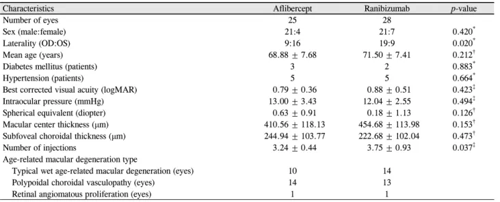
Table 1. Demographics of the study groups
Characteristics Aflibercept Ranibizumab p-value
Number of eyes 25 28
Sex (male:female) 21:4 21:7 0.420*
Laterality (OD:OS) 9:16 19:9 0.020*
Mean age (years) 68.88 ± 7.68 71.50 ± 7.41 0.212†
Diabetes mellitus (patients) 3 2 0.883*
Hypertension (patients) 5 5 0.664*
Best corrected visual acuity (logMAR) 0.79 ± 0.36 0.88 ± 0.51 0.423‡
Intraocular pressure (mmHg) 13.00 ± 3.43 12.04 ± 2.55 0.494‡
Spherical equivalent (diopter) 0.63 ± 0.91 0.18 ± 1.13 0.126†
Macular center thickness (μm) 410.56 ± 118.13 454.68 ± 113.98 0.153†
Subfoveal choroidal thickness (μm) 244.94 ± 103.77 222.68 ± 102.04 0.473†
Number of injections 3.24 ± 0.44 3.75 ± 0.93 0.037‡
Age-related macular degeneration type
Typical wet age-related macular degeneration (eyes) 10 14
Polypoidal choroidal vasculopathy (eyes) 14 13
Retinal angiomatous proliferation (eyes) 1 1
Values are presented as mean ± SD unless otherwise indicated.
OD = oculus dexter; OS = oculus sinister.
*Chi-square test; †Student’s t-test; ‡Mann-Whitney U-test.
결 과
전체 50명 53안을 대상으로 분석을 시행하였으며, 남자 40명(80%), 여자 10명(20%)이었고, 평균 연령은 70.26 ± 7.58세였다. 애프리버셉트 주입군은 25안(47.17%)이었으 며, 라니비주맙 주입군은 28안(52.83%)이었다. 치료 전 두 군 간 연령, 성별, 당뇨, 고혈압, 최대교정시력, 안압, 구면 렌즈대응치, 중심황반두께, 황반하 맥락막두께의 값은 유의 한 차이를 보이지 않았으며(p>0.05), 경과관찰 기간 동안 유리체강내 주입술 횟수는 애프리버셉트 주입군이 좀 더 적었다(p=0.037; Table 1).
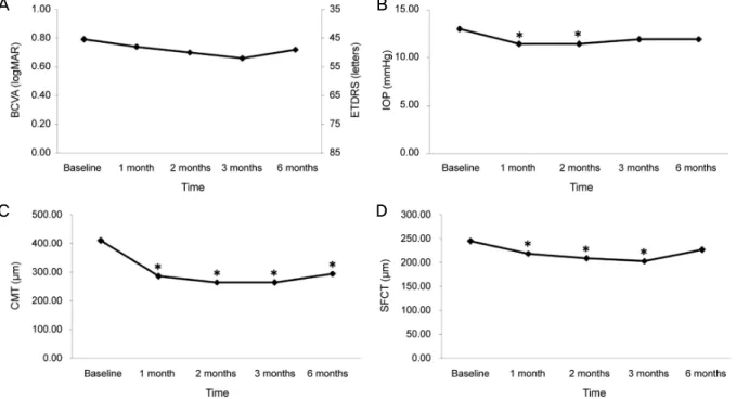
애프리버셉트 주입군의 황반하 맥락막두께는 주사 전과 첫 번째 주사 후 1개월, 2개월, 3개월, 6개월에 각각 244.94
± 103.77 μm, 219.04 ± 95.89 μm, 208.74 ± 91.03 μm, 203.64 ± 91.35 μm, 226.98 ± 96.79 μm였으며, 첫 번째 주 입술 후 3개월까지는 치료 전보다 유의하게 감소되었으며 (p<0.05), 6개월째 경과관찰 시에는 유의한 차이를 보이지 않았다(Fig. 1).
라니비주맙 주입군의 황반하 맥락막두께는 주사 전과 첫 번째 주사 후 1개월, 2개월, 3개월, 6개월에 각각 222.68 ± 102.04 μm, 210.23 ± 95.91 μm, 203.66 ± 99.39 μm, 197.27
± 100.25 μm, 210.32 ± 111.86 μm였으며, 첫 번째 주입술 후 3개월까지는 유의하게 감소되었고(p<0.05), 6개월째 경과 관찰 시에는 유의한 차이를 보이지 않았다(Fig. 2).
애프리버셉트와 라니비주맙 주입군의 황반하 맥락막두 께의 변화량을 비교해 보았다. 애프리버셉트 주입술군은 첫 번째 주입술 후 1개월, 2개월, 3개월, 6개월째 황반하 맥
락막두께가 치료 전보다 각각 평균 25.90 ± 18.87 μm, 36.20
± 26.19 μm, 41.30 ± 27.30 μm, 17.96 ± 32.82 μm 감소하였 으며, 라니비주맙 주입군은 12.45 ± 18.10 μm, 19.02 ± 16.20 μm, 25.41 ± 18.33 μm, 그리고 12.36 ± 29.86 μm 감 소하였다. 전체 경과관찰 기간 동안 황반하 맥락막두께의 변화는 첫 번째 주사 후 1개월, 2개월, 3개월에 애프리버셉 트 주입군이 라니비주맙 주입군보다 통계학적으로 의미있 게 더 많이 감소하였으며(p<0.05), 첫 번째 주사 후 6개월 에는 애프리버셉트 주입군에서 더 많이 감소하였으나 통계 학적으로 의미 있는 차이는 없었다(p=0.518; Fig. 3).
전형적인 삼출성 나이관련황반변성안에서 애프리버셉트 와 라니비주맙 주입술 후 황반하 맥락막두께의 변화량을 비교해 보았다. 애프리버셉트 주입군은 첫 번째 주사 후 1 개월, 2개월, 3개월, 6개월 후 황반하 맥막막 두께는 각각 평균 21.15 ± 22.84 μm, 31.00 ± 31.11 μm, 37.05 ± 34.64 μm, 20.20 ± 39.20 μm 감소하였으며, 라니비주맙 주입군에 서는 각각 평균 15.82 ± 20.62 μm, 22.89 ± 15.17 μm, 24.29 ± 19.85 μm, 19.39 ± 27.36 μm 감소하여 전형적인 삼출성 나이관련황반변성안의 경우 애프리버셉트 주입군 에서 황반하 맥락막두께가 더 많이 감소하였으나 두 군 간 에 통계학적으로 의미있는 차이는 없었다(p>0.05). 결절맥 락막혈관병증안에서 애프리버셉트와 라니비주맙 주입술 후 황반하 맥락막두께의 변화량을 비교해 보았다. 애프리 버셉트 주입군은 첫 번째 주사 후 1개월, 2개월, 3개월, 6개 월 후 황반하 맥막막 두께는 각각 평균 28.89 ± 16.34 μm, 40.32 ± 23.55 μm, 44.32 ± 22.83 μm, 16.79 ± 30.27 μm 감소하였으며, 라니비주맙 주입군에서는 각각 평균 7.12 ±
A B
C D
Figure 1. Changes in best corrected visual acuity (BCVA), intraocular pressure (IOP), central macular thickness (CMT), subfoveal
choroidal thickness (SFCT) after aflibercept injection. (A) BCVA was not changed significantly during the follow-up period com- pared with baseline. (B) IOP was decreased significantly 1, 2 months after aflibercept injection compared with baseline. (C) CM T was decreased significantly during the follow-up period compared with baseline. (D) SFCT was decreased significantly 1, 2 and 3 months after aflibercept injection compared with baseline. ETDRS = Early Treatment Diabetic Retinopathy Study. *Compared with baseline by repeated measure analysis of variance (ANOVA), p < 0.05.A B
C D
Figure 2. Changes in best corrected visual acuity (BCVA), intraocular pressure (IOP), central macular thickness (CMT), subfoveal
choroidal thickness (SFCT) after ranibizumab injection. (A) BCVA was not changed significantly during the follow-up period com- pared with baseline. (B) IOP was not changed significantly during the follow-up period compared with baseline. (C) CMT was de- creased significantly during the follow-up period compared with baseline. (D) SFCT was decreased significantly 1, 2 and 3 months after ranibizumab injection compared with baseline. ETDRS = Early Treatment Diabetic Retinopathy Study. *Compared with base- line by repeated measure analysis of variance (ANOVA), p < 0.05.A B
Figure 4. Mean change in subfoveal choroidal thickness (SFCT) in typical wet age-related macular degeneration and polypoidal cho-
roidal vasculopathy. (A) Mean change in SFCT in typical wet age-related macular degeneration. There was no significant difference between aflibercept and ranibizumab group (p > 0.05). (B) Mean change SFCT in polypoidal choroidal vasculopathy. SFCT was more decreased in the aflibercept injection group at 1, 2 and 3 months after initial injection (p < 0.05). *Compared by Student’s t-test, p < 0.05.Figure 3. Mean change in subfoveal choroidal thickness in
aflibercept injection and ranibizumab injection. Subfoveal choroidal thickness was more decreased in the aflibercept in- jection group. Especially, there was a statistically significant difference between the two groups at 1, 2 and 3 months after injection. *Compared by Student’s t-test, p < 0.05.13.87 μm, 13.35 ± 16.03 μm, 25.46 ± 17.57 μm, 2.15 ± 30.46 μm 감소하여 결절맥락막혈관병증안에서 황반하 맥 락막두께는 첫 번째 주사 후 1개월, 2개월, 3개월에 애프리 버셉트 주입군에서 라니비주맙 주입군에 비해 통계학적으 로 의미있게 더 많이 감소하였고(p<0.05), 첫 번째 주사 후 6개월에는 두 군 간에 의미 있는 변화량의 차이는 없었다 (p=0.222; Fig. 4).
애프리버셉트 주입군과 라니비주맙 주입군에서 첫 3회 주입술 후에 추가적인 치료를 시행한 군과 경과관찰한 군 의 황반하 맥락막두께를 비교하였다. 경과관찰 기간 중 추 가치료를 받은 경우는 애프리버셉트 주입군에서 6안(24%) 이었으며, 라니비주맙 주입군에서 13안(46.4%)이었다. 애 프리버셉트 주입군과 라니비주맙 주입군 각각 추가치료 여 부에 따라 시력, 중심망막두께, 황반하 맥락막두께를 비교 하였을 때 통계학적으로 유의한 차이는 보이지 않았다 (p>0.05; Fig. 5).
고 찰
혈관내피성장인자는 신생혈관증식에 중요한 역할을 하 는 것으로 알려졌으며,17 나이관련황반변성의 맥락막신생 혈관에 항혈관내피성장인자가 현재 주된 치료이다. 라니비 주맙은 높은 친화력으로 세포 밖의 혈관내피성장인자와 결 합하는 혈관내피성장인자에 대한 인간화 단일 클론 항체로 항원결합분절(humanized IgG1 kappa recombinant mono- clonal antibody fragment)만으로 구성되어 있으며, 모든 혈 관내피성장인자-A 동형들과 결합하여 활성을 억제시킨다.4 최근에 개발된 애프리버셉트는 인간 면역글로불린 G1의 결정가능조각(Fc) 부위에 인간 혈관내피성장인자 수용체를 결합시킨 약제로 모든 혈관내피성장인자 동형에 혈관내피 성장인자 수용체보다 더 강하게 결합하여 혈관내피성장인 자-A, B 및 태반성장인자의 작용을 억제하여 라니비주맙보 다 결합력이 높고 작용시간이 더 길다.18-21
맥락막은 혈관이 풍부하고 혈류량이 많은 조직으로 공막 갈색판과 망막색소상피 사이에 위치한다. 맥락막의 혈류는 망막색소상피와 바깥 망막에 영양물질을 공급하고 대사물 을 제거하는 역할을 하며, 맥락막은 망막색소상피와 바깥 망막을 유지시켜 정상적인 시기능을 유지하는 데 중요한 역할을 한다.22,23
Koizumi et al14의 연구에 따르면 삼출성 나이관련황반변 성 환자에게 애프리버셉트 주입 후 3개월까지 경과관찰하 였을 때 황반하 맥락막두께는 감소한다고 보고하였으며 본 연구에서도 일치하는 결과를 보였다. Yamazaki et al13은 삼 출성 나이관련황반변성 환자에게 라니비주맙 주입 후 12개 월까지 경과관찰하였을 때 치료 시작 1개월, 3개월, 6개월, 12개월 모두 치료 전보다 황반하 맥락막두께가 감소하였다 고 보고하여, 1개월 및 3개월은 본 연구와 일치하는 결과를 보였으나 6개월은 본 연구와 다른 결과를 보였다. 이는
A B
C D
E F
Figure 5. Changes in best corrected visual acuity (BCVA), central macular thickness (CMT) and subfoveal choroidal thickness
(SFCT) between only 3 monthly loading group and additional treatment after 3 loading dose group. (A) BCVA was not significantly different between the two groups after aflibercept injection. (B) BCVA was not significantly different between the two groups after ranibizumab injection. (C) CMT was not significantly different between the two groups after aflibercept injection. (D) CMT was not significantly different between the two groups after ranibizumab injection. (E) SFCT was not significantly different between the two groups after aflibercept injection. (F) SFCT was not significantly different between the two groups after ranibizumab injection.Compared by Student’s t-test. ETDRS = Early Treatment Diabetic Retinopathy Study.
Yamazaki et al13의 연구에서는 이전에 광역학치료 또는 유 리체강내 베바시주밥 주입술 등의 과거력이 있는 환자도 포함되어 본 연구와는 다른 결과가 나왔을 것으로 생각된 다. 맥락막두께가 감소하는 기전은 유리체강내 애프리버셉 트 또는 라니비주맙 주입술 후 항혈관내피성장인자가 망막 을 모든 층을 통과하여 맥락막에 가서 맥락막 신생혈관의 감소, 맥락막 혈관 투과성의 감소, 맥락막 혈관의 형태학적 변화를 초래하여 맥락막두께가 감소하였을 것으로 추정된
다.13,14 본 연구에서 치료 시작 6개월에는 치료 전과 맥락막
두께가 의미있는 차이가 없어, 유리체강내 항혈관내피성장 인자의 농도가 감소하면 맥락막의 두께의 감소는 유지가 되지 않는 것을 알 수 있다.
최근 이전 치료의 과거력이 없는 삼출성 나이관련황반변 성 환자에서 라니비주맙 또는 애프리버셉트를 1개월마다 3 회 연속 주사 후, 애프리버셉트를 주입한 환자에서 황반하 맥락막두께가 더 많이 감소하였다고 보고되었고15,16 본 연 구에서도 일치하는 결과를 보였다. 이는 애프리버셉트가 혈관내피성장인자에 대한 결합력이 높고 반감기가 길며, 혈관내피성장인자-A뿐만 아니라 혈관내피성장인자-B 및 태반성장인자를 억제하기 때문일 것이다.18-21 Julien et al24 은 원숭이 눈에 유리체강내 라니비주맙과 애프리버셉트 주 입 후 망막과 맥락막 조직에 미치는 차이를 연구하였는데, 대조군과 비교하였을 때 두 약 모두에서 맥락막모세혈관의 면적이 의미 있게 감소하였으나 맥락막모세혈관의 혈관내
피세포 두께 및 구멍의 감소, 맥락막모세혈관의 용혈이 애 프리버셉트 주사 후 더 저명하게 나타났고 이는 애프리버 셉트는 결정가능조각부위가 있으나 라니비주맙은 결정가 능조각부위가 없어 라니비주맙이 보체계 활성을 일으키지 않는 것과 관련이 있을 것으로 보고하였다. 그러나 첫 번째 주사 후 3개월 이후 경과관찰했을 때 두 약제 간에 황반하 맥락막두께의 변화를 비교한 연구 결과는 아직 없는데, 본 연구 결과 첫 주사 후 6개월에는 두 약제 간에 유의한 황반 하 맥락막두께 변화의 차이가 유지되지 않은 것을 알 수 있 다. 1개월마다 3회 연속 주사 중 애프리버셉트가 라니비주 맙보다 맥락막두께를 더 많이 감소시켜 망막색소상피를 포 함한 바깥망막의 혈류공급에 부정적인 영향을 미칠 염려가 있지만, 주사 횟수가 줄면 맥락막두께 감소량의 차이가 없 어지므로 3회 주사 후 필요시 주사하는 실제 임상에서 두 약제가 망막색소상피를 포함한 바깥망막에 미치는 영향은 비슷할 것으로 생각이 된다.
본 연구에서 결절맥락막혈관병증 환자는 애프리버셉트 주입군이 라니비주맙 주입군보다 첫 주사 후 1개월, 2개월, 3개월에 황반하 맥락막두께가 라니비주맙 주입군보다 의미 있게 더 많이 감소한 것을 확인할 수 있고, Kim et al15의 연구에서도 본 연구와 같은 결과를 보였다. 결절맥락막혈 관병증 환자에게 애프리버셉트를 주입하면 치료 효과가 더 좋은 것으로 보고되었는데, Kokame et al25은 결절맥락막혈 관병증 환자에게 라니비주맙을 주입하였을 때 결절퇴행은 33%에서 부분 혹은 완전 관해를 보였다고 보고하였고, Inoue et al26은 결절맥락막혈관병증 환자에게 애프리버셉 트를 주입하였을 때 결절퇴행은 75%에서 부분 및 완전관 해를 보였다고 보고하였다. 결절맥락막혈관병증 환자에서 맥락막 혈관 과투과성이 전형적인 삼출성 나이관련황반변 성보다 흔하다고 알려져 있으며,27 빛간섭단층촬영에서 맥 락막두께가 두꺼워져 있다고 보고되어,28 맥락막 혈관에 더 영향을 주는 치료가 결절맥락막혈관병증의 치료에 도움이 될 것이며, 애프리버셉트가 라니비주맙보다 맥락막 혈관이 상에 더 큰 영향을 주어 결절맥락망막병증의 치료에 효과 가 더 좋았을 것으로 추정할 수 있다.
애프리버셉트 주입군과 라니비주맙 주입군 각각 추가치 료 여부에 따라 황반하 맥락막두께를 비교하였을 때 추가 로 치료한 군이 치료 전 및 주사 후 경과관찰 기간 동안 황 반하 맥락막두께가 더 두꺼웠으나 추가 치료를 하지 않았 던 군과 의미 있는 차이는 없었다. 삼출성 나이관련황반변 성 환자에서 치료 전 맥락막두께가 3회 주사 후 추가 주사 필요 여부를 예측할 수 있는지 더 많은 환자에서 추가적인 연구가 필요하다.
본 연구의 제한점은 후향적 연구이며 대상환자가 라니비
주맙 주입군 28안, 애프리버셉트 주입군 25안으로 적으며, 삼출성 나이관련황반변성을 분류하기에도 대상환자 수가 적은 것과 맥락막두께는 일중 변동이 있고, 안축장, 굴절이 상, 고혈압, 당뇨, 커피의 섭취 등에 영양을 받는다고 알려 져 있는데29-32 이에 대한 보정을 하지 못했다는 것이다. 그 리고 경과관찰 기간이 6개월로 짧아 장기적인 약제의 반응 을 비교하기에 제한이 있다. 따라서 지속적인 관찰 및 추가 연구가 필요할 것으로 생각된다.
본 연구를 요약하면 삼출성 나이관련황반변성 환자에서 유리체강내 애프리버셉트 또는 라니비주맙을 1개월마다 3 회 연속 주사 후 필요할 때마다 주사를 추가하면 황반하 맥 락막두께의 변화량은 1개월마다 3회 연속 주사하는 동안 애프리버셉트 주입군이 라니비주맙 주입군보다 더 많이 감 소하였으나, 첫 주사 후 6개월에는 두 군 간에 의미 있는 차이는 없었다.
REFERENCES
1) Augood C, Fletcher A, Bentham G, et al. Methods for a pop- ulation-based study of the prevalence of and risk factors for age-re- lated maculopathy and macular degeneration in elderly European populations: the EUREYE study. Ophthalmic Epidemiol 2004;
11:117-29.
2) Friedman DS, O'Colmain BJ, Muñoz B, et al. Prevalence of age-re- lated macular degeneration in the United States. Arch Ophthalmol 2004;122:564-72.
3) Park SJ, Lee JH, Woo SJ, et al. Age-related macular degeneration:
prevalence and risk factors from Korean National Health and Nutrition Examination Survey, 2008 through 2011. Ophthalmology 2014;121:1756-65.
4) Rosenfeld PJ, Brown DM, Heier JS, et al. Ranibizumab for neo- vascular age-related macular degeneration. N Engl J Med 2006;
355:1419-31.
5) Brown DM, Kaiser PK, Michels M, et al. Ranibizumab versus ver- teporfin for neovascular age-related macular degeneration. N Engl J Med 2006;355:1432-44.
6) Avery RL, Pieramici DJ, Rabena MD, et al. Intravitreal bev- acizumab (Avastin) for neovascular age-related macular degeneration.
Ophthalmology 2006;113:363-72.e5.
7) Folk JC, Stone EM. Ranibizumab therapy for neovascular age-re- lated macular degeneration. N Engl J Med 2010;363:1648-55.
8) Lanzetta P, Mitchell P, Wolf S, Veritti D. Different antivascular en- dothelial growth factor treatments and regimens and their out- comes in neovascular age-related macular degeneration: a liter- ature review. Br J Ophthalmol 2013;97:1497-507.
9) Comparison of Age-related Macular Degeneration Treatments Trials (CATT) Research Group, Martin DF, Maguire MG, et al.
Ranibizumab and bevacizumab for treatment of neovascular age-re- lated macular degeneration: two-year results. Ophthalmology 2012;
119:1388-98.
10) Heier JS, Brown DM, Chong V, et al. Intravitreal aflibercept (VEGF trap-eye) in wet age-related macular degeneration.
Ophthalmology 2012;119:2537-48.
11) Blaauwgeers HG, Holtkamp GM, Rutten H, et al. Polarized vas- cular endothelial growth factor secretion by human retinal pigment epithelium and localization of vascular endothelial growth factor receptors on the inner choriocapillaris. Evidence for a trophic para- crine relation. Am J Pathol 1999;155:421-8.
12) Saint-Geniez M, Kurihara T, Sekiyama E, et al. An essential role for RPE-derived soluble VEGF in the maintenance of the choriocapillaris. Proc Natl Acad Sci U S A 2009;106:18751-6.
13) Yamazaki T, Koizumi H, Yamagishi T, Kinoshita S. Subfoveal cho- roidal thickness after ranibizumab therapy for neovascular age-re- lated macular degeneration: 12-month results. Ophthalmology 2012;119:1621-7.
14) Koizumi H, Kano M, Yamamoto A, et al. Short-term changes in choroidal thickness after aflibercept therapy for neovascular age-related macular degeneration. Am J Ophthalmol 2015;159:
627-33.
15) Kim JH, Lee TG, Chang YS, et al. Short-term choroidal thickness changes in patients treated with either ranibizumab or aflibercept: a comparative study. Br J Ophthalmol 2016;100:1634-9.
16) Yun C, Oh J, Ahn J, et al. Comparison of intravitreal aflibercept and ranibizumab injections on subfoveal and peripapillary choroi- dal thickness in eyes with neovascular age-related macular degeneration. Graefes Arch Clin Exp Ophthalmol 2016;254:1693- 702.
17) Ferrara N. Vascular endothelial growth factor: basic science and clinical progress. Endocr Rev 2004;25:581-611.
18) Stewart MW, Rosenfeld PJ. Predicted biological activity of intra- vitreal VEGF Trap. Br J Ophthalmol 2008;92:667-8.
19) Chappelow AV, Kaiser PK. Neovascular age-related macular de- generation: potential therapies. Drugs 2008;68:1029-36.
20) Rudge JS, Thurston G, Davis S, et al. VEGF trap as a novel anti- angiogenic treatment currently in clinical trials for cancer and eye diseases, and VelociGene- based discovery of the next generation of angiogenesis targets. Cold Spring Harb Symp Quant Biol 2005;70:411-8.
21) Papadopoulos N, Martin J, Ruan Q, et al. Binding and neutraliza-
tion of vascular endothelial growth factor (VEGF) and related li- gands by VEGF Trap, ranibizumab and bevacizumab. Angiogenesis 2012;15:171-85.
22) Linsenmeier RA, Padnick-Silver L. Metabolic dependence of pho- toreceptors on the choroid in the normal and detached retina. Invest Ophthalmol Vis Sci 2000;41:3117-23.
23) Ardeljan D, Chan CC. Aging is not a disease: distinguishing age-related macular degeneration from aging. Prog Retin Eye Res 2013;37:68-89.
24) Julien S, Biesemeier A, Taubitz T, Schraermeyer U. Different ef- fects of intravitreally injected ranibizumab and aflibercept on reti- nal and choroidal tissues of monkey eyes. Br J Ophthalmol 2014;98:813-25.
25) Kokame GT, Yeung L, Lai JC. Continuous anti-VEGF treatment with ranibizumab for polypoidal choroidal vasculopathy: 6-month results. Br J Ophthalmol 2010;94:297-301.
26) Inoue M, Arakawa A, Yamane S, Kadonosono K. Short-term effi- cacy of intravitreal aflibercept in treatment-naive patients with pol- ypoidal choroidal vasculopathy. Retina 2014;34:2178-84.
27) Sasahara M, Tsujikawa A, Musashi K, et al. Polypoidal choroidal vasculopathy with choroidal vascular hyperpermeability. Am J Ophthalmol 2006;142:601-7.
28) Chung SE, Kang SW, Lee JH, Kim YT. Choroidal thickness in pol- ypoidal choroidal vasculopathy and exudative age-related macular degeneration. Ophthalmology 2011;118:840-5.
29) Tan CS, Ouyang Y, Ruiz H, Sadda SR. Diurnal variation of choroi- dal thickness in normal, healthy subjects measured by spectral do- main optical coherence tomography. Invest Ophthalmol Vis Sci 2012;53:261-6.
30) Wei WB, Xu L, Jonas JB, et al. Subfoveal choroidal thickness: the Beijing Eye Study. Ophthalmology 2013;120:175-80.
31) Margolis R, Spaide RF. A pilot study of enhanced depth imaging optical coherence tomography of the choroid in normal eyes. Am J Ophthalmol 2009;147:811-5.
32) Lee SW, Yu SY, Seo KH, et al. Diurnal variation in choroidal thick- ness in relation to sex, axial length, and baseline choroidal thick- ness in healthy Korean subjects. Retina 2014;34:385-93.
= 국문초록 =
나이관련황반변성에서 라니비주맙과 애프리버셉트 후 맥락막두께의 변화 비교: 6개월 결과
목적: 삼출성 나이관련황반변성 환자에서 유리체강내 애프리버셉트와 라니비주맙 주입술 후 맥락두께의 변화를 비교해 보고자 한다.
대상과 방법: 삼출성 나이관련황반변성 환자 중 1개월 간격으로 3회 애프리버셉트 또는 라니비주맙 주입술 후 필요할 때마다 추가 주사를 하면서 6개월 이상 경과관찰한 50명 53안을 대상으로 후향적으로 분석하였다. 스펙트럼영역 빛간섭단층촬영을 이용하여 주사 전, 첫 번째 주사 후 1개월, 2개월, 3개월, 6개월에 황반하 맥락막두께를 측정하여 변화를 연구하였다.
결과: 황반하 맥락막두께는 애프리버셉트 주입군(25안)에서 주사 전과 첫 번째 주사 후 1개월, 2개월, 3개월, 6개월에 각각 244.94
± 103.77 μm, 219.04 ± 95.89 μm, 208.74 ± 91.03 μm, 203.64 ± 91.35 μm, 226.98 ± 96.79 μm였으며, 라니비주맙 주입군(28안) 에서는 222.68 ± 102.04 μm, 210.23 ± 95.91 μm, 203.66 ± 99.39 μm, 197.27 ± 100.25 μm, 210.32 ± 111.86 μm였다. 황반하 맥락막두께의 변화량은 첫 번째 주사 후 1개월, 2개월, 3개월에 애프리버셉트 주입군이 라니비주맙 주입군보다 의미있게 더 많이 감소 하였으며(p<0.05), 6개월에는 두 군 간에 의미 있는 차이는 없었다(p>0.05).
결론: 매달 3회 연속 주사 중에는 애프리버셉트 주입군에서 라니비주맙 주입군보다 황반하 맥락막두께가 더 많이 감소하였으나, 치료 시작 6개월 후에는 의미있는 차이는 없었다.
<대한안과학회지 2017;58(3):296-304>