J. of Korean Bone & Joint Tumor Soc.
Volume 15, Number 1, June, 2009
─ 81 ─
※통신저자: 김김 성성 수수
제주도 제주시 연동 1963-2 제주한라병원 정형외과
Tel: 064) 740-5030, Fax: 064) 743-3110, E-mail: [email protected]
* 본 논문의 요지는 2008년도 근골격계 종양 증례 토론에서 발표되었음.
슬관절 주위에 발생한 연부조직 골육종 - 증례 보고 -
제주한라병원 정형외과, 병리과*
이봉진∙김태호∙하창원*∙김성수
연부조직에 발생되는 골육종은 매우 드문 종양으로, 세계적으로 소수의 예가 보고되었으며 한국에서는 2례가 보고되었을 뿐이다. 문헌상 세계에서 최고령의 증례인 91 세 남자에서 외 상, 방사선 조사, 화골성 근염, 피부 근염 등과 관련없이 슬관절 주위에 발생한 연부조직 골육 종을 경험하였다. 절제술만으로 치료하였으며, 환자는 수술 후 1년 추시 상 생존해 있고 국소 재발이나 전이의 징후가 없으며 슬관절의 기능도 양호한 상태이다.
색인 단어: 골육종, 연부조직, 슬관절
Extraskeletal osteosarcomas are uncommon malignancies that account for about 1.2% of all soft tissue sarcomas2,3,5,9). It is an aggres- sive high grade tumor that affects adults, usually in the sixth decades of life. The prognosis is poor because of multiple recur- rences and metastases. In Korea, there were only 2 cases, the first one was occurred at the gluteal region after irradiation for the treatment of cervical carcinoma and the sec- ond one was reported in the calf muscle, in which seemed apparently to have been developed from a myositis ossificans.
We introduced an extraskeletal osteosarco- ma developing at the oldest age and having
no predisposing factors.
Case Report
A ninety-one-year-old man with a two years history of a mass around the right knee joint was presented to our hospital. He had a medication history for the control of parkinsonism. However, he had no history of trauma, irradiation, myositis ossificans, and heterotopic ossification of dermatomyosi- tis.
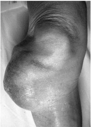
Physical examinations demonstrated a small-melon sized, firm, immobile mass at the anterolateral aspect of the right knee
joint. There were no pain, tenderness, and heating sense, but a 2×2 cm sized necrotiz- ing area in the center of the mass (Fig. 1).
The laboratory investigations revealed no specific abnormalities, plain radiographs demonstrated a soft tissue mass at the anterolateral aspect of the right knee joint, and there was no evidence of bony involve- ment.
Preoperative magnetic resonance images showed a huge, well-circumscribed, het- erogenous mixed echogenic mass which was septated and entirely encapsulated by well defined capsule in the lateral subcutaneous layer of the right knee (Fig. 2).
Preoperative whole body technetium-99m bone scans showed no specific abnormalities (Fig. 3).
The surgery was performed with a mar- ginal excision of the tumor mass and a par- tial excision of overlying skin. The tumor mass was entirely encapsulated by relatively well defined capsule, measuring 18 cm x 10.5 cm × 8 cm. The resected specimen was soft to friable and variegated with hemor- rhage and necrosis (Fig. 4A).
Microscopically, it showed malignant spin- dle cells with neoplastic osteoid formation.
The tumor cells had obviously malignant Fig. 1. Preoperative gross photograph shows a small-
melon sized mass with overlying skin necrosis.
Fig. 2. (A) T1 SE coronal MR image reveals a huge mass with heterogenous high, intermediate, and low signal inten- sities. (B) T2 SPIR coronal MR image reveals a huge mass with heterogenous high and intermediate signal intensities and septation in the lateral subcutaneous layer of the right knee joint.
A B
cytologic features, which showed significant pleomorphism and osteoid deposited in a fine, lacelike pattern (Fig. 4B-D).
After the surgery, the patient and his family refused all medical investigations and treatments including chemotherapy and radiotherapy. However, he was alive and there were no sign of local recurrence or dis- tant metastasis and functional loss during 1- year follow-up (Fig. 5).
Discussion
The diagnosis of primary extraskeletal osteosarcoma rests on three criteria : First, the presence of a uniform morphological pat- tern of sarcomatous tissue that excludes the possibility of mixed malignant mesenchymal
─ 83 ─ Fig. 3. Whole body Technetium-99m bone scans show no
abnormal uptakes.
Fig. 4. (A) Postopeartive gross photograph shows a well defined mass with encapsulation. (B) The tumor cells produce tumor osteoid and marked coagulation necrosis (Hematoxylin and Eosin stain, x 100). (C) The tumor cells have obviously malignant cytologic features, which show significant pleomorphism and striking increases in the amount and granularity of chromatin (Hematoxylin and Eosin stain, ×200). (D) Osteoid formed by the malignant cells is narrow and laid down in a lace-like pattern (Hematoxylin and Eosin stain, ×400).
A
C D
B
tumor; second, the production by sarcoma- tous tissue of malignant osteoid or bone or both; and third, the ready exclusion of osseous origin1).
Extraskeletal osteosarcoma is an uncom- mon tumor, of which only a small number of cases and studies have been reported1-10). It has been reported to constitute 1.2% of all the soft tissue sarcomas2,3,5,9) and 4.6%5,9) of all osteosarcomas.
Although osteosarcomas of bone occur chiefly during the first two decades of life, exteraskeletal osteosarcomas are rarely encountered in patients under 40 years of age5). Median age at diagnosis in several series is the sixth decades of life2,3,5,6,7,9,10)
. But, the age of our case was ninety-one
that was the oldest one in the review of lit- eratures.
Most series have found that the incidence of males and females is approximately equal for this disease10).
The lower extremity including the buttock area is the most common site of origin, con- stituting almost 69%, followed by upper extremity, trunk region including retroperi- toneum, and head and neck area2,9).
Generally, there are no specific signs or symptoms. The tumor presents as a progres- sive enlarging soft tissue mass, which is painful in about one third of the patients5,9). The duration of symptoms varies from a few weeks to several months, with a mean of 6.5 months5).
Several cases of extraskeletal osteosarcoma occurred after previous radiation thera- py1,2,3,6,9,10)
, trauma1,2,3,7,10)
including intramuscu- lar injection2,3,5,10) and fracture5), myositis ossificans2,5,8,10), and heterotopic ossification of dermatomyositis4,5). Our case was considered to a primary tumor without predisposing factors.
Simple excision is often followed by local recurrences and later, pulmonary metas- tases9). Recurrence after resection is a fea- ture of extraskeletal osteosarcomas and usu- ally occurs in more than half of the patients7). It is for this reason that a num- ber of authors have recommended wide exci- sion or radical resection as the initial opera-
tion7,9,10). Most medical centers recommend
aggressive treatments with preoperative radiotherapy or adjuvant multichemothera- py7).
Most local recurrences and distant metas- tases occur within 3 years postoperatively7). The lung are the most common site of metastasis (>80% of cases) and the resection of the metastasis can occasionally achieve a Fig. 5. Postoperative 1-year follow-up gross photograph
shows no limitation of the motion of the knee joint.
cure7). The prognosis is grave with five-year- survival rates of 25%9), 37%7).
We report the oldest patient with an extraskeletal osteosarcoma did not have pre- disposing factors. Although good results were shown on 1-year follow-up, a long term follow-up will be mandatory to verify- ing the final results.
REFERENCES
01) Allan CJ, Soule EH: Osteogenic sarcoma of the somatic soft tissues. Clinicopathologic study of 26 cases and review of literature. Cancer, 27: 1121- 1133, 1971.
02) Bane BL, Evans HL, Ro JY, et al.: Extraskeletal osteosarcoma. A clinicopathologic review of 26 cases. Cancer, 65: 2762-2770, 1990.
03) Chung EB, Enzinger FM: Extraskeletal osteosar- coma. Cancer, 60: 1132-1142, 1987.
04) Eckardt JJ, Ivinis JC, Perry HO, Unni KK:
Osteosarcoma arising in heterotopic ossification of dermatomyositis. Case report and review of the lit- erature. Cancer, 48: 1256-1261, 1981.
05) Enzinger FM, Weiss SW: Osseous tumors and tumorlike lesions of soft tissue. In: Soft Tissue Tumors, 2nd ed, C.V. Mosby, 892-905, 1988 06) Kim KH, Cho JL, Lee KH: A Case report of
extraosseous osteogenic sarcoma. J Korean Orthop Assoc, 19 : 411-415, 1984.
07) Lee JSY, Fetsch JF, Wasdhal DA, Lee BP, Pritchard DJ, Nascimento AG: A review of 40 patients with extraskeletal osteosarcoma. Cancer, 76: 2253-2259, 1995.
08) Lee SY, Jang JJ: A Case report of extraskeletal osteogenic sarcoma. J Korean Orthop Assoc, 22 : 581-586, 1987.
09) Rao U, Cheng A, Didolkar MS: Extraosseous osteogenic sarcoma. Clinicopathological study of eight cases and review of literature. Cancer, 41:
1488-1496, 1978.
10) Sordillo PP, Hajdu SI, Magill GB, Golbey RB:
Extraosseous osteogenic sarcoma. A review of 48 patients. Cancer, 51: 727-734, 1983.
─ 85 ─
Extraskeletal Osteosarcoma Around the Knee Joint - A Case Report -
Bong-Jin Lee, M.D., Tae-Ho Kim, M.D., Chang-Won Ha, M.D.*, Sung-Soo Kim, M.D.
Department of Orthopaedic Surgery, Pathology* Cheju Halla General Hospital, Jeju, Korea
An extraskeletal osteosarcoma is a rare malignancy. A small number of cases and studies have been reported in the world and only two cases have been reported in Korea. We experienced an extraskeletal osteosarcoma around the knee joint of 91-year-old male who was the oldest case in the literatures. It was developed without history of trauma, irradiation, myositis ossificans, and heterotopic ossification of dermatomyositis. This patient was treated with excision alone, how- ever he was alive and there were no sign of local recurrence or distant metastasis and functional loss during 1-year follow-up.
Key Words: Osteosarcoma, Extraskeletal, Knee
Address reprint requests to Sung-Soo Kim, M.D.
Department of Orthopaedic Surgery, Cheju Halla General Hospital 1963-2 Yeon-dong, Jeju-si, Jeju-do, Koreaorea
TEL: 82-64-740-5030, FAX: 82-64-743-3110, E-mail: [email protected]
Abstract