Progressive Transformation of Germinal Centers
in Axillary Lymph Nodes Mimicking Metastatic
Lymphadenopathy
after Breast Cancer Surgery:
A Case Report
유방암 수술 후 액와 림프절에 발생한
Progressive Transformation of Germinal Centers:
증례 보고
Sang Eun Park, MD1 , Kyu Ran Cho, MD1* , Sung Eun Song, MD1 , Ok Hee Woo, MD2 , Bo Kyoung Seo, MD3 , Jeonghyun Lee, MD4
Departments of 1Radiology and 4Pathology, Korea University Anam Hospital, Seoul, Korea
2Department of Radiology, Korea University Guro Hospital, Seoul, Korea
3Department of Radiology, Korea University Ansan Hospital, Ansan, Korea
Progressive transformation of germinal centers (PTGC) is a rarely diagnosed, benign disease of the lymph nodes that commonly manifests as chronic lymphadenopathy. PTGC may be char- acterized by single or multiple non-tender lymph nodes, and it commonly involves the cervical, axillary, and inguinal areas. Although PTGC is identified with concurrent lymphoma in some patients, it is not considered as a premalignant entity. Histopathologic diagnosis of PTGC is rarely made, and imaging findings have been reported in very few studies. We present a case of PTGC that occurred at the contralateral axillary lymph nodes and mimicked metastatic lymph- adenopathy after breast cancer surgery. We also discuss its imaging findings.
Index terms Progressive Transformation of Germinal Centers; Axilla; Lymphadenopathy;
Lymphatic Diseases
Received May 25, 2020 Revised July 13, 2020 Accepted July 28, 2020
*Corresponding author Kyu Ran Cho, MD Department of Radiology, Korea University Anam Hospital, 73 Goryeodae-ro, Seongbuk-gu, Seoul 02841, Korea.
Tel 82-2-920-5657 Fax 82-2-929-3796 E-mail [email protected] This is an Open Access article distributed under the terms of the Creative Commons Attribu- tion Non-Commercial License (https://creativecommons.org/
licenses/by-nc/4.0) which permits unrestricted non-commercial use, distribution, and reproduc- tion in any medium, provided the original work is properly cited.
ORCID iDs Sang Eun Park https://
orcid.org/0000-0002-9063-0449 Kyu Ran Cho
https://
orcid.org/0000-0002-8936-6468 Sung Eun Song
https://
orcid.org/0000-0002-9259-8294 Ok Hee Woo
https://
orcid.org/0000-0003-3953-933X Bo Kyoung Seo
https://
orcid.org/0000-0002-9512-5361 Jeonghyun Lee
https://
orcid.org/0000-0003-2041-4617
INTRODUCTION
Progressive transformation of germinal centers (PTGC) is one of the benign lymphadenop- athy that occurs in the background of reactive follicular hyperplasia. It accounts for 3.5% of chronic lymphadenopathy (1), and is assumed to be underdiagnosed in clinical practice. The imaging features of PTGC have not been reported in the previous literature. We present a case of PTGC in a breast cancer patient which mimicked metastatic axillary lymphadenopa- thy, and describe the imaging findings.
CASE REPORT
A 32-year-old female presented with a palpable breast mass in right upper outer quadrant.
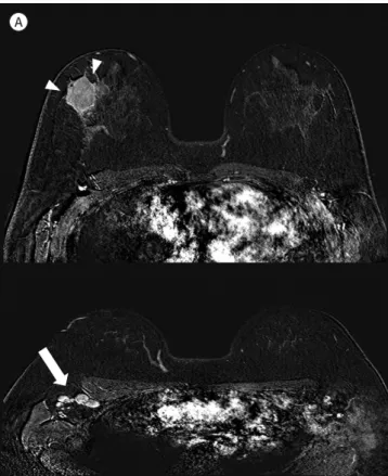
Mammography showed a round hyperdense mass with obscured margin at right breast (not shown). Breast magnetic resonance imaging (MRI) showed an irregular shaped, heteroge- neously enhancing mass in right upper outer quadrant (37 mm × 29 mm in size) on subtrac- tion image obtained 2 minutes after contrast administration. Multiple enlarged lymph nodes with cortical thickening were also present in right axilla (Fig. 1A). These imaging findings were highly suggestive of malignancy, and invasive ductal carcinoma was diagnosed by gun biopsy performed at outside hospital. PET-CT scan showed additional hypermetabolism in right supraclavicular lymph node. The patient underwent neoadjuvant chemotherapy and following breast conserving operation with radiation therapy. Histopathology was reported as no residual carcinoma of breast and regional lymph nodes.
Fig. 1. Progressive transformation of germinal centers in axillary lymphnodes after breast cancer surgery in a 32-year- old female.
A
Fig. 1. Progressive transformation of germinal centers in axillary lymphnodes after breast cancer surgery in a 32-year-old female.
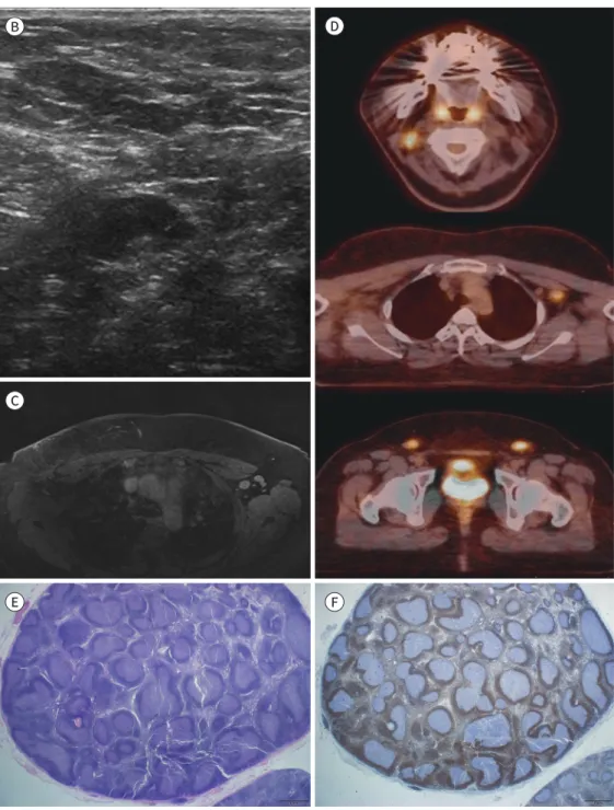
B, C. Follow-up axillary sonography (B) and MRI image (fat-saturated T1-weighted image obtained 2 min- utes after contrast administration) (C) at 6 months after conservative surgery of the right breast show multi- ple enlarged lymph nodes with cortical thickening in the left axillary area.
D. PET-CT scans show multifocal hypermetabolism in the right cervical (SUVmax 5.9), left axillary (SUVmax 3.6), and both inguinal (SUVmax right 4.4, left 5.9) lymph nodes. Metastatic lymphadenopathy is suggested.
E. Microscopic findings (hematoxylin and eosin stain, × 12.5) of excised left axillary lymph nodes shows en- larged lymph nodes with variably sized enlarged lymphoid follicles and frequent secondary germinal cen- ters. Occasionally, coalescent germinal centers are observed.
F. Bcl-2 immunohistochemical staining (× 12.5) shows a negative reaction in germinal centers.
SUVmax = maximum standardized uptake value B
C
E F
D
During clinical follow-up, palpable lymph nodes were detected in her left axilla on physi- cal examination 6 months after the surgery. On the follow-up imaging, lymph nodes with cortical thickening were newly visible at left axilla on sonography, breast MRI (Fig. 1B, C) and chest CT scan (not shown). PET-CT revealed multiple hypermetabolic lymph nodes in left ax- illa [maximum standardized uptake value (SUVmax) 3.6], right neck (SUVmax 5.9), left exter- nal iliac and both inguinal areas (SUVmax; right 4.4, left 5.9) (Fig. 1D). There was no evidence of local tumor recurrence in right breast and axilla. Because metastatic lymphadenopathy was suspected, ultrasound-guided aspiration was done for the lymph node in left axilla. However, cytology was negative for malignancy. Left axillary lymph nodes were surgically excised for the radiologic-pathologic discordance. Microscopically, the section from left axillary area showed four enlarged lymph nodes with florid follicular lymphoid hyperplasia pattern. Ger- minal centers were distended and occasionally coalescent. Immunostaining for CD3 and CD20 showed reactive zonal pattern. CD21 and Ki-67 immunostains highlighted germinal centers, but Bcl-2 immunostain showed negative reaction in lymphoid follicles. The micro- scopic and immunohistochemical findings were compatible to PTGC (Fig. 1E, F).
During 4 years of clinical and imaging follow-up, mildly enlarged lymph nodes were per- sistently noted in left axilla. No interval change was observed in lymph nodes, proving it as a benign etiology.
DISCUSSION
PTGC is a benign reactive pattern identified within a lymph node with reactive follicular hyperplasia. The affected lymphoid follicles are enlarged three to five times greater than the normal follicles, and show expansion of mantle zone. Mantle zone lymphocytes extend into the germinal centers and the distinction between the germinal center and mantle zone is lost (2). In histopathologic examination, PTGC should be differentiated from follicular lympho- ma. In cases with confusing morphology, immunostaining is the key for differential diagno- sis. Follicular lymphoma shows overexpression of bcl-2, whereas PTGC have only mild focal expression. Previous studies have suggested an association between PTGC and nodular lym- phocytes-predominant Hodgkin lymphoma (NLPHL) (3). In some reports, PTGC was more frequently observed in biopsy specimens of patients known to have NLPHL. However, PTGC is not considered as a premalignant entity. The specimen from our patient showed enlarged lymph nodes with large nodules of hyperplastic germinal centers, infiltrated by mantle zone B cells. Negative reaction for Bcl-2 immunostaining confirmed the diagnosis of PTGC.
PTGC is a pattern usually identified focally in a lymph node. The proportion of PTGC com- ponent is most commonly less than 5% of total follicles, and only few cases show PTGC in more than 20% of follicles (4). Therefore, complete excision of lymph node is required for the
ied from 43.8 to more than 60 years-old in previous studies (4, 5), quite a few young aged pa- tients are also reported. Patients often undergo multiple biopsies due to persistent lymph- adenopathy.
To our knowledge, imaging findings of PTGC are rarely reported. The sonographic feature of PTGC was reported by Talˊasiewicz et al. (6) in two cases of cervical lymphadenopathy. Ul- trasound showed hypoechoic enlarged lymph nodes without fatty hilum or increased vascu- larity. The imaging finding of fluoro-deoxy-glucgose-PET in PTGC was avid uptake of radio- tracer, as in our case (7, 8). In patients with multiple non-necrotic lymphadenopathy, differential diagnosis may include reactive hyperplasia associated with infectious, inflamma- tory diseases or lymphoma. It is hard to include PTGC in the differential diagnosis of enlarged lymph nodes, in a patient with underlying malignancy. In our patient, PTGC appeared as en- larged axillary lymph nodes with cortical thickening, mimicking metastatic lymph nodes.
However, it was somewhat unusual that metastatic lymphadenopathy occurred in contralat- eral side of primary breast cancer, without evidence of local tumor recurrence or ipsilateral lymphadenopathy.
We report a case of PTGC that involved axillary, cervical and inguinal lymph nodes after breast cancer surgery. Imaging features in this case were indistinguishable with malignant lymphadenopathy. Although suspicion for malignancy must be raised, PTGC can be includ- ed in the differential diagnosis, especially in case of lymph node enlargement in the typical location of PTGC.
Author Contributions
Conceptualization, C.K.R.; investigation, P.S.E.; methodology, C.K.R.; writing—original draft, P.S.E.;
and writing—review & editing, all authors.
Conflicts of Interest
The authors have no potential conflicts of interest to disclose.
Funding None
REFERENCES
1. Hansmann ML, Fellbaum C, Hui PK, Moubayed P. Progressive transformation of germinal centers with and without association to Hodgkin’s disease. Am J Clin Pathol 1990;93:219-226
2. Hicks J, Flaitz C. Progressive transformation of germinal centers: review of histopathologic and clinical fea- tures. Int J Pediatr Otorhinolaryngol 2002;65:195-202
3. Verma A, Stock W, Norohna S, Shah R, Bradlow B, Platanias LC. Progressive transformation of germinal centers. Report of 2 cases and review of the literature. Acta Haematol 2002;108:33-38
4. Kojima M, Nakamura S, Motoori T, Itoh H, Shimizu K, Yamane N, et al. Progressive transformation of germi- nal centers: a clinicopathological study of 42 Japanese patients. Int J Surg Pathol 2003;11:101-107 5. Özkan MC, Özsan N, Hekimgil M, Saydam G, Töbü M. Progressive transformation of germinal centers: sin-
gle-center experience of 33 Turkish patients. Clin Lymphoma Myeloma Leuk 2016;16 Suppl:S149-151 6. Talˊasiewicz K, Czachowska A, S´mialˊek-Kania K, Jaxa-Larecka D, Jagielska B. Progressive transformation of
germinal centers: an illustration of two clinical cases. Ann Hematol 2018;97:1081-1083
7. Shaikh F, Ngan BY, Alexander S, Grant R. Progressive transformation of germinal centers in children and adolescents: an intriguing cause of lymphadenopathy. Pediatr Blood Cancer 2013;60:26-30
8. Makis W, Ciarallo A, Novales-Diaz JA, Lisbona R. Progressive transformation of germinal centers in a pedi- atric patient: initial evaluation and follow-up with serial F-18 FDG PET/CT imaging. Clin Nucl Med 2011;36:
e139-141
유방암 수술 후 액와 림프절에 발생한
Progressive Transformation of Germinal Centers:
증례 보고
박상은1 · 조규란1* · 송성은1 · 우옥희2 · 서보경3 · 이정현4
Progressive transformation of germinal centers (이하 PTGC)는 드물게 나타나는 양성 림 프절 질환으로, 주로 만성적인 림프절 비대의 형태로 나타난다. 주로 한 개 또는 여러 개의 림 프절의 무통성 비대로 나타나며, 가장 흔히 침범하는 부위는 경부 림프절, 다음으로 액와부 와 서혜부 림프절로 알려져 있다. 일부 환자들에서는 조직병리에서 림프종이 함께 존재하기 도 하지만, PTGC 자체는 전암병변으로 인식되지는 않는다. 조직학적으로 PTGC로 진단되는 경우도 적지만, 이에 대한 영상의학적 소견은 거의 보고된 바가 없다. 이에 저자들은 침윤성 유방암 환자에서 수술 후 반대편 액와부 림프절 비대로 나타나 전이성 림프절로 오인한 PTGC 증례의 영상 소견을 보고하고자 한다.
고려대학교 안암병원 1영상의학과, 4병리과,
2고려대학교 구로병원 영상의학과,
3고려대학교 안산병원 영상의학과