J. of Korean Bone & Joint Tumor Soc.
Volume 12, Number 1, June, 2006
※통신저자: 안 기 찬
부산광역시 진구 개금동 633-165 인제대학교 부산백병원 정형외과학교실
Tel: 051) 890-6129, Fax: 051) 892-6619, E-mail: [email protected]
경추에 발생한 거대 세포종 - 증례 보고 -
인제대학교 부산백병원 정형외과학교실
안기찬∙정경칠∙김윤준
척추의 거대 세포종은 진단이 어려운 경우가 많고, 고도의 악성도로 인해서 수술이 불가능 한 경우가 많다. 척추의 거대 세포종은 높은 재발율과 함께 척수의 기계적 압박 가능성으로 인해 방사선 치료가 근간이었으나, 최근에는 근치적 절제술과 함께 기구를 이용한 전후방 고 정술을 시행 하여 좋은 결과를 보고 하고있다.본 정형외과학교실에서는 경부 동통을 유발하며 제3경추를 침범한 거대세포종에 대해서 근치적 절제술과 함께 후방 기기술을 통한 융합을 시 행 하였으며, 추시 관찰상 우수한 치료 결과를 보였기에 문헌 고찰과 함께 보고하는 바이다.
색인 단어: 경추, 거대 세포종, 수술적 치료, 근치적 절제술
Introduction
Giant cell tumors are potentially malig- nant and constitute about 2% of all tumors in vertebrae, except in the sacrum. Giant cell tumors affecting vertebrae are frequent- ly difficult to diagnose and are often inoper- able2). They are treated using radiation because of their high recurrence rate and the mechanical compression of the spinal cord. Dichiro and Nelson4) described tumors of the vertebra, and the affected vertebral body can be treated using radical9)or near to total excision, with anteroposterior vertebral fusion or instrumentation of the spine. We
report a case of giant cell tumor affecting the third cervical vertebra, which caused neck pain and destroyed the vertebra body and posterior arch, together with a litera- ture review. The patient was asymptomatic at the 1-year follow-up.
Case Report
The patient was a 16-year-old male whose chief complaint was neck pain. He had slipped 5 months earlier but had not recov- ered completely. Recently, the pain had worsened. His past history and familial his- tory were non-contributory.
On physical examination, he was tender over the mid-neck, but no motor weakness or sensory loss was seen on a neurologic exam.
The laboratory findings included a normal serum calcium, ALP, and phosphate.
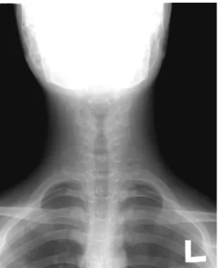
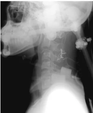
Radiologically, anteroposterior and lateral views of the C-spine showed a loss of C3 vertebral height(Fig. 1), and the lateral view showed a complete osteoclastic state (Fig. 2). No ectopic or metastatic lesions were seen on x-rays of the chest or other bones. Magnetic resonance imaging (Fig. 5) showed a medullary tumor affecting the pos- terior arch and neural canal of C3.
Microscopically, the scanning view showed increased osteoid or dense collagen (Fig. 6).
Surgery was performed under general anes- thesia, with the patient in the prone posi-
tion. A midline incision was made over the spinous process of C3. Then, the nuchal lig- ament was dissected, and we excised a soft encapsulated tumor adherent to the posterior arch of the vertebra. Remnants of the tumor adhering to the vertebral body were removed by curettage. The remnant tumor looked like whitish fat. The defect left by the tumor was filled with autologous left- side iliac bone. The C-spine from C2 to C4 was fused using an external two-wire fixa- tion method (Fig. 3 and 4). After the opera- tion, the patient’s neck pain subsided grad- ually.
Considerations
Giant cell tumors of bony tissue generally invade the epiphysis and metaphysis. There
Fig. 1. Preoperative cervical spine, anteroposterior view.
The height of the third cervical vertebra body is reduced.
Fig. 2. Preoperative cervical spine, lateral view. The complete osteoclastic state is shown, viewing the vertebral body of C3 as an upper end plate sil- houette.
are fewer than 70 reported cases of giant cell tumors in vertebrae. Dahlin3) stated that there are differences between giant cell tumors that occur in vertebrae and those found in long bones. Tumors affecting long bones are most often found in younger patients, are more common in females (3:1), and have a relatively good prognosis because the recurrence rate is low after surgical treatment. By contrast, Savini et al.7) found no fundamental differences in spinal and long bone tumors. The 67 reported examples of vertebrate tumors were uniformly distrib- uted in the cervical, thoracic, and lumbar vertebrae, and often occurred in the verte- bral body instead of the vertebral arch.
Magnetic resonance imaging is useful before surgery for both making the diagnosis and planning the surgery. Regular follow-up with magnetic resonance imaging is useful
for visualizing invasion or local recurrence of the tumor and the state of the bony-union in grafts. Diseases that should be considered in the differential diagnosis include aneurys- mal bone cyst and benign osteoblastoma. An aneurysmal bone cyst is difficult to differen- tiate radiologically because its radiologic fea- tures are similar to those of a giant cell tumor. However, giant cell tumors occur mainly in the spine, while aneurysmal bone cysts are mainly localized in the posterior vertebral arch. Microscopically, giant cell tumors are characterized by stromal cells and a cystic lesion full of blood. Benign osteoblastomas mainly affect the posterior
Fig. 3. Preoperative cervical spine, MRI sagittal image.
In the spinal canal, a moderate degree of spinal canal stenosis and cord indentation are seen.
Fig. 4. Microscopic findings. In the scanning view ( 125), the cellularity of osteoid or dense collagen is increased. On magnifying the necrotic portion, we found an osteoblastic rim of osteoid and mult- inucleated giant cells, but there was no definite nuclear atypism of stromal cells. Magnifying the canal ( 400) reveals interstitial cells with nuclei of varying size and morphology. No specific abnormal mitosis or tumor necrosis is seen.
vertebral arch and can be differentiated by the characteristic microscopic evidence of osteoid tissue. The literature review indi-
cates that giant cell tumors of the spine can be treated using radiotherapy, excision6), excision and arthrodesis, excision and arthrodesis plus radiotherapy4,5) or cryosurgery. When neurologic deficit were present, anterior decompressison and fusion were needed10). Recently, total spondylectomy of involved vetebrae with spinal shortening were carried out with excellent results11).Giant cell tumors of the vertebra have a relatively better prognosis than those of long bones. The reported recurrence rate of giant cell tumors of long bones is 40 to 50% when treated by curettaging and graft- ing of bone. The recurrence usually occurs within 2 years of surgery. As a tumor of the vertebra is difficult to resect completely owing to the anatomical features, the recur- rence rate of vertebral lesions should be much higher. However, Mirra et al.6) Fig. 5. Postoperative cervical spine, anteroposterior
view. The height of the third cervical vertebra body is increased.
Fig. 6. Postoperative cervical spine, lateral view. The third cervical vertebra is displaced posteriorly with malalignment.
Fig. 7. Follow-up after post-op. 26months cervical spine, lateral view. Fusion state was seen without recurrence.
reviewed 41 vertebral lesions and found that the recurrence rate was only 25%, regardless of the treatment modality. Our patient was a male in his mid-teens, and the tumor was limited to the cervical vertebrae. It was mainly confined to the vertebral body, with some invasion of the spinal canal seen radi- ographically and at surgery. He had no neu- rologic deficit,so we had selected posterior approach method and especially, we applied wiring method for fusion with bone graft.
Our case has been followed for 26 months after surgery and we expect a good prognosis because the lesion was removed completely without recurrence and the vertebrae was fused at follow-up period (Fig. 7).
Giant cell tumors affecting the vertebrae are very rare, especially those of the cervical spine. The tumors are often difficult to diagnose and can cause mechanical compres- sion of the spinal cord8). Owing to the opera- tive difficulty in tumorectomy and the high recurrence rate in inoperable areas, verte- bral tumors are often treated with radia- tion. We present a rare case of a giant cell tumor affecting the cervical vertebra of a 16-year-old male patient that was treated surgically, together with a brief review of the literature.
REFERENCES
11) Berman HL: The treatment of benign giant-cell
tumor of the vertebrae by irradiation. Radiology, 83:202-207, 1964.
12) Cullen: Treatment of giant-cell tumors. J Bone Joint Surg, 59-B:514, 1977.
13) Dahlin, DC: Giant cell tumor of vertebrae above the sacrum. A review of 31 cases. Cancer, 39:1350- 1356, 1977.
14) Dichiro G, Nelson KB: Soft tissue radiography of extremities in neuromuscular disease with histologi- cal correlations: Acta Radiol Diagn (Stockh), 42:65- 88, 1965.
15) Feigenberg SJ, Marcus Jr RB and Zlotecki RA:
Radiation therapy for giant-cell tumors of bone.
Clin Orthop, 41:207-216, 2003.
16) Mirra JM, Rand F, Rand R, Calcaterra T and Dawson E: Giant cell tumor of the second cervical vertebra treated by cryosurgery and irradiation. Clin orthop, 154:228-233, 1981.
17) Savini R, Gherlinzoni F, Morandi M, Neff JR and Picci P: Surgical treatment of giant cell tumor of the spine. The experience at Insituto Orthopetico Rizzoli. J Bone Joint Surg, 65-A(9):1283-1289, 1983.
18) Schwimer SR, Bassett LW, Mancuso AA, Mirra JM and Dawson EG: Giant-cell tumor of the cervi- cal spine. Am J Roentgenol, 136:63-67, 1981.
19) Stener B, Johnsen OE: Complete removal of three vertebrae for giant cell tumor. J Bone Joint Surg, 53-B(2):278-287, 1971.
10) Takayuki Shirakuni , Norihiko Tamaki , Satoshi Matsumoto and Masayasu Fujiwara: Giant cell tumor in cervical spine. Surgical Neurology, 23:
148-152, 1985.
11) K Shimizu, K Ido, K Fujio, K Tanaka and T Nakamura: Total spondylectomy and spinal short- ening for giant-cell tumour of spine. The Lancet, 348: 342, 1996.
Giant Cell Tumor of the Cervical Spine - Case Report -
Ki Chan An, M.D., Kyung Chil Chung, M.D.,Yoon Jun Kim, M.D.
Department of Orthopedic Surgery, College of Medicine, University of Inje, Busan Baik Hospital, Busan, Korea
Giant cell tumors are potentially malignant tumors in vertebrae, affecting frequently difficult to diagnose and are often inoperable. So it will be treated using radiation because of their high recurrence rate and the mechanical compression of spinal cord, but many surgeons described tumors of the vertebra, and the affected vertebral body can be treated using radical or near to total excision, with anteroposterior vertebral fusion or instrumentation of the spine. we report a case of giant cell tumor affecting the third cervical vertebra which caused neck pain and destroyed the vertebra body had treated using radical excison with fusion of posterior arch using instrumentation of the spine together with a literature review.
Key Words: Cervical spine, Giant cell tumor, Surgical treatment, Radical excision
Address reprint requests to Ki Chan An, M.D.
Department of Orthopedics, Busan Paik Hospital, Inje University school of Medicine 633-165 Gaegum-Dong, Busan Jin Gu, Busan 614-735, Korea
TEL: 82-51-890-6129, Fax: 82-51-892-6619 , E-mail: [email protected]
Abstract