Bilateral Breast Cancer in a Patient with
Neurofibromatosis Type 1:
A Case Report
제1형 신경섬유종증 환자에서 발생한 양측성 유방암: 증례 보고
Sang Hwa Woo, MD1 , Hyun Kyung Jung, MD1* , Woogyeong Kim, MD2
Departments of 1Radiology and 2Pathology, Inje University Haeundae Paik Hospital, Busan, Korea
Neurofibromatosis type 1 (NF1) is a rare neuroectodermal disease that is associated with an in- creased risk of malignancy. Here, we report a rare case of bilateral breast cancer in a 49-year- old woman with NF1 that presented as a microlobulated oval hypoechoic mass and a complex cystic solid mass on sonography. She underwent bilateral mastectomy. The masses were diag- nosed as invasive ductal carcinoma, ductal carcinoma in situ, and a malignant peripheral nerve sheath tumor. We describe the imaging findings, including ultrasonography, CT, and 18F-fluoro- deoxyglucose PET.
Index terms Neurofibromatosis Type 1; Breast; Cancer;
Malignant Peripheral Nerve Sheath Tumor; Ultrasonography
INTRODUCTION
Neurofibromatosis type 1 (NF1) is an autosomal dominant disorder that is associated with an increased risk of malignancy. Neurofibromas, gliomas, malignant peripheral nerve sheath tumors (MPNSTs), gastrointestinal stromal tumors, pheochromocytomas, juvenile myelocytic leukemia, and rhabdomyosarcomas are commonly related to NF1 (1). Several studies have suggested an association between NF1 and an increased risk of breast cancer (1-5), and women with NF1 aged under 50 years have an increased risk of breast cancer (6). Here, we describe a case of bilateral breast cancer in a 49-year-old woman with NF1.
Received May 6, 2020 Revised June 9, 2020 Accepted June 20, 2020
*Corresponding author Hyun Kyung Jung, MD Department of Radiology, Inje University
Haeundae Paik Hospital, 875 Haeun-daero, Haeundae-gu, Busan 48108, Korea.
Tel 82-51-797-0381 Fax 82-51-797-0379 E-mail drsjung@gmail.com This is an Open Access article distributed under the terms of the Creative Commons Attribu- tion Non-Commercial License (https://creativecommons.org/
licenses/by-nc/4.0) which permits unrestricted non-commercial use, distribution, and reproduc- tion in any medium, provided the original work is properly cited.
ORCID iDs Sang Hwa Woo https://
orcid.org/0000-0002-7869-5514 Hyun Kyung Jung
https://
orcid.org/0000-0001-6086-7771 Woogyeong Kim
https://
orcid.org/0000-0003-1694-6602
CASE REPORT
A 49-year-old woman with NF1 that was diagnosed at age 10 presented with a rapidly grow- ing mass in her left breast. She noticed the mass 3 years previously without evaluation, and it had begun to grow rapidly 2 months prior to her presentation. A physical examination re- vealed a firm, large mass in the left breast with underlying multiple cutaneous neurofibro- mas. Breast ultrasonography (US) (iU22; Philips Healthcare, Bothell, WA, USA) demonstrated a large complex cystic solid mass with weak internal vascularity in the left breast and a 1.2 cm hypoechoic mass with an oval shape, microlobulated margin, and increased vascularity in the right breast (Fig. 1A). A US-guided core needle biopsy with a 14-gauge needle (Stericut, TSK Laboratory, Tochigi, Japan) was performed, and the masses were diagnosed as extensive necrosis with focal lymphohistiocytic infiltration in the left breast and invasive ductal carci- noma (IDC) and ductal carcinoma in situ (DCIS) in the right breast. Contrast-enhanced chest CT showed a well-defined large mass with internal low density that did not exhibit enhance- ment on the post-contrast scan, suggesting cystic or necrotic changes (Fig. 1C). 18F-fluorode- oxyglucose (FDG) PET demonstrated focal FDG-avid uptake in the left breast with a maxi- mum standardized uptake value (SUV) of 11.8 and an SUV of 4.6 in the retroperitoneum (Fig.
1D). A US-guided core needle biopsy was performed on the retroperitoneal mass, which was diagnosed as neurofibroma (Fig. 1E).
The patient underwent bilateral mastectomy. The right mass was early breast cancer that was incidentally found on US, and as such, breast-conserving surgery (BCS) was performed.
However, radiation therapy after BCS was needed, and a left mastectomy was scheduled. Bi- lateral mastectomy was performed after consulting the patient and her family. The right mass was diagnosed as a 0.7 cm sized IDC, not otherwise specified type (Fig. 1F) with no right axillary lymph node metastasis. The IDC was of the human epidermal growth factor re- ceptor 2 (HER-2) type. On gross examination, the left breast showed a well-encapsulated solid mass with the greatest dimension of 35.0 cm. The cut surface of the mass was myxoid with multifocal hemorrhage and necrosis. Microscopically, the tumor was composed of hypercel- lular, hyperchromatic spindle, and plump cells with abundant mitoses and geographic ne- crosis (Fig. 1F). Within the tumor, there were hypocellular areas with bland-looking spindle wavy cells, suggesting a tumor originating from a benign neurogenic tumor. Immunohisto- chemical stains were positive for S-100, vimentin, CD-34, p53, and high expression of Ki-67, and negative for CD-56 and neuron-specific enolase.
After surgery, the patient received four cycles of adjuvant chemotherapy (doxorubicin 60 mg/m2/day and dacarbazine 250 mg/m2/4 days) over two months and two cycles of radiation
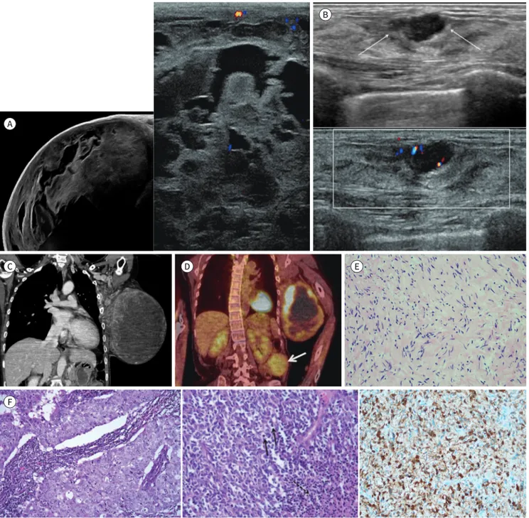
Fig. 1. Imaging and pathologic features of bilateral breast cancer in a 49-year-old woman with neurofibromatosis type 1.
A. Ultrasonography of left breast shows a large oval complex cystic and solid mass with weak internal vascularity.
B. Ultrasonography of right breast shows a 1.2 cm hypoechoic mass with an oval shape, microlobulated margin (arrows), and increased inter- nal and peripheral vascularity.
C. Coronal contrast-enhanced chest CT shows a well-defined large mass with heterogeneous enhancement.
D. 18F-FDG PET shows a focal FDG uptake in the left breast with a maximum SUV of 11.8 and retroperitoneum with an SUV of 4.6 (arrow).
E. Microscopic finding from biopsy of the retroperitoneal mass shows a hypocellular proliferation of bland stroma, consistent with neurofi- broma (H&E stain, × 200).
F. The right breast shows a poorly differentiated invasive ductal carcinoma composed of nests of pleomorphic ductal cells with tumor-infil- trating lymphocytes in the stroma (H&E stain, × 200) (left image). The left breast mass shows a highly cellular tumor composed of monomor- phic plump and spindle cells with frequent mitoses (black arrows) and geographic necrosis (black dashed arrow) (H&E stain, × 200) (middle image). The tumor cells display diffuse immunoreactivity for S-100 protein (immunohistochemical staining, × 200) (right image).
FDG = fluorodeoxyglucose, H&E = hematoxylin and eosin, SUV = standardized uptake value
A
C
F
D E
B
caused by mutation of the NF1 gene. The NF1 gene is located in the pericentromeric region of the long arm of chromosome 17, which also covers the BRCA1 gene, but the interaction between them is not clear (7). However, an association between NF1 and an increased risk for breast cancer has been suggested (1-5). The incidence of breast cancer in patients with NF1 is 3.5-times higher than that in the general population (7). The most common histopath- ological type of breast cancer in NF1 patients is IDC (8). Contrastingly, MPNST is rare, and only 16 cases of MPNST have been reported in patients with NF1 (9). Furthermore, only a few cases of bilateral breast cancer have been reported in patients with NF1 (1, 3, 4). Dursun et al. (1) and Kim et al. (3) reported metachronous bilateral breast cancers that were diagnosed as intraductal carcinoma and IDC in a 44-year-old and 46-year-old patient, respectively. How- ever, they did not report the imaging findings. Although we cannot estimate the exact timing of carcinogenesis, the patient was diagnosed with bilateral cancer, that is, she exhibited two different cancer types simultaneously. Synchronous bilateral IDCs, described as 5-cm mass- es, were previously reported in a 51-year-old patient with NF1 following mammography (4).
Owing to its rarity, imaging findings of breast cancer in patients with NF1 have rarely been reported (2, 7) Seo and Park (2) reported the imaging findings of IDC as an irregular hyper- dense mass with an indistinct margin on mammography and a hypoechoic irregular mass with an angular margin on US. DCIS was described as fine pleomorphic microcalcifications with segmental distribution on mammography, an ill-defined hypoechoic mass with micro- calcifications and irregular duct changes combined with mild skin thickening and metastatic lymph nodes on US, and as a non-mass enhancement with heterogeneous internal enhance- ment pattern on MRI (2). Bonnet et al. (5) reported that a case of MPNST in a 36-year-old wom- an presented as a large mass with scattered coarse calcifications on mammography and a hy- poechoic vascular mass with hyperechoic internal septations on US. On MRI, MPNST has been described as a hypointensity on T1-weighted imaging, a heterogeneous hyperintensity on T2-weighted imaging, and a predominantly peripheral enhancement of the mass on T1- weighted gadolinium-enhancement. In our case, IDC and DCIS appeared as a hypoechoic mass with an oval shape and microlobulated margin on US, respectively. Besides, MPNST ap- peared as a large complex cystic, solid mass.
Detecting breast cancer in a patient with NF1 is challenging because of the large number of skin neurofibromas that interfere with the physical examination and mammography (4, 6).
In addition, patients with NF1 are less likely to notice a palpable mass in the breast, mistak- ing it as a kind of neurofibroma. Therefore, the poor prognosis of breast cancer in patients with NF1 may result from late-stage diagnosis due to the delay in seeking medical care rather than a characteristic of the disease itself. Although there are not enough studies to inform
lost to follow up in 4 years and 6 months following treatment. Although we could obtain an update on the patient from a family member, our ability to evaluate the patient’s prognosis was limited.
In summary, NF1 predisposes patients to breast cancer as well as to malignant tumors.
Current breast cancer screening guidelines are not sufficient for patients with NF1. As such, NF1 patients should be aware of the possibility of developing breast cancer. If a breast mass is palpable, the mass should not be overlooked but should be evaluated by imaging.
Author Contributions
Conceptualization, J.H.K.; supervision, J.H.K.; writing—original draft, W.S.H.; and writing—review
& editing, J.H.K., K.W.
Conflicts of Interest
The authors have no potential conflicts of interest to disclose.
Funding None
REFERENCES
1. Dursun D, Aktas¸ S, Altun Z, Olgun N. Bilateral breast cancer with neurofibromatosis type 1 patient: case re- port. Eur J Breast Health 2017;13:213-215
2. Seo YN, Park YM. Association between neurofibromatosis type 1 and breast cancer: a report of two cases with a review of the literature. Case Rep Med 2015;2015:456205
3. Kim SH, Son WJ, Sin DJ, Chang MC. Bilateral metachronous breast cancer in neurofibromatosis type 1. J Korean Surg Soc 2009;76:388-391
4. Alamsamimi M, Mirkheshti N, Mohajery MR, Abdollahi M. Bilateral invasive ductal carcinoma in a woman with neurofibromatosis type 1. Arch Iran Med 2009;12:412-414
5. Bonnet SE, Kang-Chapman JK, Buckley KA, Cui X, Grignol VP, Hawley JR. Malignant peripheral nerve sheath tumor of the breast in a patient with neurofibromatosis 1. Breast J 2018;24:1066-1068
6. Maani N, Westergard S, Yang J, Scaranelo AM, Telesca S, Thain E, et al. NF1 patients receiving breast cancer screening: insights from the Ontario high risk breast screening program. Cancers (Basel) 2019;11:707 7. Sharif S, Moran A, Huson SM, Iddenden R, Shenton A, Howard E, et al. Women with neurofibromatosis 1
are at a moderately increased risk of developing breast cancer and should be considered for early screen- ing. J Med Genet 2007;44:481-484
8. Madanikia SA, Bergner A, Ye X, Blakeley JO. Increased risk of breast cancer in women with NF1. Am J Med Genet A 2012;158A:3056-3060
9. Shuayb M, Begum R. Unusual primary breast cancer - malignant peripheral nerve sheath tumor: a case re- port and review of the literature. J Med Case Rep 2017;11:161
10. Yap YS, Munusamy P, Lim C, Chan CHT, Prawira A, Loke SY, et al. Breast cancer in women with neurofibro- matosis type 1 (NF1): a comprehensive case series with molecular insights into its aggressive phenotype.
Breast Cancer Res Treat 2018;171:719-735
제1형 신경섬유종증 환자에서 발생한 양측성 유방암:
증례 보고
우상화1 · 정현경1* · 김우경2
제1형 신경섬유종증은 상염색제 우성으로 유전되는 신경외배영성 질환으로 다양한 악성 종 양이 발생할 수 있다. 하지만 유방암이 발생한 보고는 드물다. 이에 저자들은 제1형 신경섬유 종증 환자에서 발생한 양측성 유방암 증례를 영상 소견과 함께 보고하고자 한다.
인제대학교 의과대학 해운대백병원 1영상의학과, 2병리과