• 검색 결과가 없습니다.

하악에 발생한 투명세포 치성암종 : 증례 보고

N/A
N/A
Protected

Academic year: 2021

Share "하악에 발생한 투명세포 치성암종 : 증례 보고"

Copied!
6
0
0

로드 중.... (전체 텍스트 보기)

전체 글

(1)

Ⅰ. INTRODUCTION

Clear cell odontogenic carcinoma (CCOC) is often considered as a rare tumor, first described by Hansen in 1985.(1) Formerly known as clear cell odontogenic tumor, a locally aggressive benign tumor in the WHO classification of 1992,(2) it was renamed CCOC and classified among malignant tumors after the revision of this classification in 2005. It is still considered a malignant tumor in the 2017 classification.(3)

CCOC is an extremely rare neoplasm. It’s described very few in the English-language literature.(4,5) Thus, it is not easy to notice such disease. Also, treatment plan and procedures are uncertain to some clinicians.

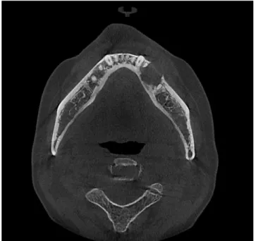
CCOC has no specific clinical and radiographic features,

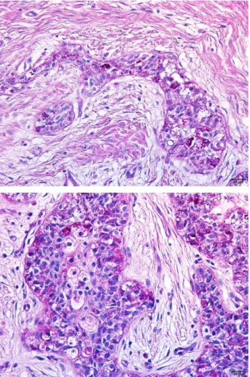
which make its diagnosis difficult. Histopathologically, CCOC is characterized by sheets and islands of vacuolated and clear cells(6) and by the presence of agglomerates of cells with eosinophilic cytoplasm. Long-term follow-up is suggested in the literature because of its potential for recurrence and distant metastases.(7)

This case report presents the histopathological and clinical features of clear cell odontogenic carcinoma excised from the mandible.

Ⅱ. CASE REPORT

A 53-year-old woman with good general health visited the oral and maxillofacial surgery department at the Seoul St.

Mary's Hospital of Catholic University of Korea, with paresthesia on #35 area. She was taking medicine for hypertension and

하악에 발생한 투명세포 치성암종: 증례 보고

이승준*, 최민호, 천승우, 이종호, 김창현, 박재억

가톨릭대학교 서울성모병원 구강악안면외과

<Abstract>

Clear Cell Odontogenic Carcinoma of the Mandible: A Case Report

Seung Joon Lee

*

, Min Ho Choi, Seung Woo Cheon, Jong Ho Lee, Chang Hyen Kim and Je Uk Park Department of Oral and Maxillofacial Surgery, Seoul St. Mary’s Hospital, The Catholic University of Korea

Clear cell odontogenic carcinoma (CCOC) is an extremely rare odontogenic neoplasm; Only a few cases have been reported in the English literatures. It displays a propensity for the mandible, most commonly presenting in the fifth to seventh decades, with a female predilection. The clinical and radiological manifestations are multiple and the diagnosis is histological. Histological feature is of large islands and strands of uniform cells with round or ovoid nuclei and clear cytoplasm. Clinically, painless swelling is the most common symptom, followed by pain, teeth mobility, and paresthesia. CCOC has a good prognosis after surgery. This case report presents the histopathological and clinical features of CCOC excised from the mandible.

Key words : Clear cell odontogenic carcinoma, CCOC, Mandible

Korean Journal of Oral and Maxillofacial Pathology 2019;43(3):97-101 ISSN:1225-1577(Print); 2384-0900(Online) Available online at http://journal.kaomp.org https://doi.org/10.17779/KAOMP.2019.43.3.006

* Correspondence: Seung Joon Lee, Department of Oral and Maxillofacial Surgery, Seoul St. Mary’s hospital, The Catholic University of Korea Tel: +82-2-2258-1781, E-mail: [email protected]

ORCID: 0000-0003-0309-7429

(2)

received hysterectomy about 3 years ago. She extracted left lower 2nd premolar (#35) about 20 years ago, so #35 was missing state.

Radiological examination showed a radiolucent mandibular

lesion extending from the left 1st premolar to the left 1st molar area (Fig. 1). On cone-beam computed tomography (CBCT), the inferior alveolar nerve canal was not destroyed yet (Fig. 2).

Initially, she noticed that there was a black spot between left lower 1st and 2nd premolar gingival area. Patient gave a history of gradual increase of that point recently. However, there were no pain and swelling of the mandible.

Also, extraoral examination showed no specific findings.

Based on the clinical findings, periapical cyst, was given as provisional diagnosis. So, cyst enucleation and biopsy was planned after left lower 1st premolar (#34) root canal treatment (RCT).

After 1 month, #34 RCT was done (Fig. 3), and then enucleation and biopsy was performed under local anesthesia. Irregularly shaped pale brown tissue fragments, measuring up to 1.7 x 1.0 cm, were confirmed. CCOC was diagnosed by biopsy result, so we explained to patient and made a decision to operate surgery under general anesthesia.

Enhanced magnetic resonance imaging (MRI) and enhanced computed tomography (CT) showed a round enhancing lesion at the bony defect site of the left mandibular body, lower premolar tooth. No metastatic lymph node or distant metastasis was detected.

Through an intraoral approach, marginal mandibulectomy from the left lower canine distal area to second molar mesial area was performed under general anesthesia. We tried marginal mandibulectomy and left lower 1st premolar and molar were also removed (Fig. 4). Marginal mandibulectomy was uneventful and the mass was removed and sent for histopathological examination (Fig. 5).

Macroscopic examination revealed, tumor size was 2.8×1.0 cm in size.

Microscopic examination of a biopsy specimen showed infiltrative tumor margin with no margin involvement. Lymphatic, vein, and perineural invasion also were absent. A diagnosis of CCOC was made on the basis of specific findings obtained Fig. 1. Panoramic radiograph at 1st visit.

Fig. 2. CBCT at 1st visit.

Fig. 3. Panoramic radiograph after #34 RCT.

(3)

by radiographs, MRI, and biopsy. The final staging was T2aN0M0 stage II.

On immunohistochemical staining, PAS showed glycogen positive, indicating clear cells (Fig. 6). PAS and DPAS stains confirmed the presence of glycogen in tumor cells.

Histology examination of the surgical specimen concluded with low mitotic activity (Ki-67 to 10%).

After 6 month, patient had no signs of recurrence and distant metastasis (Fig. 7).

Ⅲ. DISCUSSION

Clear cell odontogenic carcinoma was first described by Hansen et al. in 1985.(1) This neoplasm was initially thought to be devoid of malignant potential and classified as benign.(8) Subsequently, similar cases showing aggressive behavior, recurrent behavior and metastatic spread were reported and

then designated as CCOC by Bang et al. in 1989.(9,10) COCC is a rare tumor and difficult to make an exact diagnosis. Clinical and radiological signs are polymorphic.

The most frequent radiologic type was radiolucent. Thus, the possibility of misdiagnosis is relatively high, and then Fig. 4. Post-operative day 1 panoramic radiograph.

Fig. 5. Gross specimen

Fig. 6. PAS(+) identified glycogen positive that indicates clear cells

Fig. 7. 6 months after surgery

(4)

surgeons could operate decompression or curettage prior to pathologic examination. Radiolucent lesion should be considered to be CCOC, when with jaw enlargement and teeth mobility, in order to identify and treat patients. When symptomatic, COCC presents with swelling and tooth mobility. Paresthesia is rare and the pain is inconstant. Radiographic assessments are not specific with differentiated osteolytic images, which can invade soft tissues or cause tooth resorptions.(11) Panoramic radiographs and CT scan can only direct the diagnosis towards an aggressive tumor process. MRI reveals the invasion of soft tissues.

Histopathologically, CCOC is also difficult to diagnose. In fact, some authors thought that clear cell ameloblastomas and CCOCs might represent a clinicopathological continuum of a single neoplastic entity.(12) In addition, clear cell carcinoma and CCOC are difficult, and in some cases, impossible to distinguish morphologically and immunohistochemically, despite a different cell of origin. Bilodeau et al. suggested that location is the most important distinguishing criterion for these tumors.(13)

CCOC is a rare odontogenic tumor with female predilection and peak age incidence in the 5th and 7th decade of life.

The age was consistent with the demographic data of our patient. (14)

Published literature showed that the anterior portions of the jaws especially the mandible of females is most frequently affected.(8,15)

Braunshtein et al. reviewed the literature and found 27 cases of CCOC reported worldwide that showed a high rate of recurrence (50%) and metastasis (33%).(12) Kumar et al.

also documented a case which metastasized to the 5th lumber vertebra and hip 3 years after the initial diagnosis, thus emphasizing the need for long-term follow up.(15) Local recurrence is a common finding and cytologic atypia appears to worsen with recurrent lesions. Furthermore, CCOC can be distinguished from the clear cell variant of

calcifying epithelial odontogenic tumor because it lacks the characteristic calcification and amyloid deposition.(16)

There were no definite universal treatment protocols for CCOC in the literature because of the recurrences or metastasis.

Once the tumor is diagnosed as CCOC, resection with a wide margin is the treatment of choice, because more conservative surgery inevitably results in recurrence and/or metastasis. Adjuvant radiotherapy is a rational option for tumors that have eroded the cortex.(17) Factors such as size of the lesion, soft tissue involvement, lymph node metastasis and most importantly, the presence or absence of positive surgical margins should be considered when developing the treatment strategy.(12)

Because the evolution of CCOC is accompanied by multiple recurrences and metastasis, long-term surveillance (including head, neck, and chest imaging examinations) is mandatory.(4,18) Recurrence and metastasis were reported as late as 20 years after the first intervention.(19,20,21)

Further investigations should be conducted with oncologists to precisely define the indications of radiotherapy and/or chemotherapy in patients with CCOC.(22)

REFERENCES

1. Hansen LS, Eversole LR, Green TL, Powell NB: Clear cell odontogenic tumor–a new histologic variant with aggressive potential. Head Neck Surg 1985;8:115–123.

2. Le Charpentier Y: Classification of odontogenic tumors, (WHO, 1992). Ann Pathol 1994;14:55.

3. Wright JM, Vered M: Update from the 4th edition of the World Health Organization classification of head and neck tumours:

odontogenic and maxillofacial bone tumors. Head Neck Pathol 2017;11:68–77.

4. Zhang J, Liu L, Pan J: Clear cell odontogenic carcinoma:

Report of 6 cases and review of the literature. Med Oncol 2011;28:626

5. Yazici ZM, Mete O, Elmali Z: Clear cell odontogenic carcinoma

(5)

of the maxilla. Acta Medica (Hradec Kralove) 2011;54:122 6. Barned L, Everson JW, Reichart P(eds): World Health

Organization Classification of Tumours. Pathology and Genetics of Head and Neck Tumours. Lyon, France, IARC Press, 2005 7. Kalsi AS, Williams SP, Shah KA, Fasanmade A: Clear cell

odontogenic carcinoma: a rare neoplasm of the maxillary bone. J Oral Maxillofac Surg 2014;72: 935-938.

8. Kramer IRH, Pinborg JJ, Shear M: WHO histological typing of odontogenic tumors, 2nd edn. Springer-Verlag, Berlin, p 16, 1992

9. Brandwein M, Said-Al-Naief N, Gordon R, Urken M: Clear cell odontogenic carcinoma: report of a case and analysis of the literature. Arch Otolaryngol Head Neck Surg. 2002;128:1089–1095 10. Iezzi G, Rubini C, Fioroni M, Piattelli A: Clear cell odontogenic

carcinoma. Oral Oncol 2002;38:209–213

11. Loyola AM, Cardoso SV, de Faria PR, Servato JP, Barbosa de Paulo LF, Eisenberg AL: Clear cell odontogenic carcinoma: report of 7 new cases and systematic review of the current knowledge. Oral Surg Oral Med Oral Pathol Oral Radiol 2015;120:483–96.

12. Braunshtein E, Vered M, Taicher S, Buchner A: Clear cell odontogenic carcinoma and clear cell ameloblastoma: a single clinicopathologic entity? A new case and comparative analysis of the literature. J Oral Maxillofac Surg 2003;61:1004–1010 13. Bilodeau EA, Hoschar AP, Barnes EL, Hunt JL, Seethala RR.

Clear cell carcinoma and clear cell odontogenic carcinoma:

a comparative clinicopathologic and immunohistochemical

study. Head Neck Pathol 2011;5:101–107.

14. Ravi Prakash, Sairam, Srinivas Reddy: Clear Cell Odontogenic Carcinoma — A Rare Case Report. J. Maxillofac. Oral Surg.

2015;14(Suppl 1):S60–S63

15. Kumar M, Fasanmade A, Barret AW, Mack G, Newman L, Hyde: Metastasizing clear cell odontogenic carcinoma: a case report and review of the literature. Oral Oncol 2003;39:190–194 16. Dakiya S, Kumar R, Sarkar C, Ralte M, Shamar MC: Clear

cell odontogenic carcinoma: a diagnostic dilemma. Pathol Oncol Res 2002;8:283–285

17. Siriwardena, Tilakaratne, Rajapaksha. Clear cell odontogenic carcinoma — a case report and review of literature. Int.

J. Oral Maxillofac Surg 2004; 33: 512–514.

18. Dahiya S, Kumar R, Sarkar C, Ralte M, Sharma MC: Clear cell odontogenic carcinoma: a diagnostic dilemma. Pathol Oncol Res 2002;8:283-285.

19. Ebert CS Jr, Dubin MG, Hart CF, Chalian AA, Shockley WW:

Clear cell odontogenic carcinoma: a comprehensive analysis of treatment strategies. Head Neck 2005;27:536-542.

20. Dashow JE, McHugh JB, Edwards SP: Swelling of the anterior mandible. J Oral Maxillofac Surg 2012;70:e204-e209.

21. Swain N, Dhariwal R, Ray JG: Clear cell odontogenic carcinoma of maxilla: a case report and mini review. J Oral Maxillofac Pathol 2013;17:89-94.

22. Hadj Said, Ordioni, Benat, Gomez-Brouchet, Chossegros,

Catherine: Clear cell odontogenic carcinoma. A review. J

Stomatol Oral Maxillofac Surg. 118 2017;363–370

(6)

수치

Fig.  3. Panoramic  radiograph  after  #34  RCT.
Fig.  5. Gross  specimen

참조

관련 문서

Hypoxic cell cytotoxins Complementary cytotoxicity with radiation on euoxic and hypoxic tumor cells.. Antimetabolites Nucleotide pool perturbation Lowering

The key issue is whether HTS can be defined as the 6th generation of violent extremism. That is, whether it will first safely settle as a locally embedded group

How Defined, Effective, and Accountable Leadership Affects Alignment CII defines leadership as “the process of providing a clear, unambiguous vision for a project,

Concentration-Dependent inhibition of cell viability by adenosine in human oral fibroblast and FaDu human head and neck squamous cell carcinoma · · ·

It also suggest that cell proliferation inhibits by a novel signal transduction for adenosine effect in human FaDu hypopharynx squamous cell carcinoma cells....

Differential Expression of Desmoglein1, Desmoglein3, Epithelial Membrane Antigen, Ber-EP4 and CD10 in Basal Cell Carcinoma and Squamous Cell Carcinoma

Objective: The purpose of this study was to analyze recent trend in incidence of basal cell carcinoma and squamous cell carcinoma in patients from the Gwangju City

This is a clear indication that o-carborane functions as an electron acceptor and end-on BODIPY conformational flexibility governs the overall energy