46 www.handmicro.org 2020 by Korean Society for Surgery of the Hand,
Korean Society for Microsurgery, and Korean Society for Surgery of the Peripheral Nerve. All Rights reserved.
This is an open-access article distributed under the terms of the Creative Commons Attribution license (http://creativecommons.org/licenses/by/4.0/), which permits unrestricted use, distribution, and reproduction in any medium, provided the original work is properly cited.
Malignant tumors of the hand are rare. Squamous cell carcinoma (SCC) is the most common tumor that can develop at the nail bed. However, its prevalence is extremely rare. We report a male patient with SCC in the middle finger and provide a review of related literature. A 70-year-old male patient presented with subungal exudate of the right middle finger for 2 years. The lesion was treated by the patient himself without any relief. He visited the other hospital and underwent removal of the affected finger- nail followed by histopathological examination. An extended excision was performed to remove the lesion located in close proximity with the distal phalanx along with a portion of the phalangeal soft tissues. The tumor shows malignant squamous sheets and nests with invasive growth pattern and pleomorphism. At the 6-month postoper- ative follow-up, neither symptom relapse nor other complications were observed. Var- ious types of skin cancers, such as SCCs and malignant melanomas, can develop in the hand. However, their incidence is extremely rare. In particular, subungal lesions, which may be mistaken as fungal nail infections, require histopathological examination if they respond only slowly to treatment.
Key Words: Squamous cell carcinoma, Nail
조상에 발생한 편평상피세포암: 증례 보고
최지안1, 곽정하1, 최정환1, 임광열1, 김대철2, 윤청민1
1동아대학교 의과대학 성형외과학교실, 2동아대학교 의과대학 병리학교실
Subungal Squamous Cell Carcinoma: A Case Report
Ji-An Choi
1, Jung-Ha Kwak
1, Jeong-Hwan Choi
1, Kwang-Ryeol Lim
1, Dae Cheol Kim
2, Chung-Min Yoon
11Department of Plastic and Reconstructive Surgery, Dong-A University School of Medicine, Busan, Korea
2Department of Pathology, Dong-A University School of Medicine, Busan, Korea
서론
조상의 편평상피세포암은 조상에 생기는 가장 흔한 악성종양이지만, 매우 드문 질 환이다[1]. 그러나 조갑진균증, 조갑주위염, 화농성 육아종, 사마귀 등과 같은 질병과 혼동될 수 있어 진단이 늦게 되는 편이므로 주의를 요한다[2]. 정확한 진단은 조직검 사를 통해서만 이루어질 수 있으며, 수술적 치료가 필요하다. 이에 저자들은 우측 중 지 조상에서 조갑진균증으로 오인되어 자가치료를 해도 호전되지 않아 조직검사상 편 평상피세포암을 진단받은 환자에 대하여 수술을 시행하였다. 이에 문헌고찰과 함께 보고하는 바이다.
증례
상기 환자는 70대 남자 환자로 약 2년 전부터 우측 중지 조상에 삼출물이 발생하였 pISSN 2586-3290 · eISSN 2586-3533
Arch Hand Microsurg 2020;25(1):46-49 https://doi.org/10.12790/ahm.19.0069
Case Report
Received: November 30, 2019 Revised: December 25, 2019 Accepted: December 26, 2019 Corresponding author:
Chung-Min Yoon Department of Plastic and Reconstructive Surgery, Dong-A University school of Medicine, 26 Daesingongwon-ro, Seo-gu, Busan 49201, Korea
Tel: +82-51-240-5416 Fax: +82-51-243-5416 E-mail: [email protected] ORCID:
https://orcid.org/0000-0003-0307-5545
Arch Hand Microsurg 2020;25(1):46-49
https://doi.org/10.12790/ahm.19.0069 47
으나, 조갑진균증으로 생각하고 자가치료하다 호전이 없어 타병원 에서 조상 제거 후 조직검사를 시행하였다. 조직검사 시행 후 편평 상피세포암을 진단받고 수술적 치료를 위하여 본원 외래에 내원하 였다. 시진상 약 1 cm×1.5 cm 크기의 경계가 불분명한 가피같은 병변이 관찰되었다(Fig. 1). 촉진상 통증을 호소하였다. 외상이나 면 역억제 등 특이한 과거력은 없었다. X-ray 검사상 특이소견은 없었 으며, CT (computed tomography) 검사상으로도 피부 및 피하지
방으로만 병변이 관찰되었다. 그러나 수술 시에 병변부위는 원위 지골과 인접하여 있었다. 이에 광범위 절제술 후 원위지골 일부를 절제하고 동결절편검사를 시행하였다(Fig. 2). 동결절편 검사상 병 변부위 주위에 암세포는 관찰되지 않아 Nylon 5-0로 봉합하였다 (Fig. 3). 수술을 통해 얻어진 조직은 10% 포르말린에 고정하여 Hematoxylin & Eosin (H&E) 염색을 하였다(Fig. 4). 병리조직 검사상 침습적 성장 패턴 및 다형성을 보이는 편평상피세포암으로 확진되었다.
수술 후 6개월째 합병증은 관찰되지 않았다. 본원 혈액종양내과 와 협진하여 장기간 추적관찰을 시행할 예정이다.
이 증례보고의 출판을 위해 환자로부터 서면동의를 받았다.
고찰
편평상피세포암은 신체에 매우 다양한 부위에 발생할 수 있는 악성피부종양이지만, 조상에 발생하는 경우는 매우 드물다[1]. 국 내에서는 1990년부터 수부에 발생한 악성종양 20례를 분석한 결 과, 악성흑색종 9례, 전이암 7례, 연골육종 2례, 편평상피세포암 2 례가 보고 되었다[3]. 다른 보고에 의하면, 30여 년간 방사선사를 직업으로 한 환자의 우측 환지에서 기저편평상피세포암이 발생한 예가 있다[4]. 그러나 조상의 편평상피세포암의 증례는 국내에서 거의 보고된 바가 없다. 조상의 편평상피세포암은 본 증례와 같이 조갑진균증으로 오인하여 진단 및 치료가 늦어지는 경우가 많으므 로 주의를 요한다. 조상의 편평상피세포암은 주로 50대 남성에 호 발하는 것으로 알려져 있다. 또한 대부분의 증례에서 하나의 손가
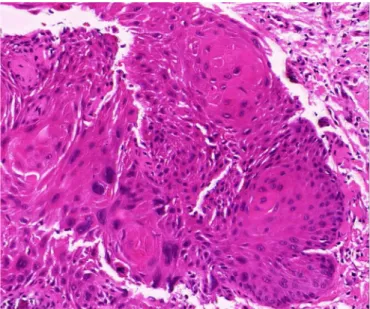
Fig. 4. Histological finding with hematoxylin and eosin staining
at original magnification ×400. The tumor shows malignant squamous sheets and nests with invasive growth pattern and pleomorphism.
Fig. 1.
Preoperative view.
Fig. 2. Intraoperative view.
Fig. 3. Immediate postoperative view.
https://doi.org/10.12790/ahm.19.0069 Ji-An Choi, et al. Carcinoma of Finger
48
락, 특히 무지에 발생하는 것으로 보고되어 있다[5]. 위험인자로는 만성감염, 방사선 노출, 인간유두종바이러스 감염, 화상 흉터, 일 광 노출, 반복되는 외상, 면역억제 및 만성 피부염 등이 있다[6]. 본 증례의 환자는 평생 농사짓던 분으로, 일광 노출이 영향을 끼쳤을 것으로 사료된다. 조상의 출혈, 궤양, 종괴, 항진균제나 항생제 치 료의 영향이 없을 경우 강하게 악성피부종양을 의심할 수 있으며, 특히 매우 빠르게 궤양병변이 증가하는 경우 편평상피세포암을 의 심해 볼 수 있다[7]. 조상의 편평상피세포암의 경우 본 증례와 같이 조갑진균증으로 오인되는 등 비슷한 양성 질환이 많아 진단이 늦 어지는 편이다. X-ray, CT, MRI (magnetic resonance imag- ing) 등이 진단에 도움은 될 수 있으나, 확진은 조직검사를 통해서 만 가능하다. 조상의 편평상피세포암은 매우 드물어 적절하고, 표 준화된 수술적 치료는 정립되어 있지 않다. 경우에 따라 광범위 절 제술에서 원위지골의 절단까지도 고려해 볼 수 있으며, 전이된 경 우 림프절 박리도 필요할 수 있다[7]. 본 증례의 경우에도 방사선학 적 검사상으로는 원위지골을 침범하지 않는 것으로 보였으나, 수 술 당시 병변에 원위지골과 인접하여 있어 원위지골 일부를 절단 하였다.
수술 후에도 드물지만 림프절 전이나 원격전이가 발생하기 때문 에 약 10년 정도 경과관찰을 해야한다[8]. 따라서 본원에서도 본 교실 및 혈액종양내과와 협진 중에 있다.
본 증례에서는 우측 중지 조상에 병변이 발생하여 조갑진균증으 로 오인한 70대 남자환자에서 조직검사 결과 편평상피세포암이 진 단 되어 본원에서 수술을 시행한 예로, 상기와 비슷한 경우가 발생 시 다른 치료에 호전이 없을 경우 최대한 빠른 조직검사가 필요할 것으로 판단된다. 또한 조상의 편평상피세포암에 대한 증례는 국 내에서 보고가 드문 증례로 생각되어 문헌고찰과 함께 보고하는 바이다.
Conflicts of Interest
The authors have nothing to disclose.
Acknowledgements
This study was supported by the research funds of Dong-A University.
References
1. Wong TC, Ip FK, Wu WC. Squamous cell carcinoma of the nail bed: three case reports. J Orthop Surg (Hong Kong). 2004;12:
248-52.
2. Tirpude BH, Bhanarkar H, Shamkuwar A, Gajbhiye A. Squa- mous cell carcinoma of the nail bed: a case report. Int Surg J.
2016;2:79-81.
3. Lee YM, Rhee SK, Song SW, et al. Malignant Tumors of the Hand. J Korean Soc Surg Hand. 201116:154-60.
4. Kim JC, Jung SG, Kim KH, Kim HL. Basosquamous carcino- ma of the hand in a radiologist with prolonged radiation expo- sure. J Korean Soc Surg Hand. 2016;21:162-6.
5. Yip KM, Lam SL, Shee BW, Shun CT, Yang RS. Subungual squamous cell carcinoma: report of 2 cases. J Formos Med As- soc. 2000;99:646-9.
6. Arumugam M. Squamous cell carcinoma of the thumb nail bed. Indian J Dermatol Venereol Leprol. 2007;73:445.
7. Sever C, Kulaci Y, Oksuz S. Subungual squamous cell carcino- ma masquerading as an onychomycosis. J Clin Anal Med.
2012;3:231-3.
8. Kouskoukis CE, Scher RK, Kopf AW. Squamous-cel carcinoma
of the nail bed. J Dermatol Surg Oncol. 1982;8:853-5.
Arch Hand Microsurg 2020;25(1):46-49
https://doi.org/10.12790/ahm.19.0069 49
조상에 발생한 편평상피세포암: 증례 보고
최지안1, 곽정하1, 최정환1, 임광열1, 김대철2, 윤청민1
1동아대학교 의과대학 성형외과학교실, 2동아대학교 의과대학 병리학교실
수부의 종양은 드문 것으로 알려져 있다. 조상의 가장 흔한 종양은 편평상피세포암이지만, 유병률 자체가 매우 드물다. 저자들은 조갑진균증으로 오인된 중지의 편평상피세포암을 경험하여 문헌고찰과 함께 보고한다. 상기 환자는 70대 남자환자로, 약 2년 전부터 우측 중지 조상에 삼출물이 발생하였으나, 조갑진균증으로 생각하고 자가치료하다 호전이 없어 타병원에서 조상 제거 후 조직검사를 시행하였다. 병변부위가 원위지골과 인접하여 있어 광범위 절제술 후 원위지골 일부를 절제하였다. 병리조직검사에서 침습적 성장 양상 및 다형성을 보이는 편평상피세포암이 관찰되었다. 수술 후 6개월간 경과관찰 결과 재발 및 합병증은 관찰되지 않았다. 수부의 피부암의 경우 악성흑색종, 편평상피세포암등 다양한 암이 발생할 수 있지만, 유병률이 흔하지 않다. 조상의 경우 조갑진균증으로 오인하기 쉬워 치유 속도가 느린 경우 반드시 조직검사가 필요할 것으로 사료된다.
색인단어: 편평상피세포암, 손톱
접수일 2019년 11월 30일 수정일 2019년 12월 25일 게재확정일 2019년 12월 26일 교신저자 윤청민
49201, 부산광역시 서구 대신공원로 26, 동아대학교 의과대학 성형외과학 교실 TEL 051-240-5416 FAX 051-243-5416 E-mail [email protected] ORCID https://orcid.org/0000-0003-0307-5545