Miyoshi형 원위부 근육병증 1례
울산대학교 의과대학 서울아산병원 재활의학교실, 병리학교실�,
고려대학교 의과대학 재활의학교실��, 연세대학교 의과대학 영동세브란스 신경과교실���
류일선∙박은하∙편성범��∙강신광�∙최영철���
– Abstract –
Miyoshi myopathy: A Case Report
Il-Sun Lew, M.D., Eun-Ha Park, M.D., Sung-Bom Pyun, M.D.**, Ph.D., Sin- Kwang Kang, M.D.*, Young-Chul Choi, M.D.***
Department of Rehabilitation Medicine, and Pathology*, Asan Medical Center, University of Ulsan College of Medicine,
Department of Rehabilitation Medicine, Korea University College of Medicine**,
Department of Neurology, Youngdong Severance Hospital, Yonsei University College of Medicine***
We report a 25-year-old man who manifested typical symptoms of Miyoshi myopathy with brief review of literatures. Miyoshi myopathy is a rare distal myopathy which develops between 15 and 30 years of age and starts from the distal muscles, especially posterior compartment of the legs. Creatine kinase (CK) level is characteristically elevated to 10~100 fold above normal range. Electromyographic findings are compatible with myopathy and muscle biopsy shows myopathic changes with non-rimmed vacuoles and absence of dysferlin protein on immunostaining.
Key Words: Miyoshi myopathy, Dysferlin, Dysferlinopathy, Distal myopathy
Address reprint requests to Sung-Bom Pyun, Ph.D.
Department of Rehabilitation Medicine, Korea University Anam Hospital 126-1, 5-ga, Anam-dong, Sungbuk-gu, 136-705, Seoul Korea
Tel: 82-2-920-6480, Fax: 82-2-929-9951, Email: [email protected]
서 론
일반적으로 근육병증은 상하지 근위부 근력 약화가 주 증상인 반면 원위부 근육병증은 원위부 근육에서 먼 저 증상이 발현되는 매우 드문 질환이다. 원위부 근육 병증은 유전방식과 임상양상에 따라 Welander형, Markesbery형, Nonaka형, Miyoshi형, Laing형 등 으로 분류되는데, 본 저자들은 특징적인 Miyoshi형의 원위부 근육병증 1예를 경험하였기에 보고하는 바이다.
증 례
증례: 25세 남자 환자로 수년 전부터 시작된 양측 하
지 위약과 비복근 근위축을 주소로 본원 재활의학과 외 래에 내원하였다(Fig. 1). 환자의 과거력에서 특이한 병력은 없었으며, 가족력에서도 특이 사항은 없었다.
내원 당시 이학적 검사에서 양측 상지 근력은 근위부와 원위부에서 모두 정상이었으며, 하지 근력은 양측 고관 절과 슬관절에서는 5/5로 정상 소견을 보였다. 양측 발 목 관절에서 족배 굴곡의 근력은 5/5로 정상 소견이었 으나, 족저 굴곡의 근력은 4/5로 약화되어 있었고 양측 비복근의 위축이 관찰되었으며 까치발 보행이 불가능하 였다. 감각은 정상이었으며, 양측 슬관절 반사는 정상 이었지만 양측 발목 반사는 나타나지 않았고, 병적 반 사는 관찰되지 않았다. 외래에서 시행한 요추부 단순 방사선 촬영에서 특이 소견은 없었으며, 요추부 자기공
명영상에서도 제 5 요추와 제 1 천추 사이의 후관절에 경도의 퇴행성 변화 소견 외에는 모두 정상이었다. 혈 액 화학적 검사에서 CK 5,329 IU/L(참고치 50~250 IU/L), LD 401 IU/L(참고치 120~250 IU/L)로 CK 수치가 매우 상승되어 있었다.
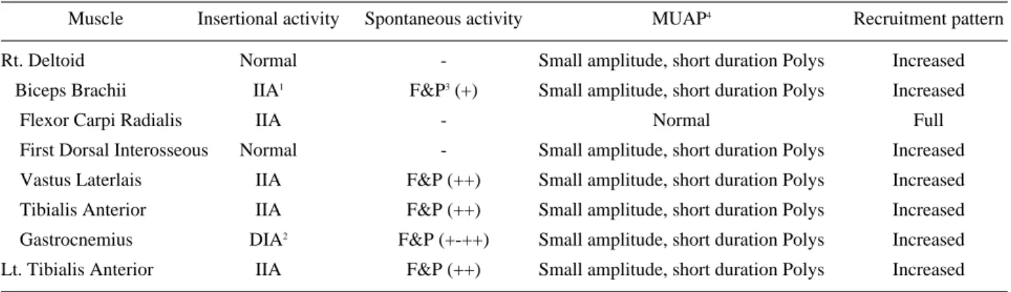
전기진단 검사에서 상하지의 운동과 감각 신경전도 검사는 정상 소견을 보였고, F-반사는 상하지에서 정상 소견을 보였지만, H-반사는 양측에서 유발되지 않았 다. 침근전도 검사에서 우측 비복근, 전경골근, 장비골 근, 대퇴 이두근에서 비정상 자발전위와 함께 작은 진 폭과 짧은 지속기간의 다상성 활동전위가 관찰되었으 며, 조기 점증양상을 보였고, 특히 우측 비복근에서는 삽입전위가 심하게 감소 되어 있는 소견을 보였다. 정 량적 침근전도 검사에서도 진폭 대 반전 비(ampl:tude/
turn ratio)가 낮아 근육병증을 시사하는 소견을 보였 다(Table 1). 좌측 대퇴 이두근과 비복근에서 근생검
을 시행하였으며, 면역형광 염색에서 좌측 대퇴 이두근 에서 Dysferlin이 관찰되지 않았고, 테두리 없는 액포 가 관찰되었으며, 다양한 크기의 근섬유와 함께 근섬유 의 괴사가 관찰되는 등 근육병증에 합당한 소견을 보였 다. 좌측 비복근에서는 근섬유는 관찰되지 않은 채 심 한 섬유화 소견만이 관찰되는 등 근위부보다 원위부에 서 심하게 침범된 소견을 보여 Miyoshi형 근육병증에 합당한 소견을 보였다(Fig. 2).
고 찰
Miyoshi형 근육병증은 1977년 Miyoshi 등에 의해 처음 보고되었으며,1 상염색체 열성으로 유전되거나 산 재성으로 발현되어, 15~30세 사이의 비교적 젊은 성인 기에 증상이 시작된다. 근력 약화는 일반적인 근육병과
Fig. 1. Anterior view (A) and posterior view (B) of the patient which shows atrophy of distal leg muscles.
Fig. 2. Pathologic findings of muscle biopsy in biceps femoris long head. (A) Mild size variation of myofibers and scattered necrotic fibers. Rimmed vacuole is not observed (H&E, ×200 magnification). (B) Absence of Dysferlin (immunofluorescence, ×200 magnification).
A B
는 달리 원위부 근육부터 약화가 나타나며 특히 비복근 에서 가장 심한 근력 약화를 보이며, CK 수치는 정상 의 10~100배 정도로 매우 높게 증가되는 것으로 알려 져 있다.2,3
원 위 부 근 육 병 증 은 현 재 까 지 밝 혀 진 바 로 는 Welander형, Markesbery형, Nonaka형, Miyoshi 형, Laing형 등이 있으며, 현재도 계속 그 표현형과 유전형이 밝혀지고 있다(Table 2).4 Miyoshi형 근육병 증이 일반적으로 성인 초기에 발병하는 것과는 달리 Welander형과 Markesbery-Griggs/Udds형은 성인 후기에 증상이 발현된다.3 또한 Welander형은 주로 상 지를 먼저 침범하며, Nonaka형과 Laing형의 경우에 는 Miyoshi형 근육병증과 유사하게 성인 초기에 발병 하지만, Miyoshi형 근육병증과는 달리 주로 무릎 이하 의 하지 근육 중 앞쪽 근육(anterior compartment)
에서 근위축이 발생하며 근생검에서 공포가 발견될 수 있고,4 CK 수치가 정상의 3~5배 이하로 상승하는 점 에서 Miyoshi형 근육병증과 구별할 수 있다. 원위부 근육병증의 아형 외에도 감별이 필요한 근육질환으로는 먼저 봉입체 근염(inclusion body myositis)을 들 수 있는데, 근 생검에서 염증세포와 테두리 있는 액포를 관찰할 수 있어 Miyoshi형 근육병증과는 다르며, 호발 부위가 상지에서는 주로 손목이나 손가락의 굴곡과 관 계된 장모지 굴곡근(flexor pollicis longus), 하지에 서는 주로 대퇴직근(restus femoris)이나 전경골근 (anterior tibialis)으로 원위부 뿐만 아니라 근위부에 서도 근위축이 발생할 수 있다는 점에서도 Miyoshi형 근육병증과 감별된다.5 그 외에도 신경질환 중 Char- cot-Marie-Tooth II 형과의 감별이 필요한데, 20대 초반부터 원위부의 근육의 약화를 보이며, 감각 신경은
Table1. Needle Electromyographic Findings
Muscle Insertional activity Spontaneous activity MUAP4 Recruitment pattern
Rt. Deltoid Normal - Small amplitude, short duration Polys Increased
Biceps Brachii IIA1 F&P3(+) Small amplitude, short duration Polys Increased
Flexor Carpi Radialis IIA - Normal Full
First Dorsal Interosseous Normal - Small amplitude, short duration Polys Increased Vastus Laterlais IIA F&P (++) Small amplitude, short duration Polys Increased Tibialis Anterior IIA F&P (++) Small amplitude, short duration Polys Increased Gastrocnemius DIA2 F&P (+-++) Small amplitude, short duration Polys Increased Lt. Tibialis Anterior IIA F&P (++) Small amplitude, short duration Polys Increased 1. IIA: increased insertional activity
2. DIA: decreased insertional activity
3. F & P: fibrillation potential & positive sharp wave 4. MUAP: motor unit action potential
Table 2. Subtypes of Distal Myopathies4
Type Inheritance Gene location Initial weakness Creatine Kinase Muscle Biopsy
Welander type Hands: finger/wrist Normal or slightly Myopathic with rimmed
Late adult onset I AD1 2p13
extensors increased vacuoles
Markesberry-Griggs type Legs: anterior Normal orslightly Myopathic with rimmed Late adult onset II AD 2q31-33
compartment increased vacuoles
Nonaka type
Early adult onset I AR2 2p1-q1 Legs: anterior slightly increased, Myopathic with compartment usually <5× normal rimmed vacuoles
Miyoshi type Legs: posterior compartment
Increased Myopathic with Early adult onset II AR 2p12-13 ; occasionally the anterior non-rimmed vacuoles and
(Dysferlin) compartment or hip girdle 10~150 × normal
vessicles
Laing type 14q11 Legs: anterior compartment, slightly increased, Myopathic
Early adult onset III AD neck flexor usually <3× normal
1. AD: Autosomal dominant 2. AR: Autosomal recessive
비교적 유지되어 있고, 신경전도 검사에서도 거의 정상 소견을 보일 수 있다는 점에서 Miyoshi형 근육병증과 유사하나 CK 수치의 증가가 없고 신경병성 침근전도 소견을 보이며, 근생검에서 근육병증 소견이 없다는 점 에서 Miyoshi형 근육병증과 감별할 수 있다. 그 외에 도 하지의 원위부에 근력약화가 나타나는 다양한 신경 근육 질환과의 감별이 필요하다(Table 3).3
Miyoshi형 근육병증의 원인에 대해서는 최근 유전자 연구에서 sarcolemma에서 dysferlin 이라는 단백이 없거나 감소되어‘dysferlinopathy’가 그 원인으로 생 각 되고 있다.7 Dysferlinopathy는 염색체 2p13.3에 위치하면서 dysferlin 단백을 encoding하는 유전자 (DYSF gene)의 돌연변이로 인해 발생하는데 돌연변 이의 유형은 결손 돌연변이(deletion), 삽입 돌연변이 (insertion) 뿐 아니라, 과오 돌연변이(missense) 혹 은 무의미 돌연변이(nonsense)등 매우 다양해서 돌연 변이 방식의 특정한 패턴이 존재하지 않는다. 또한 유 전자형(genotype)과 표현형(phenotype)사이에 중증 도에 따른 연관성이 없고, 임상적 표현형에 있어서도 가족내(intra-familial), 가족간(inter-familial)의 변 이도 상당히 다양한 것으로 알려져 있다.8
Dysferlinopathy의 임상 표현형(clinical pheno- type)은 Miyoshi형 근육병증 뿐 아니라 지대형 근이영 양증 2B(limb girdle muscular dystrophy 2B, LGMD 2B)가 있으며 동일한 DYSF 유전자의 돌연변 이가 관찰된다. 이는 Miyoshi형 근육병증과 LGMD 2B가 서로 DYSF 유전자의 대립유전자의 변형(allelic variation)이라는 사실을 나타내며,4 dysferlinopathy 가 LGMD 2B로 나타날 수도 있고, Miyoshi형 근육
병증으로 발현될 수도 있다. 더우기 DYSF의 동일한 변이가 한 가족 내에서 Miyoshi형 근육병증과 LGMD 2B의 두 가지 표현형으로 나타날 수도 있으며,9 또한 한 사람에게서 두 가지 표현형이 모두 나타날 수도 있 는 것으로 알려져 있다.10 1977년 Miyoshi 등에 의해 처음 보고된 이후 이러한 돌연변이는 대부분 일본인에 서 확인되었지만, 최근 보고에 따르면 Miyoshi형 근육 병증과 LGMD 2B 모두 전세계 어디에서도 발견될 수 있다.11
Miyoshi 형 근육병증의 예후는 비교적 양호한 것으 로 알려져 있으나, 일상 생활 동작이나 기능저하에 대 한 장기적인 예후에 관한 논문은 거의 없는 상태로 Miyoshi형 근육병증 등 원위부 근육병증에 대한 효과 적인 치료법은 아직까지 알려져 있지 않으며 corticos- teroid나 azathioprine 같은 약제가 투여되기도 하였 으나 임상적 효과를 나타내지는 못하였고,12 발목 관절 의 고정을 위한 단하지 보조기가 보행이나 계단오르기 등에 유용하게 사용될 수 있다.3
참고문헌
11. Miyoshi K, Iwasa M, Kawai H, Sasaki N, Kusaka K, Yagita M, et al: Autosomal recessive distal muscular dys- trophy-a new type of distal muscular dystrophy observed characteristically in Japan. Nippon Rinsho 1977: 35: 3922- 3928.
12. Flachenecker P, Kiefer R, Naumann M, Handwerker M, Reichmann H: Distal muscular dystrophy of Miyoshi type.
Report of two cases and review of the literature. J Neurol 1997: 244: 23-29.
13. Griggs RC, Markesbery WR: Distal myopathies. In: Engel AG, Franzini-Armstrong C, editors. Myology. New York:
McGraw-Hill, 1994, pp1246-1257.
14. Dumitru D, Amato AA, Zwarts M: Hereditary Myopathies in Dumitru D, Amato AA, Zwarts M, editors. Electrodi- agontic Medicine 2nd ed, Philadelphia: Hanley & Belfus, 2002, pp1265-1370.
15. Nonaka I, Sunohara N, Satoyoshi E, Terasawa K, Yonemoto K: Autosomal recessive distal muscular dystro- phy: a comparative study with myopathy with rimmed vacuole formation. Ann Neurol 1985: 17: 51-59.
16. Lotz BP, Engel AG, Nishino H, Stevens JC, Litchy WJ:
Inclusion body myositis. Observations in 40 patients.
Brain 1989:112: 727-747.
17. Dyck PJ: Inherited neuronal degeneration and atrophy affecting peripheral motor, sensory and autonomic neu- rons. In Dyck PJ, Thomas PK, Lambert EH, editors.
Table 3. Differential Diagnoses of the Distal Myopathies Pre- senting with Leg Weakness3
Charcot-Marie-Tooth disease (neuronal form) Myotonic dystrophy
Distal chronic muscular atrophy Inflammatory myopathies
(esp. inclusion body myositis)
Motor neuropathies (e.g., lead toxicity, porphyria) Facioscapulohumeral muscular dystrophy Scapuloperoneal syndromes
Nonspecific histology Nemaline myopathy Central core disease Centronuclear myopathy
Debranching enzyme deficiency myopathy Phosphorylase b kinase deficiency Lipid storage myopathy
Peripheral Neuropathy. Philadelphia: Saunders, 1975, pp834-855.
18. Liu J, Aoki M, Illa I, Wu C, Fardeau M, Angelini C, et al : Dysferlin, a novel skeletal muscle gene, is mutated in Miyoshi myopathy and limb girdle muscular dystrophy.
Nat Genet 1998: 20: 31-36.
19. Takahashi T, Aoki M, Tateyama M, Kondo E, Mizuno T, Onodera Y, et al: Dysferlin mutations in Japanese Miyoshi myopathy: relationship to phenotype. Neurology 2003: 60:
1799-1804.
10. Weiler T, Bashir R, Anderson LV, Davison K, Moss JA, Britton S, et al: Identical mutation in patients with limb girdle muscular dystrophy type 2B or Miyoshi myopathy
suggests a role for modifier gene(s). Hum Mol Genet 1999: 8: 871-877.
11. Nguyen K, Bassez G, Bernard R, Krahn M, Labelle V, Figarella-Branger D, et al: Dysferlin mutations in LGMD2B, Miyoshi myopathy, and atypical dysfer- linopathies. Hum Mutat 2005: 26: 165.
12. Argov Z, Sadeh M, Mazor K, Soffer D, Kahana E, Eisen- berg I, et al: Muscular dystrophy due to dysferlin deficien- cy in Libyan Jews. Clinical and genetic features. Brain 2000: 123: 1229-1237.
13. Miller RG, Blank NK, Layzer RB: Sporadic distal myopa- thy with early adult onset. Ann Neurol 1979: 5: 220-227.