Distal Myopathy with Rimmed Vacuoles Confirmed by Whole Exome Sequencing
Seong Don Seo
1†, Hyung Jun Park
2†, Hyun Seok Song
1, Hye Jin Kim
3, Jin-Mo Park
4, Young Bin Hong
2, Ki Wha Chung
3* and Byung-Ok Choi
2*
1
Department of Neurology, Kyungpook National University School of Medicine, Daegu 702-701, Korea
2
Department of Neurology, Samsung Medical Center, Sungkyunkwan University School of Medicine, Seoul 135-710, Korea
3
Department of Biological Science, Kongju National University, Gongju 314-701, Korea
4
Department of Neurology, School of Medicine, Ewha Womans University, Seoul 120-750, Korea
Received December 12, 2013 /Revised January 14, 2014 /Accepted January 28, 2014Distal myopathy with rimmed vacuoles (DMRV) or hereditary inclusion body myopathy 2 is an auto- somal recessive muscular disorder characterized by early adult-onset weakness of distal muscles and rimmed vacuoles in muscle biopsy. Mutations in the UDP-N-acetylglucosamine 2-epimerase/N-ace-tylman- nosamine kinase (GNE) gene are associated with the development of DMRV. In this study, whole exome sequencing (WES) revealed compound heterozygous GNE mutations of p.Asp176Val and p.Val572Leu in a patient with distal limb weakness. Three hundred healthy controls did not show these mutations.
All other variants found in distal myopathy-relevant genes were polymorphic. These findings con- firmed that the patient had DMRV. This work underscores the usefulness of WES in improving the molecular diagnosis of myopathy.
Key words : Distal myopathy with rimmed vacuoles (DMRV), molecular diagnosis, UDP-N-acetylglu- cosamine 2-epimerase/N-acetylmannosamine kinase (GNE), whole exome sequencing (WES)
†Authors contributed equally.
*Corresponding author
*Tel : +82-2-3410-1296, Fax : +82-2-3410-0052
*E-mail : [email protected] (Byung-Ok Choi) [email protected] (Ki Wha Chung)
This is an Open-Access article distributed under the terms of the Creative Commons Attribution Non-Commercial License (http://creativecommons.org/licenses/by-nc/3.0) which permits unrestricted non-commercial use, distribution, and reproduction in any medium, provided the original work is properly cited.
Journal of Life Science 2014 Vol. 24. No. 3. 311~317 DOI : http://dx.doi.org/10.5352/JLS.2014.24.3.311
Introduction
Distal myopathies are hereditary muscle disorders de- fined by onset of muscle weakness and atrophy in hands or feet [28]. They include a wide variety of diseases, such as Welander distal myopathy, tibial muscular dystrophy, myofibrillar myopathy, Miyoshi myopathy, and distal myo- pathy with rimmed vacuoles (DMRV). For the diagnosis of distal myopathies, a serial approach is generally used. First, patients are classified according to age of onset, inheritance pattern, and clinical course. Second, histopathological analy- sis of muscle biopsy, especially immunohistochemistry, is used.
Prior to the advance of next generation sequencing, Sanger sequencing for the coding exons of genes reported to cause the respective phenotypes was usually done to
screen for the exact causative mutation. However, the over- lapping phenotypes or clinical-genetical heterogeneities make this screening of possible candidate genes elusive [3, 25]. For instance, genes like dysferlin and myotillin are asso- ciated with both distal myopathies and limb girdle muscular dystrophies [7, 22, 23]. Recently, next-generation sequencing has made it possible to cost-effectively and rapidly sequence the protein-coding exons of the genome, by a process termed
‘whole exome sequencing (WES)’. The use of WES has iden- tified the causative genetic defect in many monogenic dis- eases including Welander distal myopathy [2, 4, 12].
The UDP-N-acetylglucosamine 2-epimerase/N-acetylmannos-
amine kinase (GNE) gene on chromosome 9p13.3 encodes the
bifunctional rate limiting enzyme for the sialic acid bio-
synthetic pathway by initiating and regulating the biosyn-
thesis of N-acetlyneuraminic acid (NeuAc) a precursor of
sialic acid [8, 10]. The GNE gene is ubiquitously expressed
and has two functional domains: the epimerase and the kin-
ase domains located in the N-terminus encoding the N-actyl-
glucosamine 2 epimerase and the C-terminus encoding the
N-acetylmannosamine kinase, respectively [14, 19]. Mutations
in GNE have been linked to not only DMRV (MIM 605820)
[27, 29] but also sialuria (MIM 269921) [13, 24]. In particular,
many mutations of GNE have been reported to be the under-
lying causes of DMRV [6, 9, 11, 14, 15, 18, 29].
In the present study, we report the clinical and genetic diagnosis of a Korean patient with undetermined distal myopathy type using WES, which revealed a pair of com- pound heterozygous mutations in the GNE gene and the myopathy type.
Materials and Methods Subjects
We enrolled a Korean family with one patient with distal myopathy and two healthy individuals (family ID: FC532, Fig. 1A). Korean healthy controls with no familial history of neuromuscular disorders (n=300) were also recruited.
Paternity was confirmed by genotyping of 15 microsatellites using the PowerPlex 16 System (Promega, Madison, WI, USA). Written informed consent was obtained from all par- ticipants according to the protocol approved by the Institutional Review Board for Ewha Womans University, Mokdong Hospital.
Clinical and electrophysiological assessments The patient was examined for mental function, cranial nerve dysfunction, motor and sensory impairments, pres- ence of contractures, deep tendon reflexes, and muscle atrophy. The strength of flexor and extensor muscles were assessed manually using the Medical Research Council (MRC) scale. Serum creatine kinase (CK) levels were measured. Nerve conduction studies (NCS) and needle elec- tromyographies (EMG) were performed by standard meth- ods [21].
Exome sequencing and identification of causative mutation
The exome for the patient (II-1) was captured using the Human SeqCap EZ Human Exome Library v3.0 (Roche/
NimbleGen, Madison, WI, USA). Captured DNA was se- quenced on the HiSeq 2000 Genome Analyzer (Illumina, San Diego, CA, USA). Sequences were mapped/aligned to the reference human genome (GRCh37, UCSC hg19) using BWA (http://bio- bwa.sourceforge.net/) via a pileup file from the BAM file. Variant calling was performed using the SAMtools (http://samtools.sourceforge.net/) and GATK programs (http://www.broadinstitute.org/gatk/). Variants were sub- mitted to ANNOVAR (http://www.openbioinformatics.
org/annovar/) for functional annotation. Single nucleotide polymorphisms (SNPs) with a quality value >20 were con-
sidered a true variant call.
Registered, novel, or uncommon variants (minor allele frequency≤0.01) in dbSNP138 (http://www.ncbi.nlm.nih.
gov), the 1000 Genomes project database (http://www.
1000genomes.org/), and Exome Variant Server (http://evs.
gs.washington.edu/EVS/) were examined. All variants pres- ent in reported myopathy genes were sorted. Candidate var- iants considered as causative were confirmed by Sanger’s sequencing method using an ABI 3100XL automatic se- quencer (Applied Biosystems, Foster City, CA, USA). Muta- tions were considered to be an underlying cause when they were detected only in the affected member of the family and not detected in more than 300 healthy controls.
In silico analysis
The affection of protein function due to amino acid sub- stitution were assessed using SIFT (http://sift.jcvi.org/) and PolyPhen2 (http://genetics.bwh.harvard.edu/pph2/); and protein stability by MUpro (http://mupro.proteomics.ics.
uci.edu/). The conservation pattern of the amino acid posi- tions were done by multiple sequence alignment of protein sequences with MEGA5 software (http://www.megasoftware.
net/). The genomic evolutionary rate profiling (GERP) scores (http://mendel.stanford.edu/SidowLab/downloads/
gerp/index.html) of the nucleotide positions were also assessed.
Results
Clinical manifestations and electrophysiological features
The proband (II-1) was a 38-year-old woman who pre- sented with slowly progressive distal muscle weakness. At the age of 35 years, she experienced frequent falling and no- ticed muscle weakness and atrophy of the distal lower limbs.
Within one year, she noticed muscle weakness of bilateral hands. She denied any other medical diseases. Family his- tory was unremarkable (Fig. 1A). When we examined her at age 45, distal muscles of upper and lower limbs were more severely affected than proximal muscles. Deep tendon reflexes are reduced. Pain and vibration sense was intact.
Serum CK level was 350 IU/L (normal range: <170/L). NCS
and EMG showed a generalized myogenic process with dis-
tal accentuation. Based on clinical, laboratory, and electro-
physiological features, she was diagnosed with distal
myopathy. However, we did not determine candidate genes
Table 1. Whole exome sequencing analysis
Items
Total yields (Gbp) Mappable reads (%) Target coverage (≥10X, %) Total SNP number Coding SNP number Total indel number Coding indel number Myopathy gene variantsa
7.95 99.489.7 101,743 22,084 8,410 31749
aFunctionally significant variants include nonsynonymous, splicing site, frameshift, stop gain, stop loss, and coding indels.
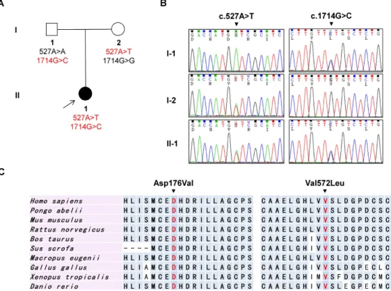
Fig. 1. Pedigree, sequencing chromatograms, and conservation profile. (A) Pedigree of the FC532 DMRV family. The proband is indicated by an arrow. Filled symbol indicates affection and open symbols indicate unaffected members. Genotypes of two
GNE
mutations are denoted at bottom of the each family member. (B) Sequencing chromatograms ofGNE
mutations c.527A>T (p.Asp176Val) and c.1714G>C (p.Val572Leu). The patient reveals both mutations, whereas the proband’s parents have only a mutation each (I-1: c.1714G>C and I-2: c.527A>T). (C) Conservation analysis results. The mutation sites were well conserved among the subset of species studied and lay in the UDP-N-acetylglucosamine 2-epimerase domain and N-acetylmannosamine kinase domain, respectively.for mutational analysis due to small-sized pedigree and non-specific clinical presentation. Therefore, we performed WES.
Identification of a compound heterozygous mis- sense mutation in GNE gene
The summary of whole exome sequencing data is outlined
in Table 1. From the exome data, 49 variants were found
in known myopathy genes (24 genes). Within these variants,
capillary sequencing analysis of the extended family mem-
bers detected a pair of compound heterozygous mutations
in GNE (NM_005476.5), c.527A>T (p.Asp176Val, paternal
origin), and c.1714G>C (p.Val572Leu, maternal origin) that
perfectly co-segregated within the family in a recessive pat-
tern (Fig. 1A, Fig. 1B). The c.527A>T (p.Asp176Val) and
c.1714G>C (p.Val572Leu) lie in the highly conserved sites
of the epimerase and kinase domains of GNE protein,
respectively. These mutations have been previously reported
to cause DMRV, and are the most common mutations in
Japanese patients [2, 22]. None of the 300 healthy controls
harbored these mutations. Both mutations were reported in
the dbSNP137 but not in 1000 Genome Database and Exome
Variant Server (EVS). All in silico predictions (SIFT,
PolyPhen2, MUpro, and GERP) yielded commendable re-
sults (Table 2) and the amino acid positions were well con-
served throughout different vertebrate species (Fig. 1C).
Table 2. Mutations of
GNE
gene in the distal myopathy patientGene Mutation
Domain GERPa
In silico
analysisbNucleotide Amino acid SIFT Polyphen2 MUpro
GNE
c.527A>Tc.1714G>C D176V
V572L GT1-UDP-GlcNAc 2-epimerase domain
Kinase domain 5.67
5.75 0.07
0.00* 1.00*
0.92* -0.11* -0.356*
aGenomic evolutionary rate profiling score
bSIFT score ≤0.05, PolyPhen2 score ~1, and MUpro scores <0 indicate a prediction of pathogenicity
*Denotes a “pathogenic” prediction
Thus, p.Asp176Val and p.Val572Leu mutations in GNE were determined as the underlying cause of our patient.
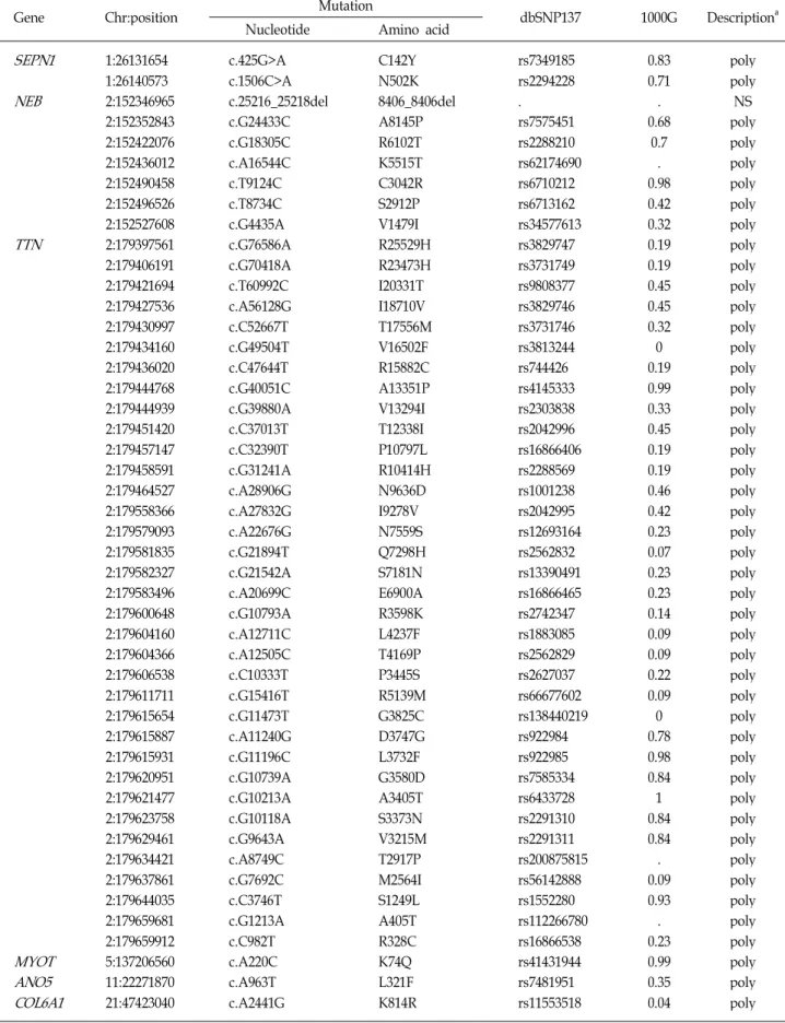
In addition to the two causative GNE mutations, many polymorphic or rare nonsynonymous variants were identi- fied in a large number of myopathy-related genes from the exome data of the proband (Table 3). However, they were not considered as underlying causes because they met at least one of the following conditions: 1) noncosegregation with affected individuals within pedigrees, 2) same variant was found in controls, or 3) inconsistency in the inheritance manner for corresponding genes.
Discussion
By WES analysis, we identified a set of compound hetero- zygous mutations at c.527A>T and c.1714G>C in the GNE gene in a patient with undetermined distal myopathy. These mutations lie in both the highly conserved bifunctional do- mains of the UDP-N-acetylglucosamine 2 epimerase/N-ace- tylmannosamine kinase enzyme: c.527A>T (p.Asp176Val) in the epimerase domain and c.1714G>C (p.Val572Leu) in the kinase domain. The co-segregation, absence of the same mu- tations in control samples, in silico predictions, and well con- served patterns leads us to affirm that the two compound heterozygous GNE mutations are the underlying cause of DMRV in this patient.
DMRV is also known as Nonaka myopathy, GNE myo- pathy, or hereditary inclusion body myopathy. It is an auto- somal recessive distal myopathy caused by the alterations in the GNE gene [5]. This disease generally develops in early adulthood and is clinically characterized by preferential in- volvement of ankle dorsiflexors [1, 20]. In addition, muscle pathology typically reveals muscle fiber atrophy with rim- med vacuoles and intracellular congophilic deposits [19].
These characteristic clinical and pathologic findings are im- portant for the initial suspicion of DMRV. However, the di- agnosis of DMRV is not always easy. Expansion of muta-
tional analysis in GNE gene has indicated that DMRV pa- tients often have an atypical clinical presentation that in- cludes proximal muscle weakness [16, 26]. Muscle biopsy is necessary for histopathological evaluation, but is a very invasive method. In addition, there have been several in- stances where patients clinically and pathologically compat- ible with DMRV were subsequently genetically diagnosed with other myopathies [25]. Our patient showed no prefer- ential involvement of ankle dorsiflexors and refused muscle biopsy. Therefore, we did not suspect her case as a DMRV before the WES analysis.
WES is an effective strategy for discovering the under- lying genetic defect in monogenic disorders because more than 90% of the pathogenic mutations of monogenic dis- orders are found in exons [2]. This technology currently has several limitations. These include shorter read lengths com- pared to the Sanger method, ambiguity in alignment, as- sembly in repetitive nucleotide regions, and large volume of data [17]. Despite these limitations, WES is an attractive strategy to diagnose genetic disease by a minimally invasive method.
In conclusion, we were able to find the exact genetic cause and designate the myopathy type of an undetermined distal myopathy patient using WES. Although no novel mu- tations were found, we were able to give a mutualistic clin- ical- genetic diagnosis without the involvement of muscle biopsy, which is otherwise an invasive method. This work underscores the usefulness of WES for the diagnosis of myopathy.
Acknowledgments
This study was supported in part by the Korean Health
Technology R&D Project, Ministry of Health & Welfare
(A120182), and by Basic Science Research Program through
the National Research Foundation (NRF) funded by the
Ministry of Education (2011-0021533), Republic of Korea.
Table 3. Nonsynonymous variants in distal myopathy-related genes from exome sequencing
Gene Chr:position Mutation
dbSNP137 1000G Descriptiona Nucleotide Amino acid
SEPN1 NEB
TTN
MYOT ANO5 COL6A1
1:26131654 1:26140573 2:152346965 2:152352843 2:152422076 2:152436012 2:152490458 2:152496526 2:152527608 2:179397561 2:179406191 2:179421694 2:179427536 2:179430997 2:179434160 2:179436020 2:179444768 2:179444939 2:179451420 2:179457147 2:179458591 2:179464527 2:179558366 2:179579093 2:179581835 2:179582327 2:179583496 2:179600648 2:179604160 2:179604366 2:179606538 2:179611711 2:179615654 2:179615887 2:179615931 2:179620951 2:179621477 2:179623758 2:179629461 2:179634421 2:179637861 2:179644035 2:179659681 2:179659912 5:137206560 11:22271870 21:47423040
c.425G>A c.1506C>A c.25216_25218del c.G24433C c.G18305C c.A16544C c.T9124C c.T8734C c.G4435A c.G76586A c.G70418A c.T60992C c.A56128G c.C52667T c.G49504T c.C47644T c.G40051C c.G39880A c.C37013T c.C32390T c.G31241A c.A28906G c.A27832G c.A22676G c.G21894T c.G21542A c.A20699C c.G10793A c.A12711C c.A12505C c.C10333T c.G15416T c.G11473T c.A11240G c.G11196C c.G10739A c.G10213A c.G10118A c.G9643A c.A8749C c.G7692C c.C3746T c.G1213A c.C982T c.A220C c.A963T c.A2441G
C142Y N502K 8406_8406del A8145P R6102T K5515T C3042R S2912P V1479I R25529H R23473H I20331T I18710V T17556M V16502F R15882C A13351P V13294I T12338I P10797L R10414H N9636D I9278V N7559S Q7298H S7181N E6900A R3598K L4237F T4169P P3445S R5139M G3825C D3747G L3732F G3580D A3405T S3373N V3215M T2917P M2564I S1249L A405T R328C K74Q L321F K814R
rs7349185 rs2294228 . rs7575451 rs2288210 rs62174690 rs6710212 rs6713162 rs34577613 rs3829747 rs3731749 rs9808377 rs3829746 rs3731746 rs3813244 rs744426 rs4145333 rs2303838 rs2042996 rs16866406 rs2288569 rs1001238 rs2042995 rs12693164 rs2562832 rs13390491 rs16866465 rs2742347 rs1883085 rs2562829 rs2627037 rs66677602 rs138440219 rs922984 rs922985 rs7585334 rs6433728 rs2291310 rs2291311 rs200875815 rs56142888 rs1552280 rs112266780 rs16866538 rs41431944 rs7481951 rs11553518
0.83 0.71 . 0.68
0.7 . 0.98 0.42 0.32 0.19 0.19 0.45 0.45 0.32 0 0.19 0.99 0.33 0.45 0.19 0.19 0.46 0.42 0.23 0.07 0.23 0.23 0.14 0.09 0.09 0.22 0.09 0 0.78 0.98 0.84 1 0.84 0.84 . 0.09 0.93 . 0.23 0.99 0.35 0.04
poly poly NS poly poly poly poly poly poly poly poly poly poly poly poly poly poly poly poly poly poly poly poly poly poly poly poly poly poly poly poly poly poly poly poly poly poly poly poly poly poly poly poly poly poly poly poly
aPol: polymorphic; NS: nonsegregated with affected individual
References
1. Argov, Z. and Yarom, R. 1984. "Rimmed vacuole myopathy"
sparing the quadriceps. A unique disorder in Iranian Jews.
J Neurol Sci
64, 33-43.2. Bamshad, M. J., Ng, S. B., Bigham, A. W., Tabor, H. K., Emond, M. J., Nickerson, D. A. and Shendure, J. 2011.
Exome sequencing as a tool for Mendelian disease gene discovery.
Nat Rev Genet
12, 745-755.3. Cho, A., Hayashi, Y. K., Monma, K., Oya, Y., Noguchi, S., Nonaka, I. and Nishino, I. 2013. Mutation profile of the GNE gene in Japanese patients with distal myopathy with rim- med vacuoles (GNE myopathy).
J Neurol Neurosurg Psychiatry
doi: 10.1136/jnnp-2013-305587. [Epub ahead of print].4. Choi, B. O., Koo, S. K., Park, M. H., Rhee, H., Yang, S. J., Choi, K. G., Jung, S. C., Kim, H. S., Hyun, Y. S., Nakhro, K., Lee, H. J., Woo, H. M. and Chung, K. W. 2012. Exome sequencing is an efficient tool for genetic screening of Charcot-Marie-Tooth disease.
Hum Mutat
33, 1610-1615.5. Eisenberg, I., Avidan, N., Potikha, T., Hochner, H., Chen, M., Olender, T., Barash, M., Shemesh, M., Sadeh, M., Grabov-Nardini, G., Shmilevich, I., Friedmann, A., Karpati, G., Bradley, W. G., Baumbach, L., Lancet, D., Asher, E. B., Beckmann, J. S., Argov, Z. and Mitrani-Rosenbaum, S. 2001.
The UDP-N-acetylglucosamine 2-epimerase/N-acetylmann- osamine kinase gene is mutated in recessive hereditary in- clusion body myopathy.
Nat Genet
29, 83-87.6. Eisenberg, I., Grabov-Nardini, G., Hochner, H., Korner, M., Sadeh, M., Bertorini, T., Bushby, K., Castellan, C., Felice, K., Mendell, J., Merlini, L., Shilling, C., Wirguin, I., Argov, Z. and Mitrani-Rosenbaum, S. 2003. Mutations spectrum of GNE in hereditary inclusion body myopathy sparing the quadriceps.
Hum Mutat
21, 99.7. Hauser, M. A., Horrigan, S. K., Salmikangas, P., Torian, U.
M., Viles, K. D., Dancel, R., Tim, R. W., Taivainen, A., Bartoloni, L., Gilchrist, J. M., Stajich, J. M., Gaskell, P. C., Gilbert, J. R., Vance, J. M., Pericak-Vance, M. A., Carpen, O., Westbrook, C. A. and Speer, M. C. 2000. Myotilin is mu- tated in limb girdle muscular dystrophy 1A.
Hum Mol Genet
9, 2141-2147.8. Hinderlich, S., Stasche, R., Zeitler, R. and Reutter, W. 1997.
A bifunctional enzyme catalyzes the first two steps in N-ace- tylneuraminic acid biosynthesis of rat liver. Purification and characterization of UDP-N-acetylglucosamine 2-epimer- ase/N-acetylmannosamine kinase.
J Biol Chem
272, 24313- 24318.9. Kayashima, T., Matsuo, H., Satoh, A., Ohta, T., Yoshiura, K., Matsumoto, N., Nakane, Y., Niikawa, N. and Kishino, T. 2002. Nonaka myopathy is caused by mutations in the UDP-N-acetylglucosamine-2-epimerase/N-acetylmannos- amine kinase gene (GNE).
J Hum Genet
47, 77-79.10. Keppler, O. T., Hinderlich, S., Langner, J., Schwartz-Albiez, R., Reutter, W. and Pawlita, M. 1999. UDP-GlcNAc 2-epi- merase: a regulator of cell surface sialylation.
Science
284, 1372-1376.11. Kim, B. J., Ki, C. S., Kim, J. W., Sung, D. H., Choi, Y. C.
and Kim, S. H. 2006. Mutation analysis of the GNE gene in Korean patients with distal myopathy with rimmed vacuoles.
J Hum Genet
51, 137-140.12. Klar, J., Sobol, M., Melberg, A., Mabert, K., Ameur, A., Johansson, A. C., Feuk, L., Entesarian, M., Orlen, H., Casar-Borota, O. and Dahl, N. 2013. Welander distal myo- pathy caused by an ancient founder mutation in TIA1 asso- ciated with perturbed splicing.
Hum Mutat.
34, 572-577.13. Leroy, J. G., Seppala, R., Huizing, M., Dacremont, G., De Simpel, H., Van Coster, R. N., Orvisky, E., Krasnewich, D.
M. and Gahl, W. A. 2001. Dominant inheritance of sialuria, an inborn error of feedback inhibition.
Am J Hum Genet
68, 1419-1427.14. Liewluck, T., Pho-Iam, T., Limwongse, C., Thongnoppa- khun, W., Boonyapisit, K., Raksadawan, N., Murayama, K., Hayashi, Y. K., Nishino, I. and Sangruchi, T. 2006. Mutation analysis of the GNE gene in distal myopathy with rimmed vacuoles (DMRV) patients in Thailand.
Muscle Nerve
34, 775-778.15. Malicdan, M. C., Noguchi, S., Nonaka, I., Hayashi, Y. K.
and Nishino, I. 2007. A Gne knockout mouse expressing hu- man GNE D176V mutation develops features similar to dis- tal myopathy with rimmed vacuoles or hereditary inclusion body myopathy.
Hum Mol Genet
16, 2669-2682.16. Motozaki, Y., Komai, K., Hirohata, M., Asaka, T., Ono, K.
and Yamada, M. 2007. Hereditary inclusion body myopathy with a novel mutation in the GNE gene associated with proximal leg weakness and necrotizing myopathy.
Eur J Neurol
14, e14-15.17. Ng, S. B., Turner, E. H., Robertson, P. D., Flygare, S. D., Bigham, A. W., Lee, C., Shaffer, T., Wong, M., Bhattacharjee, A., Eichler, E. E., Bamshad, M., Nickerson, D. A. and Shendure, J. 2009. Targeted capture and massively parallel sequencing of 12 human exomes.
Nature
461, 272-276.18. Nishino, I., Malicdan, M. C., Murayama, K., Nonaka, I., Hayashi, Y. K. and Noguchi, S. 2005. Molecular patho- mechanism of distal myopathy with rimmed vacuoles.
Acta Myol
24, 80-83.19. Nonaka, I., Noguchi, S. and Nishino, I. 2005. Distal myo- pathy with rimmed vacuoles and hereditary inclusion body myopathy.
Curr Neurol Neurosci Rep
5, 61-65.20. Nonaka, I., Sunohara, N., Ishiura, S. and Satoyoshi, E. 1981.
Familial distal myopathy with rimmed vacuole and lamellar (myeloid) body formation.
J Neurol Sci
51, 141-155.21. Oh, S. J. 2003. Clinical electromyography: nerve conduction studies, 3rd eds., Lippincott Williams & Wilkins, USA.
22. Park, H. J., Hong, J. M., Suh, G. I., Shin, H. Y., Kim, S. M., Sunwoo, I. N., Suh, B. C. and Choi, Y. C. 2012. Heterogene- ous characteristics of Korean patients with dysferlinopathy.
J Korean Med Sci
27, 423-429.23. Selcen, D. and Engel, A. G. 2004. Mutations in myotilin cause myofibrillar myopathy.
Neurology
62, 1363-1371.24. Seppala, R., Lehto, V. P. and Gahl, W. A. 1999. Mutations in the human UDP-N-acetylglucosamine 2-epimerase gene define the disease sialuria and the allosteric site of the
초록:Rimmed vacuole을 가진 원위부 근육병증의 전체 엑솜 서열분석을 이용한 유전적 원인 규명 서승돈
1†박형준
2†송현석
1김혜진
3박진모
4홍영빈
2정기화
3*최병옥
2*(
1경북대학교 의과대학 신경과 ,
2성균관대학교 의과대학 신경과 ,
3공주대학교 생명과학과 ,
4이화여자대학교
의과대학 신경과 )
Rimed vacuole을 가진 원위 근육병(distal myopathy with rimmed vacuoles, DMRV)은 제2형 유전성 봉입체
근육병으로도 불리며 초기 성인기에 발병하여 원위부의 근력약화를 보이는 임상양상과 rimmed vacuole의 근육
병리소견을 특징으로 하는 상염색체 열성의 근육병이다 . 이러한 DMRV의 원인은 UDP-N-acetylglucosamine 2-epi- merase/N-acetylmannosamine kinase (GNE) 유전자의 돌연변이임이 밝혀져 있다. 저자들은 원위부 근력약화를 호소
하는 환자에서 전체 엑솜 염기서열분석을 이용하여 GNE 유전자의 복합 이형접합성 돌연변이(p.Asp176Val 및
p.Val572Leu)를 확인하여 DMRV를 진단할 수 있었다. 본 연구는 근육병의 정확한 분자진단에 있어서 전체 엑솜
염기서열분석의 유용성을 보여주었기에 이를 보고하는 바이다 .
enzyme.
Am J Hum Genet
64, 1563-1569.25. Shi, Z., Hayashi, Y. K., Mitsuhashi, S., Goto, K., Kaneda, D., Choi, Y. C., Toyoda, C., Hieda, S., Kamiyama, T., Sato, H., Wada, M., Noguchi, S., Nonaka, I. and Nishino, I. 2012.
Characterization of the Asian myopathy patients with VCP mutations.
Eur J Neurol
19, 501-509.26. Sim, J. E., Park, H. J., Shin, H. Y., Nam, T. S., Kim, S. M.
and Choi, Y. C. 2013. Clinical characteristics and molecular genetic analysis of Korean patients with GNE myopathy.
Yonsei Med J
54, 578-582.27. Tomimitsu, H., Ishikawa, K., Shimizu, J., Ohkoshi, N., Kanazawa, I. and Mizusawa, H. 2002. Distal myopathy with rimmed vacuoles: novel mutations in the GNE gene.
Neurology
59, 451-454.28. Udd, B. 2010. in
Disorders of voluntary muscle
, pp. 323-340, 8th eds. (Ed.: George Karpati, D. H.-J., Kate Byshby, Rober C. Griggs), Cambridge University Press, New York.29. Yabe, I., Higashi, T., Kikuchi, S., Sasaki, H., Fukazawa, T., Yoshida, K. and Tashiro, K. 2003. GNE mutations causing distal myopathy with rimmed vacuoles with inflammation.